

 PDF
PDF ePub
ePub Citation
 Print
Print


Fuchs corneal dystrophy (FCD) is a bilateral, slowly progressive disease causing focal excrescences of Descemet's membrane, or so-called guttata, which commonly appears during the fifth to seventh decades of life [1-3]. FCD occurs predominantly among women and is inherited in an autosomal dominant fashion with incomplete penetrance [4,5]. The exact pathogenic mechanism of progressive endothelial cell loss has not yet been determined. Currently, evidence is emerging that decreased expression of anti-apoptotic genes or aquaporin genes in the cornea may be involved in the pathogenesis of FCD [6-9]. Secondary stromal edema due to progressive endothelial cell loss often leads to loss of vision.
With regard to time-dependent changes, endothelial cell density normally decreases with age by 0.6% per year in the normal population [10,11]. Some diseases, including angle closure glaucoma, uveitis, and pseudoexfoliation syndrome are involved in more progressive endothelial cell loss than usually occurs [12,13]. In addition, intraocular surgery is also a critical factor involved in progressive endothelial cell loss, considering that the rate of endothelial cell loss increases to 2.5% per year with cataract surgery and 4.2% per year with penetrating keratoplasty [10,14,15].
It is unclear how fast endothelial loss or dysfunction progresses during the long-term natural course of FCD. This is very concerning when cataract surgery is required. Seitzman et al. [16] suggested that a preoperative pachymetry measurement of less than 640 µm indicated tolerable endothelial loss, resulting in the promotion of visual rehabilitation after cataract surgery in FCD patients. Afshari et al. [17] reported that cataract extraction advanced the time of penetrating keratoplasty by a mean of 3.2 years in a 30-year observational study in FCD patients. The effect of cataract surgery on the natural course of endothelial cell changes in FCD patients should be determined in a quantitative manner. This information will help to determine which approach would be better for FCD patients who need cataract surgery.
Thus, for the first time, we evaluated the natural course of long-term endothelial cell changes in FCD patients. We also investigated the effect of phacoemulsification on the annual rate of change in endothelial indices in FCD patients.
Materials and Methods
We retrospectively reviewed the medical records of 34 patients who were diagnosed with FCD at Seoul National University Hospital from 1994 to 2010. FCD was diagnosed when the patients showed central guttata in both eyes on examination by a slit-lamp and specular microscope, without any history of ocular inflammation. Among these patients, we selected 16 patients who had been followed up for more than 1 year and classified them into 3 groups: group A, patients with no ocular surgery (16 eyes in 8 patients); group B, patients who had undergone phacoemulsification only (7 eyes in 4 patients); and group C, patients who had undergone penetrating keratoplasty with simultaneous extracapsular cataract extraction (2 eyes in 2 patients) or phacoemulsification afterwards (2 eyes in 2 patients). The follow-up period ranged from 12.3 months to 109.9 months (group A, 18.2 to 109.9 months; group B, 12.3 to 45.8 months; and group C, 41.7 to 85.1 months). Every patient in group C had a diagnosis of bullous keratopathy due to FCD. Age- and sex-matched non-FCD patients who underwent phacoemulsification and penetrating keratoplasty were also included to compare exponential changes in endothelial cell count (ECC) between non-FCD and FCD patients (8 eyes in 7 patients; 6 with viral keratitis, 1 with band keratopathy, and 1 with bullous keratopathy). Endothelial cell density, polymegethism (coefficient of variance, CV), pleomorphism (hexagonality), and pachymetry were measured by experienced ophthalmic technicians using a specular microscope (Noncon ROBO CA; Konan Medical, Hyogo, Japan) and ultrasonic pachymeter (Pocket II; Quantel Medical, Clermont-Ferrand, France). We investigated the annual change in FCDs using a non-linear mixed model with the SAS ver. 9.1 (SAS Institute, Cary, NC, USA). We evaluated the exponential rate of endothelial cell and pachymetry change in each group by exponential curve fitting regression analysis using the SPSS ver. 17.0 (SPSS Inc., Chicago, IL, USA). The Kruskal-Wallis method was used for comparing demographic findings between groups. A non-linear mixed model was used in group A, which included a sufficient number of patients to show statistical significance.
Results
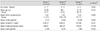
There were 3 men and 13 women among the 16 patients. Ages ranged from 22 to 82 years (62.32 ± 13.10 years, mean ± standard deviation), and the mean follow-up period was 3.88 ± 2.25 years. The mean endothelial cell density and pachymetry measurements of the 16 patients at initial examination were 1,413.91 ± 735.42/mm2 and 576.56 ± 74.04 mm, respectively. There were no statistical differences in age, sex, initial endothelial cell density, and initial pachymetry measurements among the 3 groups (Table 1).
First, we investigated the natural course of FCD among patients who had not undergone surgery. Non-linear mixed models for time-dependent changes in these patients showed that only pachymetric data tended to increase with statistical significance (p= 0.001), with a mean follow-up of 4.15 years (Table 2). The changes in ECC, CV, and hexagonality were not significant during these periods using the aforementioned model. This suggests that functional deterioration progresses over a period of 4 years without noticeable morphologic changes in endothelial cells.
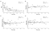
Next, we evaluated whether long-term endothelial changes were worse when FCD patients underwent cataract surgery only or penetrating keratoplasty with cataract surgery. Regression analysis showed that the mean rates of annual endothelial cell loss were 0.82%/yr, 20.39%/yr, and 29.27%/yr in groups A, B, and C, respectively, and the rate in group C showed statistical significance (p < 0.05) (Fig. 1A). Thus, only the patients who underwent penetrating keratoplasty and cataract surgery showed exponential endothelial cell loss over time. The mean rates of pachymetric change were -1.81%/yr, +1.21%/yr, and +1.89%/yr in groups A, B, and C, respectively, showing no statistical significance (Fig. 1B). The mean rates of change in CV were +1.94%/yr, +1.21%/yr, and +0.48%/yr in groups A, B, and C, respectively, showing no statistical significance (Fig. 1C). The mean rates of hexagonality change were -1.04%/yr, -8.06%/yr, and -1.19%/yr in groups A, B, and C, respectively, showing no statistical significance (Fig. 1D). There was no significant change in the logarithm of the minimum angle of resolution (logMAR) visual acuity of group A during the follow up period (p = 0.100, Wilcoxon rank-sum test). However, there was significant improvement of logMAR visual acuity in group B after surgery (from 0.642 to 0.235, p = 0.043, Wilcoxon rank-sum test). The logMAR visual acuity in group C showed a high tendency to deteriorate after surgery, but did not show any statistical significance (from 0.429 to 1.494, p = 0.068, Wilcoxon rank-sum test). Taken together, morphologic changes and functional deterioration were not aggravated in an exponential manner in either non-operated FCD patients or those with cataract surgery, but in those with penetrating keratoplasty combined with cataract surgery.
Finally, we checked the exponential changes in endothelium parameters in age and sex-matched non-FCD patients (mean age non-FCD patients, 59.0 ± 6.3 vs. FCD patients, 68.5 ± 5.5, p = 0.298; sex distribution, p = 0.428) who underwent penetrating keratoplasty and cataract surgery to indirectly compare these changes with those in FCD patients (Fig. 2). The mean rate of endothelial cell loss was 19.43% per year (with statistical significance) in non-FCD patients and 29.27% per year in FCD patients. Thus, endothelial density was exponentially reduced in both FCD and non-FCD patients who underwent penetrating keratoplasty and cataract surgery. The mean rates of pachymetric change were +0.13% per year with no significance in non-FCD patients and +1.89% per year in FCD patients. The mean rates of CVs change were +1.21% per year without any significance in non-FCD patients and +0.48% per year in FCD patients. The mean rates of hexagonality change were -0.48% per year without any significance in non-FCD patients and -1.19% per year in FCD patients. This implies that morphologic changes and functional deterioration were not exponentially worse in either group. No patients experienced corneal stromal edema during the study period.
Discussion
Our retrospective long-term follow-up data showed that endothelial density did not significantly decrease over the course of four years in middle-aged FCD patients, while changes in pachymetric corneal thickness appeared to increase during the same period. This suggested that endothelial function progressively deteriorated during this period, although the density and morphologic changes were not severely advanced. This outcome supports Seitzman's suggestion that the pachymetric index should be given more importance when determining whether cataract surgery should be performed in FCD patients [16].
In terms of the semi-quantitative view using exponential curve fitting regression analysis, the mean endothelial cell density loss rate was 1.4-times higher (0.82%/yr) in middle-aged FCD patients (mean age, 62 years) compared to that in the normal population (0.5%/yr) during a mean follow-up of 4.15 years [10]. In addition, the natural course of changes in endothelial density and morphology in FCD patients did not follow an exponential curve; this can provide relief for providers and patients during the long-term follow-up of patients in their fifties.
Another interesting finding was that cataract surgery did not significantly worsen the natural course of FCD patients, although the annual decrease in ECC increased. In addition, visual acuity significantly improved an average of 2 years after the phacoemulsification. This may be explained from the clinical impression that the majority of Korean patients had guttata confined to the central cornea, while those of Caucasians are distributed around the entire cornea. Relatively intact peripheral corneal endothelial cells may have facilitated endurance of the mechanical stress of phacoemulsification. Meanwhile, FCD patients who underwent both penetrating keratoplasty and cataract surgery experienced exponential decreases in endothelial density similar to those in non-FCD patients. Although we could not directly compare the loss rate between the groups due to small numbers, the annual decrease rates in endothelial density were higher in FCD patients than in non-FCD patients.
Whenever cataracts that disturb vision develop in FCD patients with moderate-to-low ECC densities, ophthalmologists have to decide whether cataract surgery should be immediately performed or postponed until keratoplasty is required. The better option for visual rehabilitation in FCD patients has long been controversial; it is unclear whether cataract surgery alone or cataract surgery combined with keratoplasty should be performed [18-20]. Our data indicated how fast the natural course of ECC changes progress in FCD patients with the intervention of cataract surgery. Compared with combined cataract surgery and keratoplasty, cataract surgery alone appeared to have some benefit in providing less astigmatic visual quality; nevertheless, cataract surgery alone exacerbated ECC loss. In addition, considering that no exponential changes were aggravated after performing cataract surgery alone, cataract surgery would be a preferable option in FCD patients compared to an approach of "wait-and-do" penetrating keratoplasty combined with cataract surgery.
The present study has two major limitations, namely the small population size and the wide variance in follow-up periods among the groups. Although these limitations weaken the statistical power of the present study, we believe it may be worthy of notice considering that it is a first report of long-term quantitative observation of endothelial cell change in FCD patients. Further prospective well-controlled studies with larger patient groups are needed.


 XML Download