

 PDF
PDF ePub
ePub Citation
 Print
Print


Conjunctivochalasis is defined as redundant, loose, nonedematous, inferior bulbar conjunctiva interposed between the globe and the lower eyelid [1], occurring in patients with ocular irritation, epiphora, blurred vision, dry eye, and subconjunctival hemorrhage [1-3]. First-line treatments include topical preservative-free artificial tears, steroids, and antihistamines, with surgery as necessary if symptoms do not resolve. The primary surgical method consists of crescent-shaped resection and re-approximation of redundant inferior bulbar conjunctiva [1]; several studies have demonstrated the general efficacy of this method [2,4]. However, this approach has several limitations, resulting in the development of modified surgical techniques, including amniotic membrane graft transplantation [5,6], scleral fixation of the redundant conjunctiva with stitches [7], and superficial cauterization using bipolar devices [8].
More recently, a surgical approach using a high-frequency radiowave electrosurgical unit was described [9]. This method minimizes heat dissipation and resultant cellular alteration, reducing postoperative pain and scar formation and facilitating wound healing [10,11]. These authors reported that this electrosurgical system produced successful results in conjunctivochalasis, with a shorter operative time but comparable clinical outcomes to standard surgical methods.
However, in practice, these investigators often encountered complaints of early postoperative pain in electrocauterized patients who had conjunctival epithelial defects in the electrocauterized area in the early postoperative days. We hypothesized that, despite good clinical outcomes, the high temperatures generated by the electrosurgical unit may induce greater than anticipated thermal damage in the subconjunctival tissue and overlying epithelial cells, inducing an inflammatory response. Therefore, we compared postoperative inflammation in an experimental conjunctivoplasty model utilizing high frequency radiowave electrocauterization with a procedure using simple excision and suturing.
Materials and Methods
Animals
Ten male New Zealand White rabbits, each weighing between 2.5 and 3.0 kg, were used in all experiments. Prior to surgery, the rabbits were anesthetized with an intramuscular injection of zolazepam (10 mg/kg) and xylazine hydrochloride (10 mg/kg). At the end of the study, all rabbits were sacrificed by an intravenous injection of 5 mL of a solution of sodium pentobarbital (65 mg/mL).
The study protocol (no. 11-0118) was approved by the institutional animal care and use committee of the Seoul National University Hospital. The study adhered to the principles of laboratory animal care (National Institutes of Health publication no. 85-32, revised 1985) and the requirements of the Association for Research in Vision and Ophthalmology statement on the use of animals in ophthalmic and vision research.
Animal model
The right eye of each rabbit underwent electrocauterization, whereas the left eye underwent excision and suturing. For each eye, we excised or electrocauterized an area of inferior bulbar conjunctiva, 1 mm in width and 6 mm in length, which was located 2 mm from the limbus.
Electrocauterization was performed as described previously [9]. Briefly, subconjunctival coagulation was performed using a high frequency radio wave electrosurgical unit (Ellman Surgitron 4.0 Dual RF; Ellman International, Hewlett, NY, USA) and a fine-needle electrode (004 Super Fine, Ellman International) at an intensity that would not char the conjunctiva. The output power in the coagulation mode was set at 1 to 2, corresponding to settings of 0.5 to 1 in the original description. Each eye underwent approximately 10 to 20 subconjunctival coagulations in a horizontal direction.
The corresponding conjunctival area was excised using Vannas scissors in the contralateral eye, followed by re-approximation and suturing with 10-0 Nylon, taking care to avoid tension and overrides of each conjunctival margin. Suture material was removed after 14 days. No topical or systemic medication was administered postoperatively to any animal.
Clinical outcomes
All eyes were examined by slit-lamp biomicroscopy and photographed at each follow-up examination. Baseline photographs were taken at the end of the surgery and after 5, 7, 14, and 21 days.
Cytokine quantification
Tears were collected using the Schirmer I method with filter paper (Schirmer tear test; Haag-Streit AG, Koeniz, Switzerland) [12] on the day of surgery and 5 and 21 days later, prior to all procedures, including intramuscular injection of anesthetics. Since cytokine concentrations in tears alter during the course of a day [13], we collected all tear samples from 9 to 11 AM in the morning to exclude any diurnal variations. Concentrations of interleukin (IL)-1β (CSB-E06900Rb; Cusabio Biotech, Hubei, China) and tumor necrosis factor (TNF)-α (DY5670; R&D Systems, Minneapolis, MN, USA) in tears were determined using an enzyme linked immunosorbent assay (ELISA). All samples were assayed in triplicate to ensure reproducibility of the data.
Histopathologic analysis
After 21 days, the rabbits were anesthetized, and rectangular sections of inferior conjunctiva, including the previously operated area at the center, were excised with Vannas scissors and placed onto nitrocellulose paper (Merck Millipore, Bedford, MA, USA) without anatomical distortion. The sections were fixed in Davison's solution for 24 hours, dehydrated in a series of graded alcohol solutions over the next 24 hours, and tissues were embedded in paraffin. Five micrometer sections were obtained with a microtome and stained with hematoxylin-eosin. Inflammatory cells were counted in five randomly selected fields (×200) of each hematoxylin-eosin stained slide. All animals were euthanized after harvesting conjunctival tissue.
Results
Clinical outcomes
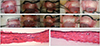
All eyes that underwent electrocauterization demonstrated smooth conjunctiva without any inflammation or scarring after 5 days. However, mild to moderate edema were observed in the eyes that underwent excision and suturing, until suture removal 14 days post-procedure. Moreover, scarring persisted in these eyes until euthanization at 21 days postoperatively (Fig. 1). Fluorescein staining under blue light revealed no evidence of epithelial damage in any eye.
Cytokine quantification
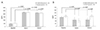
The baseline concentrations of TNF-α and IL-1β in tears were similar in the two groups (p > 0.05, Mann-Whitney U-test). In both groups, TNF-α concentration was significantly elevated after 5 days compared with baseline concentrations, decreasing thereafter but remaining significantly higher than baseline until 21 days post-operation (p = 0.005 and p = 0.005, respectively, in cauterization group; p = 0.005 and p = 0.007, respectively, in excision group; Wilcoxon signed rank test) (Fig. 2A). However, there were no significant differences in TNF-α concentration at each time point (p > 0.05, Mann-Whitney U-test) (Fig. 2A). In contrast, IL-1β concentration in tears did not change significantly in either group over time and did not differ between the two groups at each time point (p > 0.05, Mann-Whitney U-test and Wilcoxon signed rank test) (Fig. 2B).
Histopathologic analysis
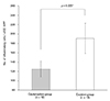
Similar to the clinical findings, our histological analysis revealed that stromal infiltration of inflammatory cells, consisting predominantly of neutrophils, was significantly higher in the eyes that underwent excision than those that underwent electrocauterization (190.4 ± 32.3 and 125.4 ± 16.9, respectively; p = 0.035, Mann-Whitney U-test) (Figs. 1 and 3).
Discussion
Our in vivo animal study demonstrated, contrary to our initial hypothesis, that electrocauterization for conjunctivoplasty tended not to be excessively inflammatory, being comparable to simple suturing and excision in terms of inflammation.
The conventional surgical technique in patients with conjunctivochalasis consists of removing crescent-shaped conjunctiva and closing the incision with a continuous suture [1,2,4]. Despite the success of this method, its limitations resulted in the development of modified techniques, including a recent approach using a high frequency radiowave electrosurgical unit [9]. Electrocauterization, however, risks scarring of Tenon's capsule, which could result in severe postoperative sequelae, including fornix shortening, diplopia, or conjunctival damage. High frequency radiowaves may encounter resistance when passing through tissue, producing heat that could boil intracellular water, increase intracellular pressure, and result in cell lysis [10]. Although heat dissipation and cellular alteration have been reported to be minimal in high frequency radiowave surgery, we often observed conjunctival epithelial defects on the first postoperative day, which lasted a few days, associated with complaints of pain during the early postoperative period. Therefore, we hypothesized that this heat may induce greater inflammation than expected, resulting in epithelial defects and thermal damage to underlying Tenon's tissues.
In the present rabbit model, we found that electrocauterization yielded better clinical outcomes than excision plus suturing, although chemosis was similar in the two groups within 5 days. Since the high frequency radiowave surgical unit generates high temperatures, we expected epithelial damage to the directly treated area of the conjunctiva during the acute postoperative period. Contrary to our clinical observations, biomicroscopy with fluorescein staining under blue light revealed no evidence of epithelial damage in rabbit on the first postoperative day. This implied that collateral tissue damage may be lower than expected with high frequency radiowave electrosurgical units, or possibly that there is more rapid epithelial healing in rabbits than in humans, as the wound size is smaller and wound healing may be more hyperproliferative in rabbits than in humans. Thus, this rapid epithelial healing in a rabbit model may affect the inflammation in the subtenon, differing from the human clinical situation.
To assess whether inflammation was associated with clinical outcomes, we also measured the concentration of tear inflammatory cytokines IL-1β and TNF-α, as well as evaluating histology. IL-1 is an important mediator of inflammation and immunity, inducing the expression of other important inflammatory cytokines, including IL-6, IL-8, TNF-α, and granulocyte-macrophage-colony stimulating factor. IL-1β also stimulates the proinflammatory response of conjunctival epithelial cells, and this response is augmented in the presence of TNF-α [14]. TNF-α is secreted by inflammatory leukocytes, including monocytes and eosinophils, as well as by epithelial cells. Moreover, TNF-α secreted in response to inflammation may upregulate the expression of adhesion molecules and chemokines, which are important in the activation and migration of other inflammatory leukocytes [14,15]. Therefore, we hypothesize that IL-1β and TNF-α are the primary inflammatory cytokines in eyes with postoperative inflammation following surgery for conjunctivochalasis. Additionally, we would have liked to investigate the level of substance P as well; however, an antibody against rabbit substance P is not available. We found that the baseline concentrations of IL-1β in rabbit eyes that underwent electrocauterization and surgery were 170.06 and 187.12 pg/mL, respectively, similar to concentrations in eyes of normal healthy humans (12.9 to 227.4 pg/mL) [13,16-18]. While the baseline concentrations of TNF-α were similar in rabbit eyes that underwent electrocauterization and surgery (490.26 and 565.08 pg/mL, respectively), these concentrations were higher than the TNF-α concentration in normal human tears (18.8 pg/mL) [13]. Following either electrocauterization or surgery, the concentration of IL-1β did not change significantly over time. In contrast, the concentrations of TNF-α in both groups increased significantly at 5 days postoperatively and remained higher than baseline through 21 days post-operation. These findings indicate that IL-1β expression by epithelial cells is not altered significantly with either type of conjunctivochalasis surgery. This may be due to the absence of epithelial defects in both groups, which would serve to minimize the epithelial wound healing process that results in IL-1β expression. Since TNF-α expression was enhanced in both groups without a change in IL-1β expression, the TNF-α likely derives from non-epithelial cells, including the reflex arc of the trigeminal and facial nerves, which are involved in the corneal wound healing process [19] but is not induced by IL-1β.
Finally, histologic examination confirmed that postoperative inflammation was significantly lower in eyes that underwent high frequency radiowave surgery than in eyes that underwent conventional excision surgery. Taken together with ELISA data, electrocauterization tends to be less inflammatory than simple excision and suturing or is at least comparable to that technique.
In conclusion, our results demonstrated that electrocauterization for conjunctivoplasty using high frequency radiowave electrosurgery may be advantageous in terms of inflammation compared with the conventional excision and suture technique.
 XML Download