Journal List > Korean J Ophthalmol > v.26(6) > 1024955

Kim, Kim, and Kee: Optic Disc Atrophy in Patient with Posner-Schlossman Syndrome

Abstract

A 32-year-old man with blurred vision in the right eye and headache presented with anterior uveitis, an intraocular pressure (IOP) of 60 mmHg, an open angle, no visual field defects, and normal optic nerve. He had a history of five previous similar attacks. In each of the previous instances, his anterior uveitis and high IOP were controlled with antiglaucoma medications and topical steroids. However, at the fifth attack, his optic disc was pale and a superior paracentral visual field defect was shown. Brain magnetic resonance image studies were normal. This case represents that a recurrent Posner-Schlossman syndrome (PSS)-induced optic disc atrophy likely due to ocular ischemia caused by a recurrent, high IOP. Although PSS is a self-limiting syndrome, we should manage high IOP and prevent ischemia of the optic nerve head by treating with ocular antihypertensive medications.

Posner-Schlossman syndrome (PSS) is a self-limiting and benign condition characterized by unilateral, recurrent episodes of markedly elevated intraocular pressure (IOP) with mild idiopathic anterior uveitis, open angles, no visual fields defect, and normal optic discs [1]. However, an association between primary open-angle glaucoma (POAG) and PSS, with about 45% of PSS patients having an accompanying POAG, was shown [2,3]. Another complication of PSS, nonarteritic anterior ischemic optic neuropathy (NAION) was also reported [4,5]. We report a case of optic disc pallor which is different from glaucomatous optic neuropathy and NAION occurring after recurrent episodes of marked elevated IOP secondary to PSS.

Case Report

The patient was seen in 1996 at the age of 32, with blurred vision in the right eye and headache. He had no previous medical history, including diabetes and hypertension or trauma history. He was using no systemic medications. His vision was 0.9 in the right eye, 1.0 in the left eye. There was mild epithelial edema of the cornea with a few keratic precipitates and no cells seen on slit-lamp examination of the right eye. The IOP was 60 mmHg in the right eye. Gonioscopy showed an open angle. The left eye was normal. He had five episodes of PSS crises over the next 14 years, with a highest IOP of 60 mmHg. Anterior segment inflammation and elevated IOP were controlled after few weeks of treatment with antiglaucoma medication and topical corticosteroids. This was his treatment course after each PSS event.
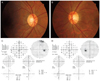
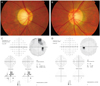
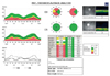
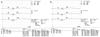
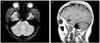
At his first medical examination in 1996, his cup/disc ratio was noted to be 0.4 in the right eye and 0.4 in the left eye. Visual fields were normal (Fig. 1A and 1B). After his fifth attack, in 2010, his uncorrected visual acuity was 1.0 in both eyes and a relative afferent papillary defect of right eye was observed. Color vision was normal. His right optic disc was pale and the field loss in the right eye had progressed to a superior paracentral scotoma (Fig. 2A and 2B). Retinal nerve fiber thickness of the right eye was generally reduced (Fig. 3). There was P100 delay of the right eye in the visual evoked potential test (Fig. 4). There were no abnormal findings on brain magnetic resonance image (Fig. 5).

Discussion

PSS is a syndrome associated with recurrent anterior segment inflammation and elevated IOP. After an attack, IOP and facility of aqueous outflow return to normal. The etiology of PSS is unknown, although abnormal vascular process, autonomic defect, allergic conditions, variation of developmental glaucoma, association of HLA-Bw54, cytomegalovirus, and herpes simplex virus have been suggested [6-8].
There were case reports of PSS and NAION [4,5]. The presumed mechanism causing NAION was decreased perfusion to the optic disc secondary to the rise in intraocular pressure. Risk factors for NAION such as old age, small cup to disc ratio, and hypertension were considered when treating PSS patients.
This is case of optic disc pallor in a patient with recurrent anterior segment inflammation and high intraocular pressure. Optic disc perfusion is directly proportional to mean arterial pressure and inversely proportional to IOP [9]. We had not found something that explained optic disc pallor excluding high IOP. Decreased optic nerve perfusion by recurrent high IOP might induce optic disc pallor.
In conclusion, PSS could cause optic disc changes such as glaucomatous change and ischemic change. Thoughtful attention and aggressive IOP control are required especially in PSS patients with prolonged periods of high intraocular pressure and recurrent attacks.

Figures and Tables

Fig. 1
At that time of first attack, optic discs (A,B) and visual fields (C,D) of both eyes appeared normal. GHT = glaucoma hemifield test; VFI = visual field index; MD = mean deviation; PSD = pattern standard deviation.
kjo-26-473-g001
Fig. 2
After his fifth attacks, the right optic disc (A) was pale and the left optic disc (B) showed no change. The field defect in the right eye (C) had progressed to a superior paracentral scotoma and that of the left eye (D) had no change. GHT = glaucoma hemifield test; VFI = visual field index; MD = mean deviation; PSD = pattern standard deviation.
kjo-26-473-g002
Fig. 3
The retinal nerve fiver thickness of right eye was generally reduced compared with that of the left eye. RNFL = retinal nerve fiber layer; TEMP = temporal; SUP = superior; NAS = nasal; INF = inferior; OD = right eye; OS = left eye; Imax = inferior maximum; Smax = superior maximum; Tavg = temporal average; Navg = nasal average; Max-Min = the difference value between maximum and minimum thickness; Savg = superior average; Iavg = inferior average.
kjo-26-473-g003
Fig. 4
Visual evoked potential showed P100 delay in the right eye (A) compared with that of the left eye (B).
kjo-26-473-g004
Fig. 5
Brain magnetic resonance image showed no abnormal findings (A,B).
kjo-26-473-g005

Notes

This article was presented in part as a poster at the 104th autumn meeting of Korean Ophthalmological Society, Goyang, Korea, November 2010.

No potential conflict of interest relevant to this article was reported.

References

1. Posner A, Schlossman A. Syndrome of unilateral recurrent attacks of glaucoma with cyclitic symptoms. Arch Ophthal. 1948. 39:517–535.
2. Raitta C, Vannas A. Glaucomatocyclitic crisis. Arch Ophthalmol. 1977. 95:608–612.
3. Kass MA, Becker B, Kolker AE. Glaucomatocyclitic crisis and primary open-angle glaucoma. Am J Ophthalmol. 1973. 75:668–673.
4. Kim R, Van Stavern G, Juzych M. Nonarteritic anterior ischemic optic neuropathy associated with acute glaucoma secondary to Posner-Schlossman syndrome. Arch Ophthalmol. 2003. 121:127–128.
5. Irak I, Katz BJ, Zabriskie NA, Zimmerman PL. Posner-Schlossman syndrome and nonarteritic anterior ischemic optic neuropathy. J Neuroophthalmol. 2003. 23:264–267.
6. Hirose S, Ohno S, Matsuda H. HLA-Bw54 and glaucomatocyclitic crisis. Arch Ophthalmol. 1985. 103:1837–1839.
7. Teoh SB, Thean L, Koay E. Cytomegalovirus in aetiology of Posner-Schlossman syndrome: evidence from quantitative polymerase chain reaction. Eye (Lond). 2005. 19:1338–1340.
8. Yamamoto S, Pavan-Langston D, Tada R, et al. Possible role of herpes simplex virus in the origin of Posner-Schlossman syndrome. Am J Ophthalmol. 1995. 119:796–798.
9. Hayreh SS. Anterior ischemic optic neuropathy. Arch Neurol. 1981. 38:675–678.
TOOLS
Similar articles