Abstract
Trisomy 14 mosaicism is a rare chromosomal abnormality with distinct and recognizable clinical features. We report a patient with presumed retinal dystrophy having diffuse retinal pigment epithelial abnormalities, which has not been previously reported in association with trisomy 14. This case expands the clinical spectrum of this rare entity.
Trisomy 14 mosaicism has been associated with multiple ophthalmologic abnormalities, including hypo- and hypertelorism, downward slanting of the palpebral fissure, blepharoptosis, and microophthalmia [1-4]. This is the first report of congenital retinal pigment epithelial abnormalities in association with this syndrome.
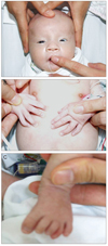
This female infant was the 3,200 g product of a 38-week gestation delivered by spontaneous vaginal delivery to a 30-year-old mother. The mother's pregnancy was uncomplicated, and the family history was unremarkable. At birth, the baby was noted to have a prominent forehead, narrow palpebral fissure, hypertelorism, and a broad nose (Fig. 1A). Skeletal survey showed bilateral hypoplastic first ribs, right-sided fifth finger clinodactyly, and prominent long phalanges of the fourth digits on both feet (Fig. 1B and 1C). She was suspected to have a chromosomal anomaly and was referred to the ophthalmology department for evaluation of possible ocular abnormalities. On examination at two weeks of age, she had weak pupillary response to light but no blink or avoidance response to strong light. With regard to the horizontal length of the palpebral fissure, the right and the left were 1.7 and 1.6 cm, respectively, which was considered short. Telecanthus and hypertelorism were also present; intercanthal distance was 2.8 cm, and interpupillary distance was 4.3 cm (Fig. 2). Slit-lamp examination of both eyes revealed normal anterior segments, and dilated fundus examination revealed normal optic nerves and retinal vasculature. Retinal pigment epithelial mottling and atrophy in the macula of both eyes with diffuse retinal pigmented epithelium drop-out in the mid and periphery were observed (Fig. 3). After a three-month lapse from the original examination, fix and follow behavior was not consistent in either eye, and there were no interval changes to the lesions in the retina. Continued observation after this was no longer possible due to follow-up loss.
An echocardiogram demonstrated a small ventricular septal defect and patent ductus arteriosus, and an abdominal ultrasound showed a normal sized liver and kidneys with normal echogenicity. Chromosomal analysis of peripheral blood revealed a mosaic complement with trisomy 14 (47,XX,+14) and a normal female complement (46,XX) (Fig. 4).
Neither parent had a significant past medical history nor a family history of ophthalmologic issues, and both had normal fundus findings. Also, the results of chromosomal analysis were normal in peripheral blood.
Although complete trisomy 14 is not compatible with postnatal life, trisomy 14 mosaicism has been diagnosed in newborns and children with multiple congenital anomalies [1,2]. This chromosomal abnormality was first described in 1970, and there are several characteristic features which may enable the clinician to suspect this diagnosis prior to obtaining a chromosome analysis [1]. The most common features are growth and psychomotor retardation, dysmorphic craniofacial features such as abnormal or low-set ears, micrognathia, cleft or highly arched palate, short neck, and congenital heart and genitourinary abnormalities [2]. Ophthalmologic features include hypo- and hypertelorism, downward slanting of the palpebral fissure, blepharoptosis, deep-set eye, eversion of the eyelids, a "evanescent translucent film over the eyes," and microophthalmia [1-4].
Retinal pigmentary dystrophy was not previously described in any reports of this chromosomal abnormality. In other chromosomal abnormalities such as trisomy 21 (Down's syndrome) and trisomy 18 (Edward's syndrome), retinal pigment epithelium abnormalities are reported to be a clinical feature. In trisomy 21, focal hyperplasia of the retinal pigment epithelium has been reported, and retinal depigmentation is reported to be a clinical feature in trisomy 18 [5,6].
In mosaic trisomy 14, several other more unusual features have been described but are seen in a smaller percentage of patients. One such feature is abnormal skin pigmentation, which is a common finding in individuals with mosaic chromosomal abnormality. In trisomy 14 mosaicism, hyper- and hypopigmentation of the skin have been reported, so chromosome 14 is supposed to be related to pigmentation of the epithelium. The patient we describe did not exhibit abnormal skin pigmentation at birth, yet this was evident by three months [7,8].
Howard et al. [9] have reported a patient who had a ring 14 with a terminal deletion but no retinal pigmentation. They suggested that a region on chromosome 14 proximal to q32.2 might be involved in controlling these changes.
Further electrophysiologic testing would be helpful to determine if there are any abnormalities of either the rods or cones in association with trisomy 14 mosaicism, though neither has been previously reported. Unfortunately, electroretinogram testing was not possible in this patient. Yet, the lesion of the retina persisted for three months; lesions both in the periphery as well as in the posterior pole should be considered as retinal dystrophy. We expand the clinical spectrum of this rare entity with dysmorphic features and ocular abnormalities to include retinal dystrophy.
Figures and Tables
 | Fig. 1Photographs of the general appearance in mosaic trisomy 14 syndrome at 2 weeks of age. The patient had a prominent forehead, narrow palpebral fissure, and broad nose (A), right-sided fifth finger clinodactyly (B), and prominent long phalanges of the fourth digits on both feet (C). Informed consent was received from parents of the patient. |
 | Fig. 2Photographs of external ocular appearance in mosaic trisomy 14 syndrome at 2 weeks of age. The patient had a narrow palpebral fissure with hypertelorism and telecanthus. |
References
1. Johnson VP, Aceto T Jr, Likness C. Trisomy 14 mosaicism: case report and review. Am J Med Genet. 1979. 3:331–339.
2. Fujimoto A, Allanson J, Crowe CA, et al. Natural history of mosaic trisomy 14 syndrome. Am J Med Genet. 1992. 44:189–196.
3. Lynch MF, Fernandes CJ, Shaffer LG, Potocki L. Trisomy 14 mosaicism: a case report and review of the literature. J Perinatol. 2004. 24:121–123.
4. Von Sneidern E, Lacassie Y. Is trisomy 14 mosaic a clinically recognizable syndrome? Case report and review. Am J Med Genet A. 2008. 146A:1609–1613.
5. Ginsberg J, Ballard ET, Buchino JJ, Kinkler AK. Further observations of ocular pathology in Down's syndrome. J Pediatr Ophthalmol Strabismus. 1980. 17:166–171.
6. Ginsberg J, Bove K, Nelson R, Englender GS. Ocular pathology of Trisomy 18. Ann Ophthalmol. 1971. 3:273–279.
7. Tunca Y, Wilroy RS, Kadandale JS, et al. Hypomelanosis of ito and a 'mirror image' whole chromosome duplication resulting in trisomy 14 mosaicism. Ann Genet. 2000. 43:39–43.
8. Kuster W, Konig A. Hypomelanosis of Ito: no entity, but a cutaneous sign of mosaicism. Am J Med Genet. 1999. 85:346–350.
9. Howard PJ, Clark D, Dearlove J. Retinal/macular pigmentation in conjunction with ring 14 chromosome. Hum Genet. 1988. 80:140–142.


 PDF
PDF ePub
ePub Citation
 Print
Print


 XML Download