

 PDF
PDF ePub
ePub Citation
 Print
Print


Pupillary dilation is an essential component of ophthalmologic tests and treatments. Magnitude of dilation depends on the sphincter muscle of the pupil, controlled by parasympathetic nerves, and on the dilator muscle of the pupil, controlled by sympathetic nerves.1,2 The parasympathetic antagonist tropicamide and the sympathetic agonist phenylephrine are frequently used to achieve dilation in the clinical setting.
In studying these two eye drops, Siderov and Nurse3 reported that twice the normal dose of 0.5% tropicamide achieved a larger pupil size than a single dose. Krumholz et al.4 reported no significant difference in pupil size among 3 different eye drops regimens: 0.5% tropicamide and 2.5% phenylephrine, 0.5% tropicamide and 1.25% phenylephrine, and 0.25% tropicamide and 1.25% phenylephrine. Since diluted eye drops may still have clinically significant pupillary dilation effect, they may still be useful when combined with eye drops having a different mechanism of action.
The current study evaluated the mydriatic effect of different types of eye drops and investigated their efficacy through delivering multiple drops of a single drug and through combining eye drops with different mechanisms of action.
Materials and Methods
We studied 100 eyes in 50 patients requiring pupillary dilation as part of their visit to the Department of Ophthalmology at Dongsan Medical Center, Keimyung University, between July and September 2007. Given the possible side effects of phenylephrine, patients with a history of cardiac disease, high blood pressure, ophthalmologic surgery possibly affecting the pupil size, eye trauma, diabetes, or more than 0.25 mm of anisocoria before pupillary dilation were excluded from the study.
The subjects were divided into 3 groups. Group 1 received one drop of 1% tropicamide (Mydriacyl®, Alcon Co.,USA) in the right eye and one drop of 2.5% phenylephrine (Mydfrin®, Alcon Co., USA) in the left eye. Group 2 received two drops of 1% tropicamide in the right eye, delivered 5 minutes apart, and one drop each of 2.5% phenylephrine and 1% tropicamide in the left eye, delivered 5 minutes apart. Group 3 received two drops of 2.5% phenylephrine in the right eye and one drop each of 2.5% phenylephrine and 1% tropicamide in the left eye, delivered 5 minutes apart. To monitor mydriatic effect by age, each group was divided into a subgroup less than 14 years of age (A) and a subgroup greater than 15 years of age (B). There were 10 patients in group 1, and 20 patients each in groups 2 and 3. Group 1 included 5 patients in both the subgroup A and B, group 2 included 10 patients in both the subgroup A and B, and group 3 included 10 patients in both the subgroup A and B.
To prevent the dilution of the eye drops through tearing, 0.5% proparacaine hydrochloride (Alcaine®, Alcon Co., USA) was delivered to each eye 5 minutes before administering the dilating drugs. All eye drops were placed within the conjunctival sac of the lower eyelid, and patients were directed to close their eyes approximately 1 minute after drop delivery to prevent loss of medication through the punctum in the conjunctival sac.
The pupil size was photographed by a single evaluator using a Zeiss FF 450 camera (Carl Zeiss Meditec, Jena, Germany) before the use of eye drops and at 5, 10, 15, 18, 21, 24, 27, 30, 35, and 40 minute after eye drops administration. For calculating pupil size, the VISUPAC program was used. Three repeated measurements were taken within an interval of a few seconds at each time point, and the mean value of the maximum horizontal diameter and the maximum vertical diameter of the pupil was recorded as the pupil size. Through applying built-in illumination grade II and flash 17, the measurement conditions for all test subjects were the same. Pupil size greater than 6 mm was considered to be clinically effective for ophthalmological testing. A maximum pupil size less than 6.0 mm was regarded as ineffective pupillary dilation.5,6
For statistical analysis, SPSS 12.0 was used to perform Wilcoxon signed ranks test. Informed consent was acquired from all patients using a form approved by the Institutional Review Board of the above medical center.
Results
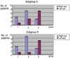
Patient age ranged from 3 years to 41 years, with a mean of 15.7 years. Thirty patients were male, and 20 patients were female. The mean pupil sizes before eye drops administration were 4.26±0.41 mm, 4.06±0.6 mm, and 4.0±0.49 mm in groups 1, 2, and 3, respectively (Table 1). The maximum pupil size was defined as the maximum pupil size recorded during the 40 minutes following eye drops administration, the time needed to reach the maximum pupil size was similar in all groups examined (Table 2). In 8 of the 10 patients in group 1, the maximum pupil size was larger in the 1.0% tropicamide eye compared to the 2.5% phenylephrine eye (p=0.016). Sixteen patients in group 2 had a larger pupil in the two-fold 1.0% tropicamide eye than in the combined 2.5% phenylephrine and 1% tropicamide eye (p=0.081). Fourteen patients in group 3 had a larger pupil in the combined 2.5% phenylephrine and 1% tropicamide eye than in the two-fold 2.5% phenylephrine eye (p=0.009). Figure 1 compares the maximum pupil sizes among the groups. No significant difference was found on comparison of the subgroup A and B within the 3 groups.
Seven eyes achieved a maximum pupil size less than 6.0 mm: 3 eyes in group 2 (B), 2 eyes in group 3 (A), 1 eye in group 1 (B), and 1 eye in group 2 (A). One patient in group 2 (B) experienced pupil diameter less than 6.0 mm in both eyes. Investigation demonstrated that this patient was wearing soft contact lenses in both eyes at the time of eye drops administration. This situation did not confound the statistical analysis.
Discussion
Tropicamide 1.0% and phenylephrine 2.5% eye drops are frequently used for the purpose of cycloplegic refraction, for fundus examination and photocoagulation, and for preventing the formation of posterior iris synechiae in uveitis. Dilation is also essential in preparation for several intraocular procedures, such as cataract extraction. Phenylephrine is a sympathetic agonist that shows mydriatic effect through direct action on sympathetic nerve receptors located on the pupillary dilator muscle of the iris. Phenylephrine rarely causes complications in the cardiovascular and autonomic nervous system.7,8
We undertook this study to determine whether tropicamide, with a different mydriatic mechanism than phenylephrine, is capable of achieving enough pupillary dilation as an alternative eye drops.
Twa et al9 and Pop et al10 used a computer program to determine pupil size after photographing the anterior segment. This proved to be a precise and economical test method with higher repeatability than pupil measurement using the Colvard pupillometer or ruler. The current study also measured pupil size using photographs of the anterior segment.
Jung and Kim11 measured the pupil size every 5 minutes for 90 minutes after treatment with 1.0% tropicamide and 2.5% phenylephrine to compare pupillary dilation in patients with diabetic retinopathy. They reported that larger maximum pupil size was achieved through using 2.5% phenylephrine. This suggests a reduced effect of anticholinergic mydriatic drugs as a result of the neuropathy of the sympathetic innervation caused by the derangements in the sympathetic nervous system. The current study compared the pupillary dilation effects of two different types of eye drops in a normal population, regardless of age. We found larger maximum pupil size in eyes administered 1.0% tropicamide.
In 16 patients in group 2, we found that the pupil size of the eye treated with multiple drops of 1.0% tropicamide was larger than that of the eye treated with combined use of 2.5% phenylephrine and 1% tropicamide. It is difficult to conclude that multiple drops of 1.0% tropicamide are more effective than combination eye drops due to the lack of a statistically significant difference between the groups.
Smith and Smith12 reported that eye drops with anticholinergic effect block the parasympathetic nervous system and paralyze contraction of the sphincter muscle of the pupil, leading to an unmasking of the sympathetic nervous system effect on the pupillary dilator muscle. In the current study, the combined use of 2.5% phenylephrine and 1% tropicamide in group 3 resulted in larger pupil size compared to two-fold treatment with 2.5% phenylephrine. Therefore, it is believed that combined use of 1% tropicamide and 2.5% phenylephrine was more effective than use of 2.5% phenylephrine alone.
Concerning limitations of the current study, there were statistically significant differences among the 3 groups, but the differences were mostly among patients that achieved a pupil size larger than 6.0 mm. This is an acceptable size for conducting general fundus examination. Therefore, the differences were not significant from a clinical perspective. Furthermore, since the study population was small, a statistically significant analysis could not be made when analyzing mydriatic effect by age. The current study followed the pupil size for 40 minutes after eye drops administration. Future complementary studies will need to consider patients that show a delayed medication effect beyond 40 minutes.
In summary, 1.0% tropicamide eye drops showed better mydriatic effect than 2.5% phenylephrine eye drops, and combination 1% tropicamide and 2.5% phenylephrine were found to be superior to multiple drops of 2.5% phenylephrine in achieving pupillary dilation. Although combination eye drops were found to be inferior to multiple drops containing 1% tropicamide, the difference between the two groups was not statistically significant. Nevertheless, it is believed that combination eye drops will produce a better outcome in enhancing the mydriatic effect than multiple drops of single eye drops.


 XML Download