Journal List > Korean J Ophthalmol > v.23(4) > 1024688

Kim, Lee, Lee, and Yoon: Valsalva Retinopathy Associated With an Oratorical Contest

Abstract

A 17-year-old man presented to us with a chief complaint of decreased visual acuity accompanied by central scotoma. There was nothing unusual in his medical history other than a recent oratorical contest. At the time of initial diagnosis, the corrected visual acuity was 20/20 in the right eye and 20/100 in the left eye. No significant findings were apparent on ophthalmic evaluation. On fundoscopy, there was a dumbbell-shaped macular bleed with a well-defined margin in the left eye. The clinical course was closely monitored along with drug therapy. Four weeks post presentation, the pre-retinal hemorrhage had nearly resolved. On fluorescein angiography, no significant findings were observed. In the left eye, the corrected visual acuity had improved to 20/25. Valsalva retinopathy is a pathology that occurs when a sudden increase in intra-thoracic pressure or abdominal pressure occurs in an otherwise healthy person. Here we report a case of Valsalva retinopathy occurring following an oratorical contest along with a review of the relevant literature.

Valsalva retinopathy, first described by Duane in 1972, is caused by a sudden increase in intra-thoracic or abdominal pressure. Most cases have been reported to follow a benign course where the visual acuity is recovered as the hemorrhage is absorbed.1
Valsalva retinopathy may occur during various activities, including aerobic excercise,2 excessive sexual intercourse,3 blowing a bubble,4 prostate surgery,5 dental surgery,6 vomiting, as a result of constipation in pregnant women,7 carrying heavy loads, receiving endotracheal intubation, severe coughing,8 colonoscopy,9 and LASIK surgery.10 In Korea, valsalva retinopathy has been reported to occur as a result of lifting a barbell,11 blowing into a rubber glove,12 and strenuous exercise.13

Case Report

A 17-year-old man presented to us with a chief complaint of the sudden onset of decreased visual acuity, accompanied by central scotoma, which developed in the left eye. There was no significant past medical or surgical history. No relevant ophthalmologic history was noted. At the time of initial diagnosis, the corrected visual acuity was 20/20 in the right eye and 20/100 in the left eye. Intraocular pressure was 13 mmHg bilaterally. A slit lamp exam demonstrated no significant findings in the anterior segment including the cornea, anterior chamber, and anterior vitreous body.
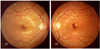
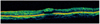
On fundoscopy, there was a dumbbell-shaped macular bleed with a well-defined margin in the left eye (Fig. 1). Optical coherence tomography demonstrated that a highly reflective shadow was present in the macular area (Fig. 2). There was nothing unusual in the patient history other than a recent oratorical contest. The clinical course was closely monitored, and drug therapy was initiated.
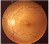
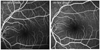
One month after presentation, the patient had an improved uncorrected visual acuity of 20/20 in the right eye and 20/25 in the left eye. On fundoscopy, the pre-retinal bleeding observed at the time of initial diagnosis had resolved (Fig. 3). Fluorescein angiography demonstrated no significant findings (Fig. 4).

Discussion

The Valsalva maneuver decreases venous return to the heart by exhaling against a closed glottis; this leads to an increase in abdominal and intrathoracic pressures. The decreased venous markedly lowers cardiac output. Inspiration with a closed glottis increases venous return. This markedly increases the cardiac output within several minutes, thus increasing arterial pressures.1
The venous system of the head and neck lack a functional valvular system. Therefore, if abdominal or intrathoracic pressures are suddenly raised with a closed glottis, this pressure increase would be directly transferred to the vascular system of the head and neck.
This increase in arterial pressure has effects on the eye. A marked increase in intraocular pressure provokes spontaneous rupture of capillary vessels surrounding the central fossa. This can result in a loss of visual acuity in otherwise healthy eyes.14 The resulting retinal hemorrhage spontaneously resolves within several weeks to several months in most cases and is not accompanied by notable complications.15
In the current case, retinal bleeding occurred in a 17-year-old man who had no significant past history, immediately following an oratorical contest. Presumably, an excessive amount of oration caused a sudden increase in venous pressure, resulting in valsalva retinopathy.

Figures and Tables

Fig. 1
A fundus photo (OU) obtained at the initial examination. This photo of the left eye demonstrates a preretinal hemorrhage at the fovea.
kjo-23-318-g001
Fig. 2
Optical coherence tomography (OCT) finding (OS) at the initial examination. OCT findings demonstrate a high reflectivity lesion at the macula, consistent with a preretinal hemorrhage.
kjo-23-318-g002
Fig. 3
A fundus photo (OS) obtained four wk after the initial presentation. The previous preretinal hemorrhage on the macula has mostly resolved.
kjo-23-318-g003
Fig. 4
Fluorescein angiography findings (OS) four wk after the initial presentation. The fluorescein angiography was unremarkable.
kjo-23-318-g004

Notes

This paper was presented as a poster at the 97th Korean Ophthalmologic Society Meeting.

References

1. Jones WL. Valsalva maneuver induced vitreous hemorrhage. J Am Optom Assoc. 1995. 66:301–304.
2. Roberts DK, MacKay KA. Microhemorrhagic maculopathy associated with aerobic exercise. J Am Optom Assoc. 1987. 58:415–418.
3. Friberg TR, Braunstein RA, Bressler NM. Sudden visual loss associated with sexual activity. Arch Ophthalmol. 1995. 113:738–742.
4. Georgiou T, Pearce JA, Taylor RH. Valsalva retinopathy associated with blowing balloons. Eye. 1999. 13:686–687.
5. Fanin LA, Thrasher JB, Mader TH, Truxal AR. Valsalva retinopathy associated with transrectal prostate biopsy. Br J Urol. 1994. 74:391–392.
6. Krepler K, Wedrich A, Schranz R. Intraocular hemorrhage associated with dental implant surgery. Am J Opthalmol. 1996. 122:745–746.
7. Callender D, Beirouty ZA, Saba SN. Valsalva hemorrhagic retinopathy in a pregnant woman. Eye. 1995. 9:808–809.
8. Duanne TD. Valsalva hemorrhagic retinopathy. Trans Am Oph Soc. 1972. 70:298–313.
9. Oboh AM, Weilke F, Sheindlin J. Valsalva retinopathy as a complication of colonoscopy. J Clin Gastroenterol. 2004. 38:793–794.
10. Moshfeghi AA, Harrison SA, Reinstein DZ, Ferrone PJ. Valsalva-like retinopathy following hyperopic laser in situ keratomileusis. Ophthalmic Surg Lasers Imaging. 2006. 37:486–488.
11. Jung EY, Kim IJ, Lee EC. A case of recurrent valsalva retinopathy associated with exercising a barbell. J Korean Ophthalmol Soc. 2004. 45:1040–1044.
12. Lee CH, Kim HK, Kim HC. Valsalva Retinopathy Associated with Blowing up of a Rubber Glove. J Korean Ophthalmol Soc. 2002. 43:1095–1099.
13. Kim DK, Lee WJ, Cho MS, Kim JS. A case of atypical valsalva retinopathy after strenuous exercise. J Korean Ophthalmol Soc. 2005. 46:915–920.
14. Gass JD. Stereoscopic Atlas of Macular Diseases: Diagnosis and Ttreatment. 1997. Vol. 2:4th ed. St Louis: Mosby;737–774.
15. Choi SW, Lee SJ, Rah SH. Valsalva retinopathy associated with fiberoptic gastroenteroscopy. Can J Ophthalmol. 2006. 41:491–493.
TOOLS
Similar articles