

 PDF
PDF ePub
ePub Citation
 Print
Print


Ophthalmic manifestations of multiple myeloma are rare.1 Among these, neuro-ophthalmic manifestations are the commonest form of ophthalmic presentation, most frequently appearing as dysfunction of cranial nerves 2, 3 or 6. Sixth cranial nerve palsy is the most common cranial of these; however, it has rarely been described as the initial presenting feature of multiple myeloma.2,3 Herein, we report a case of fluctuating sixth nerve palsy as the first manifestation of multiple myeloma.
Case Report
A 63-year-old woman complained of horizontal diplopia of two weeks duration. She reported that the diplopia did not have a fixed pattern and was not aggravated by fatigue. There was no past ophthalmic or medical history of note.
Upon first examination, her corrected visual acuity was 20/20 in both eyes. Her external ocular examination, intraocular pressures, and pupillary light reactions were all normal. The extraocular motility examination revealed abnormal abduction of the left eye, which never reached beyond the midline of the orbit. She had a left esotropia of 50-prism diopters (PD) at nearin primary position, 80PD in the left gaze, and 30PD in the right gaze (Fig. 1). The right ocular movement was not disturbed in any direction. One day later, she could abduct the left eye beyond the midline and had a left esotropia of 35PD at near in primary position, 50PD in the left gaze, and 14PD in the right gaze (Fig. 2), which showed improved abduction of the left eye (Fig. 2).
Two weeks later, the motility examination again revealed a complete inability to abduct the left eye beyond the midline. Systemic examination was unremarkable.
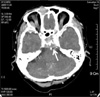
Serum acetylcholine receptor binding antibody and Tensilon test were negative. A computed tomography (CT) scan of the brain revealed multiple, enhancing, soft tissue, mass-like lesions involving the left cavernous sinus and the apex of both petrous bones (Fig. 3).
Serum protein electrophoresis revealed two monoclonal components; IgAκ and free κ light chains. Bence-Jones protein was detected in urinary protein electrophoresis (1.4 g/l, with 83% of free κ light chains). Bone marrow biopsy revealed a hypercellular marrow with diffuse infiltration of atypical plasma cells, which accounted for about 60% of the total marrow cellularity. On the basis of these findings, a diagnosis of IgAκ-type multiple myeloma was made.
Because of financial concerns, the patient refused treatment.
Discussion
Fluctuating weakness of extraocular muscles is known to be associated with myasthenia gravis. Previous reports have also documented fluctuating cranial nerve palsy due to an expanding basilar artery aneurysm,4 cerebellar astrocytoma,5 and cavernous sinus meningioma.6 However, we are not aware of any previous English-language reports of a case of fluctuating sixth nerve palsy associated with multiple myeloma, nor could we find any such references in a computerized search utilizing MEDLINE.
The mechanism of sixth cranial nerve palsy in multiple myeloma may include the following: hematologic effect, compression, meningeal metastasis, or direct infiltration of the nerve itself.1 One possible explanation is that the mobility of this nerve within the cavernous sinus probably allowed for the fluctuation of symptoms in this patient. According to the movement of the sixth cranial nerve, the degree of compression might vary. Alternatively, it is assumed that the cystic lesion would present in the neighborhood of sixth cranial nerve. A fluctuating sign may be attributed to the varying pressure inside cystic lesion.
The best assessment of the cavernous region is achieved by magnetic resonance imaging (MRI).7 Unfortunately, MRI of brain was not performed in this case because of financial concerns.
Generally, the prognosis of multiple myeloma associated with intracranial lesions is poor. Various treatments have been tried, including intravenous or intrathecal chemotherapy and cranial irradiation. However, responses are usually partial and short-lived, with results achieved only after irradiation.7
In summary, this fluctuating sixth cranial nerve palsy is a first manifestation of multiple myeloma. We suggest that multiple myeloma should be included in the differential diagnosis of any fluctuating sixth cranial nerve palsy. Although ophthalmic signs are rare and generally occur late in the course of multiple myeloma, they can still be its first signs.


 XML Download