

 PDF
PDF ePub
ePub Citation
 Print
Print


Some patients with posterior blowout fractures have been identified who exhibit hypertropia on the affected side and also appear to exhibit significant limitations in downgaze. However, the mechanism underlying hypertropia in orbital floor fractures remains a matter of some controversy. This motility disturbance can be explained by paralysis of the inferior rectus (IR) muscles1 or by orbital floor fractures which are located posterior to the equator of the globe.2-3
Nardi4 suggested that hypertropia subsequent to blowout fractures may be attributable to the fused common sheath of the inferior rectus (IR) and inferior oblique (IO) muscles. Therefore, if the IR loops downward after a blowout fracture, it may drag and stretch the IO, resulting in hypertropia.
We are unaware of any previous reports on IO entrapment, or on the results of IO-weakening surgery subsequent to posterior blowout fracture in the literature. Here, we describe a patient who manifested hypertropia and excyclotropia associated with a fixed IO following a posterior blowout fracture.
Case Report
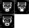
A 51-year old man presented to our hospital suffering from vertical and torsional diplopia after reduction of a blowout fracture at another hospital one year ago. The man described seeing double images separated vertically, and the right image was slightly slanted toward his nose. No head position abnormalities or facial asymmetry were noted. A thin-section coronal CT scan revealed multiple fractures in the right facial bone, including the orbital floor, medial wall, and zygomatic arch, and also a metallic screw fixed to the inferior orbital walls. We detected no fracture line on the anterior orbital floor, but we did note that a fracture remained on the posterior orbital floor (Fig. 1A,1C). Also, the portion of the right inferior oblique muscle in front of the equator was vertically reoriented. The medial portion of the inferior oblique muscle was not traced on a coronal CT scan (Fig. 1A, 1B).
The alternate prism cover test indicated a 14PD right hypertropia (RHT), 4PD esophoria (E) in the primary gaze, 18PD RHT, 4PD E in levoversion with the head turned to the right, and 6PD RHT, 4PD E in dextroversion, with the head turned to the left. The patient was orthophoric in upgaze, and 35PD RHT, 10PD E in downgaze. We also noted a mild V pattern. The Bielschowsky head tilt test generated a negative response, and we recorded an 8PD RHT on tilting the head to the right shoulder, and 6PD RHT on tilting the head to the left shoulder.
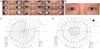
Underaction of the right inferior rectus (RIR) and right superior oblique (RSO) muscles associated with downgaze limitations were evident, but IO overaction of the right eye was determined to be minimal. We noted no overaction or limitation of the right superior rectus (RSR) or contralateral IO (Fig. 2A). The patient's binocular single visual field was limited in the right superior temporal quadrant (Fig. 2C). The double Maddox rod test (DMRT) revealed a10-degree extorsion in the right eye. Foveal extorsion was also found to be minimal in the right eye.
Under general anesthesia, the patient underwent an RIO 14 mm recession and an RIR 3 mm resection. Results of the ntraoperative forced duction test were not remarkable, and only a mild loosening of the RIR was noted. The technique for the 14 mm recession of the IO was as described by Parks.5 The RIR resection was also performed because the approach to the IR was relatively easy, following the IO recession in the forniceal incision, and because the anterior portions of the IR might have been relatively paretic, as was mentioned previously.3
Two weeks after the surgery, 4PD esophoria only was noted in the patient's primary gaze. The vertical and torsional diplopia was eliminated completely in the primary position. Mild horizontal diplopia remained. However, this also disappeared one month after surgery (Fig. 2B) and the patient's field of single binocular vision was expanded in the primary gaze and in the downgaze. (Fig. 2D).
Discussion
Seiff and Good3 explained two possible mechanisms for hypertropia in posterior blow out fractures. First, the increased angle of contact of the IR is attributable to an inferior looping of the muscle itself. The second mechanism relevant to hypertropia is a relative anchorage of the muscle to the fracture, resulting in a Faden-like effect. These effects create a relative IR paresis, and cause the SR of its antagonist to generate hypertropia. Our patient exhibited none of the restrictions or overactions of the RSR and contralateral LIO which had been exhibited in the preoperative version and binocular single visual field tests. Also, they noted that the tethering of IO, as proposed by Nardi,4 should induce torsional diplopia, but this did not occur in any of their patients.
In cases involving IR paresis after a posterior blowout fracture, fundus intorsion is normally evident, because the IR is an extorter.6 However, manifested extorsions such as those described in our study, might be quite exceptional, and might be explained by co-existent SO palsy.7
Our patient manifested no ocular torticollis, had a negative response on a Bielschowsky head tilt test, and exhibited no RSO loosening on the intraoperative forced duction test, all of which are telltale signs of SO palsy.8,9 However, true SO palsy could not be excluded, as it does not necessarily require toricollis to be present and although mild, there is in fact a difference in right hypertropia on head tilt. Moreover, a slack SO tendon would be expected only if the palsy was congenital, not acquired.10 Therefore, we believe that the fixed IO in the floor fracture might disturb the manifestation of co-existent SO palsy, as evidenced by the observations of anomalous head posture for fusion, distinct IO overaction, and the positive Bielschowsky head tilt test. We assume that the torsional symptoms manifested by our patient might be attributable to SO paresis combined with entrapment of a portion of the anterior torsional fiber of the RIO into the fracture site. We also believe that the SO paresis observed in our patient might indicate true palsy, as the torsional diplopia was so severe.
In conclusion, Nardi's hypothesis for the major role of IO in hypertropia following blowout fractures might not have been confirmed by our case . If the connective tissue of the IO muscle or muscle itself has become entrapped in the orbital floor, the involved eye would be unable to be lowered due to the restriction of the SO on downgaze, and definitive IOOA would not be noted. We suggest that the entrapment of the IO in the orbital floor following a blowout fracture may alter the manner in which SO palsy manifests or may induce a restrictive SO paresis.
 XML Download