

 PDF
PDF ePub
ePub Citation
 Print
Print


The levator palpebrae superioris muscle, which originates from the orbital margin of the small wing of the sphenoid bone in the annulus of Zinn, runs along the superior wall of the orbit and travels anteriorly parallel to the superior rectus muscle. After passing through Whitnall's ligament it becomes an aponeurosis and attaches anteriorly to the lower surface of the superior tarsal plate.1
Whitnall's ligament, which is formed by a collection of levator muscle sheaths at the level of aponeurotic change of the levator muscle, attaches to the muscle sheath surrounding the tendon of the superior rectus muscle and the medial side of the trochlea and to the lateral orbital margin medial to the orbital lobe of the lacrimal gland. Some believe that Whitnall's ligament may help support the levator palpebrae, but Anderson and Beard2 have shown experimentally that Whitnall's ligament has no supportive relationship with the levator palpebrae. Whitnall's ligament also supports the upper eyelid, lacrimal gland, and superior orbit, and serves as an anatomical landmark for the superior border of the levator aponeurosis (LA).
The LA and Whitnall's ligament are important landmarks in ptosis surgery, especially upper levator surgery, and differences in anatomical structure may affect surgical results. The distance between the insertion of the levator palpebrae and Whitnall's ligament has been measured as 14-20 mm in previous reports,2,3 but there are no precise standard values for Asians.
Using cadavers, we investigated the clinical significance of the anatomical structures of the LA and Whitnall's ligament and the relationships between the insertion of the LA and adjacent structures.
Materials and Methods
We examined 20 eyeballs from ten cadavers with no history of orbital trauma. Subjects with lesions in the orbit, deformation of the head or face, or significantly decreased orbit volume were excluded. No cadavers were fixed.
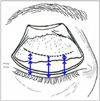
We performed exenteration of the orbit to investigate the relationship between the insertion of the LA and adjacent structures. After resecting the periosteum surrounding the orbit by penetrating deeply through the skin using a number 15 blade, we continued subperiosteal dissection towards the orbital apex. Internal orbital structures containing periosteum were totally resected from the orbital apex without trauma. After resecting the skin and the orbicularis oculi parallel to the upper eyelid, we observed the LA by resecting the orbital septum and orbital fat. After finding Whitnall's ligament, which is located between the levator palpebrae muscle and aponeurosis, we noted its tension, density, and shape. We observed the relationship between the superior rectus muscle and the levator palpebrae muscle and measured the transverse width of the levator palpebrae muscle at the point of orbital apex and Whitnall's ligament, respectively. We observed fibrotic change or fatty invasion of the levator muscle after confirming the shape of the LA where it attached to the tarsal plate. We used three points (medial 1/3, center, lateral 1/3) to divide the LA into three parts so that we could measure the distance between the margin of the eyelid and the border of the upper tarsal plate from the origin of the upper tarsal plate (Fig. 1).
Results
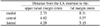
Demographic data is shown in Table 1. Among the ten subjects, there were six males and four females and the mean age at death was 66.8 years (range: 40-83 years). The levator palpebrae muscle, which was resected from the orbital apex, runs through the anterior orbit to form the LA, which becomes fibrous at the level of Whitnall's ligament. Whitnall's ligament extends laterally, dividing the lacrimal gland into two lobes, and connects medially to the trochlea.
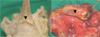
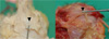
Regarding Whitnall's ligament, 12 eyes had clear, white fibrotic bands (Fig. 2), four eyes had thin bands with reduced tension (Fig. 3), and in four eyes we could not identify the precise shape of the band (Fig. 4).
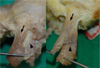
The levator palpebrae muscle typically borders the LA near Whitnall's ligament. Viewed superiorly, the levator muscle travels to the anterior orbit along the superior rectus muscle, forming an 'X' shape (Fig. 5). The border of the two muscles was observed according to position. At the nasal border, there was a filmy connection between the fibers, and after dissection of this connection, it was possible to separate the two muscles the temporal border, on the other hand, was easily dissected as there were no such connections (Fig. 6).
LA attachment to the tarsal plate was observed in two forms: the nasal dehiscence form in 13 eyes (65%) and the parallel attachment form in seven eyes.
The distances from the eyelid margin to the LA insertion were 8.31 mm medially, 5.57 mm centrally, and 5.15 mm laterally. The distances from the upper border of the tarsal plate to the LA insertion were 2.75 mm medially, 4.82 mm centrally, and 4.29 mm laterally (Table 2). The width of the levator palpebrae muscle in the orbital apex averaged 4.74 mm and the average width at the point of Whitnall's ligament was 11.15 mm, about a 2.42-fold increase.
Discussion
In 1910 Whitnall first described Whitnall's ligament as a muscle sheath that gathers into a thick band and becomes a ligament near where the levator palpebrae becomes an aponeurosis.4,5 In 1959, Fink defined this structure as the "superior transverse ligament".6 There are several theories regarding the function of Whitnall's ligament. One view believes that Whitnall's ligament supports the upper eyelid with its suspensory shape and supports the ligament of the levator palpebrae, while another theory hypothesizes that Whitnall's ligament functions as a fulcrum which changes the horizontal direction of the levator palpebrae to the vertical direction of the LA.2,7 When Whitnall's ligament functionally fails due to damage or aging, the levator palpebrae muscle becomes prolonged and sunken, thereby decreasing the elevating force of the eyelid.8 It is therefore important to correctly recognize the anatomy of Whitnall's ligament duringupper eyelid surgery to prevent damage to Whitnall's ligament. It is also important to know the length from Whitnall's ligament to the insertion of the LA when resecting the levator palpebrae muscle.
However, there has been little research done regarding Whitnall's ligament. Codere et al.9 dissected 20 cadavers and described Whitnall's ligament as a sleeve which surrounds the levator palpebrae muscle they postulated that Whitnall's ligament acts as a mobile fulcrum that converts the anterior-posterior vector force of the levator to a superior-inferior direction during eyelid movement. Cho et al.10 reported weak Whitnall's ligaments in 21 of 263 eyes with ptosis that had undergone levator resection. In our study, we found various shapes of Whitnall's ligament: 12 eyes had a definite band-like structure, four eyes had a weak string shape, and four eyes had an undifferentiated structure. So, we found weak or undifferentiated Whitnall's ligaments in eight of the 20 eyes (40%), which is a higher proportion than reported in the previous studies. A possible reason for this result is age-related changes to the upper eyelid, as the mean age at death of the subjects in our study was 66.8 years, 13.2 years higher than that of a former study. Changes to the LA and weakness of Whitnall's ligament may both contribute to the progression of senile blepharoptosis.
The levator muscle and the superior rectus muscle are crossed in an X-shaped fashion and linked medially by a thin fibrous membrane due to medial surface contact between the two muscles. This crosslink may lead to significant changes of the eyelid following superior rectus surgery we postulate that resection of the levator muscle and muscle sheath adhesion is needed. Although the upper lid can theoretically retract following superior rectus surgery, this is rarely seen clinically. In this study, we found no connection between the superior rectus muscle and the levator muscle except on the medial side.
The levator palpebrae muscle gradually changes to aponeurosis, and appears as a white fibrous strand at the level of Whitnall's ligament. The levator palpebrae muscle is divided into anterior and posterior parts 10-12 mm above the upper tarsal margin. The posterior part is the Muller muscle and is attached to the tarsal plate. The anterior part joins with the orbital septum at the level of the lid crease. A few fibers are attached to the skin lying across the orbicularis. However, most of the fibers strongly attach to the lower three mm of the tarsal plate or weakly attach to the upper 2-3 mm of the tarsal plate.2 In a study of Caucasian and Korean cadavers, Jeong et al.11 reported that the insertion of the LA is 3-4 mm from the eyelid margin in Caucasians and 2-4 mm in Koreans. Ushino Haramoto et al.12 reported that at about 2-3 mm above the upper margin of the tarsal plate, the LAis converted to thin and transparent fascia and attaches to the tarsal part of the orbicularis oculi.
Although many anatomical studies of the levator palpebrae have been performed, its insertion site has not been clearly defined. We measured the insertion point of the levator aponeurosis medially, centrally, and laterally, with reference to the upper lid margin and upper margin of the tarsal plate. The distance from the insertion of the LA to the upper margin of the tarsus or upper lid margin was shortest in the medial measurement. Morphologically, medial dehiscence was observed in 13 eyes, while parallel adhesion was observed in seven. The higher proportion of medial dehiscence was considered the result of involutional change, as the subjects had an average age at death of 66.8 years.
In this study, we observed various shapes of Whitnall's ligament and its relationship with adjacent structures. The main limitations of this study were that most of the cadavers were older, and that cadaveric studies are not always consistent with studies in vivo. Despite these limitations, this study provides a comprehensive description of the anatomy of Whitnall's ligament and the LA in Korean subjects and the results will be useful for levator muscle surgery.


 XML Download