Intravitreal bevacizumab injection and photodynamic therapy (PDT) with Verteporfin are effective for the treatment of choroidal neovascularization (CNV).1,2 Recently, combined PDT with Verteporfin and intravitreal bevacizumab injection has shown a synergistic effect and might reduce the need for retreatment and cyclic injections of bevacizumab.3
Single treatments of PDT and intravitral bevacizumab injection are considered relatively safe and effective.4,5 However, PDT may cause visual disturbances, subretinal hemorrhage, suprachoroidal hemorrhage and retinal pigment epithelial tear, and there are reported complications following intravitreal bevacizumab injection such as retinal pigment epithelial tear and acute glaucoma.6-11
Chung et al.12 reported a case of retinal detachment with macular hole following combined PDT and intravitreal bevacizumab injection but the safety of combined treatment with these two modalitiesis not well established.
We herein present a case of serous retinal detachment after combined PDT and intravtreal bevacizumab injection in subfoveal CNV.
Case Report
A 53-year-old woman presented with metamorphopsia in her right eye. She was diagnosed with subfoveal CNV secondary to age-related macular degeneration (AMD) and the best-corrected visual acuity of the right eye was 20/50. PDT with Verteporfin was performed according to the standard protocol of the Treatment of Age-Related Macular Degeneration with Photodynamic Therapy Investigation and repeated after 5 months.13 The visual acuity of the involved eye did not change.
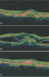
Fifteen months after the second PDT, fluorescein angiography of the affected eye revealed persistent subfoveal CNV and the best corrected visual acuity remained 20/50 (Fig. 1A, 2A). Combined PDT according to the same standard protocol and 1.25 mg intravitreal bevacizumab injection was performed on the same day. One day after the treatment, the patient's vision had decreased to counting fingers. Fundus examination and optical coherence tomography (OCT) showed serous retinal detachment involving the macula (Fig. 2B).
On post-treatmentday two, the patient was initiated on an oral steroid (Triamcinolone® 24 mg #1) to complete a two week course. One week into steroid therapy, the visual acuity remained the same, but OCT findings of the right eye showed decreased serous retinal detachment (Fig. 2C). The vision slightly improved to 20/200 upon completion of the two week steroid course and fluorescein angiography showed decreased leakage of fluorescein dye (Fig. 1B).
Discussion
There have been several reports about serous retinal detachment after PDT treatment.7,9,14 Mennel et al.7 quantified transient serous retinal detachment in CNV after PDT using OCT. Kim et al.9 also suggested that serous retinal detachment might develop following PDT but might also resolve spontaneously several days following treatment. However, there is neither a published report of serous retinal detachment after the combined use of PDT and intravitreal bevacizumab injection nor following intravitreal bevacizumab injection alone.
Several mechanisms may be responsible for subretinal fluid accumulation after combined treatment with PDT and intravitreal bevacizumab injection. The PDT may induce fluid leakage from the CNV itself or damage of retinal pigment epithelium causing an alteration of the blood-retinal barrier.7,9 Bevacizumab may cause instability of the intraretinal and subretinal fluid and subsequent serous retinal detachment by rapidly modulating the permeability and activity of the CNV.10,12
In our patient, there was no evidence of increased subretinal fluid after the first two treatments of PDT alone and the PDT protocol was the same for all three treatments. Occurrence of the serous retinal detachment just after combined PDT and intravitreal bevacizumab injection suggests the interaction of these two modalities as a possible causal role. However, the definitive causation of serous retinal detachment in PDT alone, intravitreal bevacizumab injection alone or combined treatment cannot be determined.
A two week course of oral steroids was used to manage the serous retinal detachment after the combined treatment of PDT and intravitreal bevacizumab injection. The acute inflammatory reaction following either modality may be relieved by systemic steroids. Kim et al.9 injected Triamcinolone® into the vitreous in a patient with serous retinal detachment following PDT, whereas, we elected to use a more conservative management with an oral steroid.
In conclusion, combined PDT and intravitreal bevacizumab injection can be associated with serous retinal detachment which may cause sudden visual loss. Additional studies are needed to establish the standard protocol, safety and complications regarding the combined treatment of PDT and intravitreal bevacizumab injection.


 PDF
PDF ePub
ePub Citation
 Print
Print


 XML Download