

 PDF
PDF ePub
ePub Citation
 Print
Print


Intraocular foreign bodies (IOFBs) resulting from penetrating ocular injuries are usually detected at the first visit. However, the presence of IOFBs may not always be readily identified and symptoms may only become apparent after a prolonged period of time.1-3 IOFBs resulting from mowing activity are more prevalent in males, and in Korea, accidents are more frequent from June to September.4 In many cases, the foreign bodies are found to be metallic in nature. Metallic foreign bodies can penetrate the cornea and induce intraocular inflammation and traumatic cataract at the vitreoretina.5-8 Intralenticular foreign bodies are not and located uncommon following penetrating eye injuries (~5-10%).9 They generally present with a decrease in visual acuity resulting from aggravation of a traumatic cataract. However, they are rarely found to remain in the crystalline lens for many years without causing visual impairment.10,11
In Korea, an asymptomatic intralenticular metallic foreign body has never been reported. In this report, we present a case of an asymptomatic intralenticular metallic foreign body in place for 6 months and we review the relevant literature.
Case Report
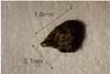
A 66-year-old male visited our ophthalmology department because of decreased visual acuity in his left eye of 10 days duration. The patient had a past history of ocular trauma to the left eye while mowing his lawn 6 months before his visit. He had not been treated because he did not feel any discomfort. On presentation, his best-corrected visual acuities (BCVAs) were 1.0 and 0.4 in the right and left eye, respectively. The intraocular pressure was 11 mmHg in the right eye and 14 mmHg in the left eye. In the right eye, the anterior segment did not show any obvious abnormalities. In the left eye, corneal opacity, which did not involve the visual axis, an incidental intralenticular metallic foreign body, and lens opacity were found. The posterior lens capsule was intact (Fig. 1). There was no inflammation in the cornea or anterior chamber. Funduscopy showed no definite abnormalities in either eye. Emergent surgical removal was performed. Under local anesthesia, capsulorrhexis of the anterior lens capsule and hydrodissection and hydrodelineation of the lens were performed via a corneal incision site. With viscoelastic aid, the intralenticular foreign body was elevated into the anterior chamber. The foreign body was then removed with foreign body forceps. Phacoemulsification and posterior chamber intraocular lens implantation was then performed. The implanted IOL was an AcrySof™ (SA60AT, Optic 6.0 mm, Length 13.0 mm, Alcon, USA). The diameter of the removed foreign body was about 1 mm and it was identified to be metallic by a magnet. It was thought to be a part of the lawn mower blade (Fig. 2). At 3 days postoperatively, the BCVA of the left eye was 1.0. There were no specific findings except trace cell reactions in the anterior chamber. At 17 days postoperatively, the BCVA of the left eye was 1.0. There was no inflammation in the anterior chamber and the IOL was well-seated (Fig. 3).
Discussion
The frequency of IOFBs following penetrating eye injuries is approximately 40% and the incidence of intralenticular foreign bodies is approximately 5% to 10%.9,10,12 In many cases, the lens becomes opaque and extraction is required for visual rehabilitation.12 However, stable visual function without significant cataract formation has been described in some cases.13,14
In many cases, IOFBs induce direct ocular injury and need to be surgically removed. Retained IOFBs can cause complications such as endophthalmitis, cataract, retinal detachment and siderosis bulbi, and lens-induced glaucoma.15,16
However, some IOFBs can be retained without any symptoms.1-3 Ahn2 suggests that the reason some IOFBs can be retained for an unusually long period of time is that these IOFBs are encapsulated by a thin membrane. Lin et al.3 reported an occult plastic intravitreal foreign body retained for 30 years that was removed by chance during a cataract operation. Dhawahir-Scala and Kamal11 reported an intralenticular foreign body which had been retained for 60 years. Hwang et al.17 reported a case of lens particle glaucoma induced by a retained IOFB that had been in the anterior chamber for 20 years. Ahn2 reported a case of noninfectious endophthalmitis caused by an IOFB that had been retained in the posterior wall of the left eye for 16 years. However, there is no report of a retained intralenticular foreign body in Korea.
The cause of cataract development after injury cannot always be identified, although it is usually a result of a capsular and epithelial rift.18 The healing capacity of the anterior lens capsule, in contrast to the posterior capsule, is well documented and is thought to result from the presence of the subcapsular epithelium. If the capsule defect is small, epithelial proliferation rapidly restores its continuity, limiting the free passage of ions and fluid that may result in progressive cataract formation.19,20 In our case, the size of the intralenticular foreign body was 1 mm and the capsular break was small enough to heal spontaneously. We believe that the intralenticular foreign body was retained and stable because of encapsulation, although there was no pathologic confirmation of this. The visual axis was not involved with the injured capsule or foreign body. We believe that the reasons stated above explain why the patient did not experience any ocular discomfort for 6 months despite the presence of a intralenticular metallic foreign body.
Management of intralenticular metallic foreign bodies is often conservative until intraocular inflammation or cataracts develop.21 Small intralenticular foreign bodies which do not involve the visual axis can be removed by an intraocular magnet.22 If the risk of surgically removing the foreign body is less than the risk of leaving it undisturbed in the eye, early removal should be considered. Nonetheless, the decision to proceed with surgery should be based on various factors including the size and chemical composition of the foreign body and the potential for infection.23
If visual acuity is compromised by cataract formation induced by an intralenticular foreign body, the standard management is removal of the foreign body, phacoemulsification, and IOL implantation (tri-combined operation).9,24,25 In our case, we successfully performed a tri-combined operation. Progressive cataract formation is not inevitable, as there have been documented cases of localized lens opacities with stable visual function in the presence of small, embedded lenticular foreign bodies.13,26 However, we suggest that early surgical removal of the foreign body become the treatment of choice, especially with recent surgical advances that enable safe removal of the foreign body with good visual results.
Penetrating ocular injuries caused by embedding of metallic foreign bodies during lawn mowing activities are common in Korea. As such, even in the absence of symptoms, patients with a history of lawn mowing should be thoroughly examined and asked about ocular trauma. When a retained intralenticular foreign body is found, it should be removed, because a long-persisting intraocular foreign body can cause intraocular complications.


 XML Download