

 PDF
PDF ePub
ePub Citation
 Print
Print


Many investigators had tried to correlate optical coherence tomography (OCT) findings and the functional outcome after successful macular hole surgery.1-3 Several recent reports have indicated that the features of the photoreceptor layer seen on OCT represent the integrity of the photoreceptor layer, and these features could be a key for the functional outcome.4-7 These reports have suggested that the returning back or centripetal migration of the photoreceptors after macular hole surgery are responsible for vision recovery.5,8 It was also reported that visual acuity often continues to improve for two or more years after surgery9 and that the long-term reorganization of the photoreceptor layer is a possible mechanism affecting this outcome.
We report on three cases that showed the reorganization of the photoreceptor layer as being concurrent with the continuing improvement of visual acuity after closure of the macular holes.
Case Report
The OCT scans from patients whose visual acuity increased gradually after successful closure of macular hole surgery were analyzed retrospectively. The vertical and horizontal scans with 5 mm lengths were obtained by a skilled technician. The identical locations of the serial scans for each patient were confirmed based on the OCT findings and the video images that were taken simultaneously with the OCT scans. The scanned images were enhanced as described previously.6 In brief, the raw files were exported to Adobe Photoshop® (version 7.0) and the reflectivity level was optimized as follows. The lowest level below which at least 0.1% of data points were detected was set to be 0 or the darkest in the new scale; the highest level above which at least 0.1% of raw data points were present was set at 255 or the brightest in the new scale. As a result of using this new scale, we were able to use most of the information present in each image. The dimensions of the images were changed to reproduce the actual dimensions of the scans.
Case 1
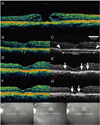
A 67-year-old woman who had complained of decreased vision of the left eye for one month was diagnosed with a stage 3 macular hole (Fig. 1A) and had visual acuity of 20/80. She underwent pars plana vitrectomy, triamcinolone acetonide-assisted internal limiting membrane (ILM) peeling and fluid-gas exchange.
The postoperative OCT scans obtained at four weeks demonstrated the photoreceptor layer with 1820 µm wide disorganization (Fig. 1B and C). Visual acuity was 20/100. At six weeks, a macula spared retinal detachment with multiple breaks was found. Pars plana vitrectomy, laser photocoagulation and fluid-gas exchange were performed. At six months, visual acuity was 20/40 and the OCT studies disclosed reorganization of the photoreceptor layer at multiple locations along the macula (Fig. 1D and E). Slight disorganization of the photoreceptor layer was noted under the center of the fovea. By 1 year, the patient's visual acuity had improved further to 20/25 and the photoreceptor layer of the fovea had been reorganized on the OCT images (Fig. 1F, G).
Case 2
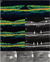
A 64-year-old woman who had complained of having a four-month period of visual disturbance was diagnosed with a stage 4 macular hole in the right eye (Fig. 2A) and had a visual acuity of 20/250. She underwent pars plana vitrectomy, ILM peeling, and fluid-gas exchange.
The postoperative OCT scans obtained at two weeks demonstrated 1870 µm wide disorganized features in the outer retina (Fig. 2B and C) and the patient had visual acuity of 20/125. By four months, visual acuity had improved to 20/50. The OCT scans revealed partial reorganization of the photoreceptor layer, the signal of which appeared as a broken line (Fig. 2D and E). At postoperative 15 months, the photoreceptor layer had been reorganized further and had only a small residual defect (Fig. 2F and G). Her visual acuity had improved to 20/40.
Case 3
A 32-year-old woman who had experienced decreased vision of the right eye for six months was diagnosed with a stage 2 macular hole (Fig. 3A) and had visual acuity of 20/80. She underwent pars plana vitrectomy, ILM peeling and fluid-gas exchange.
At postoperative two weeks, her visual acuity was 20/100 and the OCT scans revealed disorganization of the photoreceptor layer under the fovea (Fig. 3B and C). The disorganization was measured 1260 µm wide. After six months, visual acuity was 20/80 and the OCT studies revealed partly organized photoreceptor layer, which had some signal discontinuity and appeared as a broken line (Fig. 3D and E).
One year after the surgery, visual acuity was 20/40 and the discontinuity of the photoreceptor layer had been resolved on the OCT (Fig. 3F and G).
Discussion
Ko et al. suggested that the highly backreflecting signal of the photoreceptor layer returns when the photoreceptors return to their anatomical position after macular hole surgery.5 They have noted some discontinuity of the photoreceptor layer and have proposed that this discontinuity represents the residual photoreceptor impairments. Moshifeghi et al. reported two cases in which the macular hole was repaired successfully with a persistent outer retinal defect, which was resolved at five to six months postoperatively.8 They suggested that the partial migration of the photoreceptor into the center of the hole during the healing process would resolve the defect.
These theories, however, are insufficient to explain the reorganization of the photoreceptor layer shown in our cases. The OCT images that were taken in the early postoperative period demonstrated broad disorganization of the photoreceptor layer, which underwent a gradual reorganization over a period of one or more years. Unlike the cases of centripetal migration, the weak signal of the photoreceptor layer at multiple locations was an early indication of the reorganization of the photoreceptor layer in our cases. Due to the discontinuity, the signal of the photoreceptor layer looked like a broken line. As vision improved, the signal became more distinct and the discontinuity was resolved. These findings suggest that local reorganization of the photoreceptor takes place after anatomical restoration.
In the second case, the photoreceptor layer had a residual defect in spite of the reorganization. This defect would prevent further improvement of visual acuity.4 Extent of the defect was reported to be correlated with postoperative visual acuity.7
It is important to perform serial scans at a consistent, predetermined location to verify these results. In spite of repeated scanning by a skilled technician, minimal deviation of the scans could not be avoided due to the following several reasons. As the macula lacks major vessels, shadowing of the vessels cannot serve as a reliable reference. The horizontal temporal vessels are not reliable references for the location of a vertical scan. In an unpublished study (in peer-review), the authors found that there was a long-term change of the foveal thickness after macular hole surgery. Analysis of the foveal contour may be a crude reference, but requires a cautious interpretation, because a minimal anterior-posterior movement of the eye ball during a scan would change of the foveal shape considerably. We also used the video images that were taken simultaneously with the scans (Fig. 1 to 3, H to J). However, the resolution of the video image is low and there is a little delay from scanning OCT to taking the image.
Nonetheless we think that our hypothesis is still valid, because the reorganization occurred in area of 1 mm or more which is broader than the foveal depression.
It is not clear if the changes in the photoreceptor layer features shown in these cases are usual in the eyes that show the continuing improvement of vision after closure of the macular hole. However, reorganization of the photoreceptor layer seems to play a role in long-term visual recovery after closure of macular holes. To understand the mechanism, further studies involving more cases using a higher resolution OCT are necessary.

 XML Download