

 PDF
PDF ePub
ePub Citation
 Print
Print


Orbital complications associated with intranasal sinus surgery are well known in the field of otolaryngology.1-4 During the past decade, endoscopic sinus surgery (ESS) has been developed, and improved the surgical technique for the chronic paranasal sinusitis.5
However, ESS can leads to a greater potential for damage to the orbital tissues because it carries the risk of complications as a result of the narrow surgical field, difficulty in mastering the technique and the necessity of a practiced hand.6 The intimate anatomic relationship between the paranasal sinuses and the other structures including the cribriform plate, skull base and lamina papyracea, can increase the risk of injury to the periocular tissue during ESS.7 Orbital complications include periorbital ecchymosis with an injury of the lamina papyracea, retrobulbar hematoma, ocular motor defect with extraocular muscles (EOM) injury, optic nerve injury, nasolacrimal duct injury, orbital abscess or emphysema, etc.4,8-12
Mark13 and Flynn14 reported exotropia (XT) with deficient eye movement after traditional intranasal sinus surgery. Among the damage to the EOM, damage to the medial rectus (MR), inferior rectus (IR) and superior oblique have been reported. The incidence of complications related to the MR, including muscle contusion, hematoma, the entrapment of bony defects, paralysis and transection, are most common.8,12-13,15-17
When the muscle transection has been identified by orbital imaging of computed tomography (CT) or magnetic resonance imaging (MRI), decisions about the optimal surgical procedures are difficult and quite challenging. Moreover, the results of corrective surgery are unpredictable.12,15 Thacker et al reported that 7 of 11 patients (63%) with surgical correction for strabismus after ESS required two or more surgical procedures.12 They suggested that the management of these complications are quite complicated.
This study evaluated the effect of vertical rectus muscles (VRM) transposition for a large-angle XT caused by a transection of the MR as a complication of ESS.
Materials and Methods
Four consecutive patients who underwent surgery for a large XT and a lack of adduction after ESS were enrolled in this study. Their CT or MRI of the orbit revealed a complete transection of the MR.
Each patient underwent comprehensive ophthalmic examinations, including an ocular motility and diplopia test. The deviating angles were determined using an alternate prism cover test in six cardinal gazes and head tilt positions.
The range of adduction deficits were recorded at the version test using the following scales; 0 indicated full adduction, which means the free arrival of medial limbus at medial canthal area, -4 indicates that the medial limbus could not past midline, -8 indicates a fixed XT, which means the lateral limbus reached the lateral canthal area with an inability to move medially. The adduction deficits were subdivided into -1 to -8. An adduction of -1, -2, -3, -4 indicated that the eye could rotate nasally from the midline to 75%, 50%, 25%, 0% of full adduction, respectively, and -7, -6, -5 meant that the eye could reach the point of the one fourth, two fourths, three fourths on the line from the lateral canthus to the midline.
A forced duction test (FDT) was performed to determine the secondary contracture of the antagonist lateral rectus muscle (LR) and adjacent soft-tissue. Both eyes were tested in order to determine the degree through a relative comparison with the sound eye. A forced generation test (FGT) also was carried out with the subjects being instructed to adduct the eye while the examiner applied traction to the eye in the abducted direction using toothed forceps.
In all patients, VRM transposition was performed based on the time interval from injury, presence of secondary contracture of the LR and adjacent soft-tissue, and the surrounding tissue-adhesion.
In 2 patients with a 3 months duration after the injury and minimal secondary contracture of LR, full-tendon VRM transposition of the Foster type was performed. Two patients, who had a long duration of 11 months and 36 months after the injury, showed severe contracture of the LR and severe surrounding tissue-adhesion. The augmented Hümmelsheim operation was performed concurrently with an ipsilateral LR recession.
Full-tendon VRM transposition of the Foster type18 was performed. The full-tendons of the two vertical recti were disinserted and transposed towards the insertion site of the MR. They were reattached to the sclera whilst maintaining the distance from the limbus to the new insertion of the transposed muscle equivalent to the original insertion to preserve the spiral of Tillaux. A single posterior fixation suture was then placed at the sclera of 8mm posterior to each upper and lower corner of MR insertion, fixing 25% of the belly of each transposed muscle adjacent to the MR to augment the procedure (Fig. 1A).18
Our augmented Hümmesheim procedure involved half-splitting of each VRM 15 mm posterior to the insertion, detaching the two medial halves from the scleral insertions and transposing them to the MR by pulling each half through the undersurface of the severed MR. The half-splitting medial portions of the VRM were made a cross to transposed the end of half of the superior rectus muscle (SR) to near the inferior corner of MR insertion and the end of half of the IR to near the superior corner of MR insertion (X-type Hümmesheim operation) (Fig. 1B).19 The patients were followed up for more than 1.5 years.
Results
The visual acuity was not decreased in the injured eye of all patients. The anterior segment and fundus were normal. Three of the 4 patients had an injury to the MR of the right eye and the other had an injury to the MR of the left eye. All patients had a lack of contractility at FGT on MR. At the FDT in adduction, two patients with early surgery had mild resistance and the other two patients showed severe limitations toward the nasal side. The preoperative adductions ranged from -5 to -8. The postoperatively adductions improved to between -3.5 to -5.
Table 1 shows the patients' characteristics. The preoperative angle of XT was 40Δ - 85Δ, which improved to 0Δ in 3 patients and 25Δ XT in 1 patient after surgery.
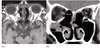
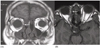
The orbital CT or MRI revealed a bony defect in the medial wall as well as a transection of the MR posterior to the globe in all patients.
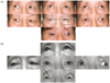
Patient 1 (a 66-year-old man) (Fig. 2 and 3) and patient 2 (a 54-year-old woman) showed 40Δ XT in the primary position and -4 adduction in the right eye. These 2 patients underwent a full-tendon VRM transposition of Foster 3 months after ESS. They obtained orthophoria without diplopia in the primary, left, up and down gazes even one year after surgery. Adduction of each right eye was improved to -3.5.
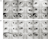
The initial visit of patients 3 (a 30-year-old man) (Fig. 4 and 5) and 4 (a 63-year-old woman) was 11 months and 3 years after ESS, respectively. In patient 3, a large XT of 85 Δ was shown and the eyeball was not moved nasally with a severe lack of adduction (-8). The FDT in adduction was strongly positive due to the secondary contracture of the antagonist LR and adjacent massive adhesion.
The patient underwent an X-type augmented Hümmelsheim procedure (Fig. 1B) coupled with an 8 mm recession of the contractured LR using an anterior ciliary artery preservation at the same time. Postoperatively, the patient became orthophoric without diplopia in the primary, left, up and down gazes, and the eye was moved to the midline. He has been remained orthophoria for 2.5 years after surgery.
Patient 4 had a history of a previous recession of the LR in the left eye at 6 months after ESS. However, the 70Δ XT and adduction deficit persisted. The patient visited one of authors 2.5 years later. On the FDT, severe resistance (-7) was present, which indicated massive adhesion posterior to the eyeball. She also underwent an X-type augmented Hümmelsheim procedure combined with a rerecession of the previously recessed LR using a hang-loose suture. Thereafter, the eyeball was maintained in the adducted position by a traction suture for seven days to maximize the effect of a hang-loose recession. Postoperatively, the adduction of the right eye was improved to -5 but the 25Δ XT remained. Despite of the residual 25Δ XT and -5 of adduction, the patient was satisfied with the results. More surgery will be needed to correct the residual XT.
With the exception of the mild chemosis of patient 1 immediately after surgery, which subsided using topical steroid drops, there was no other complication in any patient such as anterior ischemia including severe chemosis, corneal hazing or an anterior chamber cell reaction over the follow-up period.1-9,16 All the patients were satisfied with their postoperative results.
Discussion
Injury to the EOM after ESS includes contusion, hematoma, entrapment to the adjacent structures, paralysis, direct laceration and complete transection. Various types of ocular motility defects can present as XT, or esotropia (ET) or vertical deviation depending on the nature of the injury.12,15
Huang et al15 examined the clinical characteristics of 30 cases of MR injuries after ESS. They reported 2 types; 1) the type associated with the complete transection of the midportion of the MR showing marked adduction deficits and no mechanical restriction at the FDT with a large-angle XT, and 2) a grossly intact or contused muscle with only moderate to small-angle XT or ET according to the extent of the orbital soft-tissue entrapment or adhesion. The latter type was positive at the FDT on both adduction and abduction. When a lack of MR function is present without anatomical entrapment or transection of the muscle itself, immediate surgery is not suggested because the adduction might have considerable recovery with even full function through a several-month period.12,16,20
None of the patients in this study had any hematoma that could overshadow the other injuries in the orbital cavity and the transection of the midportion of MR could be clearly determined by an assessment of both the axial and coronal images in orbital CT or MRI.
When a large-angle XT is induced, the surgical strategy includes VRM transposition procedures to replace the lost MR function, anterior orbitotomy with a recovery of the functional MR stump, antagonist LR weakening procedures (recession of the LR, botulinum A toxin injection into the LR, extirpation with attachment of the LR to the lateral orbital wall), and nasal periosteal globe fixation.12,21
Combination surgery of VRM transposition and LR recession has been performed for the correction of XT caused by a lack of adduction. Olitsky and Brooks22 reported a patient with a large-angle XT after ESS, who obtained orthophoria in the primary position after a botulinum A toxin injection into the LR and a modified Hümmelsheim procedure to the MR accompanied with a 7 mm resection of the transposed portion of each VRM 2 weeks after ESS. Thacker et al12 also reported 4 patients with similar injuries, who underwent a full-tendon or half-tendon VRM transposition with botulinum A toxin injection of the LR or LR recession, and showed XT less than 8Δ, postoperatively.
Lee et al16 reported a case of a transected MR after ESS with 65Δ XT. Exotropia of 20Δ remained through a 12 mm LR recession as the first procedure and Hümmelsheim operation at the second procedure. Most surgeons prefer two-step surgery in order to reduce the risk of the anterior segment ischemia.
The LR recession alone results in considerable undercorrection of the large-angle XT and continues to progress the contracture of the LR and adjacent soft-tissue while awaiting transposition as a second procedure. Therefore, performing a VRM transposition first in 2 step surgery might be better because it can produce less undercorrection than when performing a LR recession first, which can allow an adjustment of the recession amount.
In patients 1 and 2, there was only mild mechanical restriction (-1) on the FDT 3 months after ESS. The full-tendon VRM transposition was conducted as a first operation. They have been orthophoric over a 6-month postoperative period and additional surgery was not required. In patients 3 and 4, who had a severely limited adduction on FDT due to secondary LR contracture and adjacent scarring, X-type augmented Hümmesheim operations were carried out concurrently with the larger LR recessions. The correction amount was as large as 85Δ and 45Δ respectively. Patient 4 showed 25Δ of residual XT postoperatively due to the contractured LR with adjacent scarring despite the large re-recession of the previously recessed LR. Therefore, another operation will be needed. Overall, early surgical management should be performed before the contracture of LR and adjacent soft tissue scarring.
When the MR is completely transected, the purpose of reattaching the lacerated ends include correcting the XT, improving the adduction function and reducing the risk of anterior segment ischemia by avoiding additional VRM transposition.15,23 It is seldom possible to examine the MR more posteriorly in the orbit through a conjuncival incision during conventional strabismus surgery.20 An orbital exploration may be indicated if the remaining posterior stump of MR is quite long and the patient presents within a few days after injury.12,15 However, the posterior stump may retract posteriorly, necessitating orbital surgery
The current use of new powered cutting and aspiration instruments containing a rotating blade in ESS potentiates the risk of catastrophic complications.9 MR injury from the powered cutting and aspirating instrumentation is hardly ever surgically reparable through an orbital exploration.24 Muscle recovery is not always possible because of the muscle destruction or severe damage converting into scar tissue.
Thacker et al12 mentioned that it could be possible to reattach the MR to the globe by recovering through an anterior orbitotomy if the remaining posterior stump is longer than 20 mm, and showed contractility on multipositional MRI scanning. However, an attempt to recover the stump was indicated in 4 patients and the following results were not promising. Although marked improvement in primary ocular alignment was achieved after a MR posterior stump was reattached to the globe, there was rarely a recovery of the adduction function.12,15,23,25 Four patients in this study also obtained good ocular alignment through VRM transposition without any attempts to recover the MR stump.
Likewise, Huang et al15 performed suturing of the remaining anterior and posterior MR remnants through an orbital exploration in 5 patients with a MR transection, along with a botox injection into the antagonist LR. In three out of 4 patients, early surgical intervention within 3 weeks after the injury improved the primary ocular alignment. However, the severely restricted adduction persisted. In the other 1 case of early surgery, the additional VRM transposition was performed later, with only moderate improvement in ocular alignment. One patient had a late orbital exploration 4 weeks after the injury and the surgery was more difficult because of the extensive orbital soft-tissue scar formation. In that latter case, the VRM transposition were performed as a secondary procedure but with little improvement in ocular alignment.
In this study, VRM transposition was performed as the first procedure in 4 patients with MR transection. Three of them had orthophoria without diplopia in all gazes. They may ignore the second image of the injured exo-eye. One patient with a transposition procedure 3 years after ESS, had a residual 25Δ XT. In this patient, the surgery was less effective due to the marked contracture of the LR and severe adhesion with surrounding tissues for a long duration.
The VRM carries the risk of anterior segment ischemia when it is performed concurrently with a LR recession.26-28 Varying techniques including half-tendon transposition, preservation of the anterior ciliary artery and a botox injection into the antagonist LR, are a useful tools for reducing the risk of anterior segment ischemia.22,29,30 Half-tendon transposition such as the Hümmelsheim or a Jensen procedure have been devised to preserve a portion of the anterior ciliary artery circulation. However, it might not be as effective as a full-tendon transposition.16,29,30
The X-type Hümmelsheim procedure was used to augment the effect of the half-tendon transposition. The nasal half-tendons of the VRM were pulled and crossed under the MR and sutured at the other ends of the MR insertion.19 A large correction amount of XT with large recession of ipsilateral LR was achieved. In patient 3 with 85Δ XT, the anterior ciliary artery preservation procedure associated with an X-type augmented Hümmelsheim procedure and a recession of the ipsilateral LR was carried out to avoid anterior segment ischemia.
The fact that the right eye was involved in 3 out of 4 patients is noteworthy. In previous reports, the right eye was involved in 73% of case of ESS.12,17 The reason why right orbital complications occurred more commonly is that the instrumentation tip in the right nasal cavity is directed to the lamina papyracea while a right-handed surgeon usually operates from the patient's left side in ESS.31
In conclusion, the management of a transected MR and resultant XT after ESS is challenging due to the acquired adduction deficit and intractable diplopia. The surgical strategies in the transposition procedures should be selected based on the time of presentation after ESS, the adjacent adhesion, the amount of exotopia, mechanical restriction at FDT and the VRM functions at presentation.
VRM transposition procedures are effective in correcting XT secondary to a MR transection and aligning the eyes. VRM transposition alone is a useful method within short duration after injury before the formation of severe LR contracture and soft-tissue scarring. The X-type augmented Hümmelsheim operation coupled with an ipsilateral LR recession is effective when large-angle XT is associated with severe contracture of the LR and massive adhesion with the adjacent tissue after a long duration.
These results with a review of previous articles are expected to provide useful information for devising a suitable treatment plan and estimating the prognosis. However, more study with a larger number of patients and a longer follow-up will be needed.


 XML Download