

 PDF
PDF ePub
ePub Citation
 Print
Print


The incidence of consecutive esotropia developing after exotropia surgery has been reported to be 6-20%.1-6 Generally, comitant consecutive esotropia with small deviation disappears spontaneously with time. However, appropriate treatments are required for the cases with diplopia persisting more than 2 weeks after surgery, increase of deviation, limitation of eye movement, or the risk of developing amblyopia.7
As a non-surgical management, alternate occlusion or prism may be prescribed to reduce overcorrection, and instillation of miotics or the correction of refractive error may be attempted. However, surgical managements should be considered for the cases whose esotropia over 14 Prism diopter (PD) at distance persisting more than 6 months after surgery, the cases who do not wish to undergo conservative treatments, the cases whose deviation does not change or increase, the cases whose diplopia is persistent due to lateral incomitancy, and the cases with impairment of abduction.7 Nevertheless, there are no guidelines that clarify the appropriate surgical methods and amounts for consecutive esotropia.
We examined the postoperative change of deviation and the surgical method of patients who underwent surgical correction for consecutive esotropia, and from this study, the authors want to provide some helps of surgical correction for consecutive esotropia.
Materials and Methods
This study was performed on 13 patients who underwent surgery for consecutive esotropia developed after exotropia surgery from January 2001 to December 2006 and who are available for follow up longer than 6 months. Based on the medical records of the subjects, the deviation and surgical methods at the time of exotropia surgery were investigated. During the follow up period, the postoperative development of amblyopia, the effect of occlusion therapy, surgical methods for consecutive esotropia, the deviation angle immediately after surgery, the change of deviation were examined.
Deviation was measured by the alternate prism cover test in all patients at near (33 cm) and far (6 m) using a fixation target. Refractive errors over 1.0 diopter (D) (spherical equivalent) were corrected prior to exotropia surgery.
Results
At the time of exotropia surgery (the first surgery), the mean age of the subject group was 9.2 years (4-19 years). Seven patients of the group were male (53.8%) and 6 were female (46.2%). The average amount of exodeviation at the time of the first surgery was 27.1PD (18-40PD) based on the far fixation. None of the patients had amblyopia. Five patients were emmetropic and five were myopic. Hyperopia was found in 3 patients. The average refractive error was -0.53D (-3.5~+2.0D: spherical equivalent). All refractive errors greater than 1.0 diopter were corrected prior to the first surgery.
The exotropia was intermittent in all cases. Eight patients showed the basic type, three showed the divergence excess type, and two patients had the convergence insufficiency type. There was no pseudodivergence excess type found with the patch test and +3.00 spherical lens test. None of the cases showed lateral incomitancy. Bilateral lateral rectus muscle recession was performed in all 13 cases.
Alternate occlusion was performed in 13 cases (100%) 2 weeks after the development of consecutive esotropia. The alternate occlusion was performed for 5 to 8 hours, (mean 6.2 hours) in a day, for 1 to 3 months, (mean 1.8 months). However, in 3 cases (23.1%), no change of deviation was found at the end of the alternate occlusion. In 4 cases (30.8%), the esodeviation increased by 3 to 8PD (mean 4.5PD). In 6 patients (46.2%) the esodeviation decreased by 4 to 10PD (mean 5.2PD). None of the cases developed amblyopia. Nine patients (69.2%) complained of diplopia, and base-out Fresnel prism was prescribed to 4 of the 9 patients.
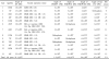
The surgical result of consecutive esotropia is summarized in Table 1. Surgery for consecutive esotropia (second surgery) was performed 7-25 months (mean 15.3 months) after the first surgery. The mean age of the subject group at the time of second surgery was 9.8 years (6-20 years), and the mean esodeviation was 21.1PD (16-30PD). The medial rectus muscle recession was performed in 10 cases, and among them, 8 patients received bilateral recession, and 2 patients received unilateral recession. For the remaining 3 patients, unilateral lateral rectus muscle advancement was performed. The surgical amount was determined based on the deviation at the time of the second surgery according to the "Cooper's dictum". On postoperative day one, 2 patients (15.4%) of the subject group showed orthophoria, and 10 patients (76.9%) showed deviations within 8PD. Deviations measured 6 month after the surgery were within 8PD in 12 patients (92.3%). The average follow up period was 17.1 months (6-32 months). Ten patients (76.9%) showed deviation within 8PD at the last follow up, and the surgical success rate was favorable. However, the mean deviation of the subjects immediately after surgery was 1.2PD esodeviation, 0.9PD esodeviation one month after surgery, 2.4PD exodeviation 6 months after surgery, and 4.7PD exodeviation at the last follow up, showing a tendency to progress to exodeviation as the follow up period increased (Table 1).
Discussion
The incidence of consecutive esotropia after exotropia surgery has been reported to be 6-20%.1-6 Comitant consecutive esotropia with a small deviation (10-15PD) disappears spontaneously with time in most cases. Therefore, a small deviation can be observed for 2 weeks without any treatments. However, for the cases whose diplopia are persistent or who do not show improvement in deviation after 2 weeks of observation, development of amblyopia should be prevented by the reduction of the deviation and maintaining fusion.7
As a non-surgical management of consecutive esotropia, alternate occlusion or prism may be prescribed to reduce overcorrection and to maintain fusion, and instillation of miotics or the correction of refractive error may be attempted. In our study, alternate occlusion was performed on all 13 cases. Unchanged or increased deviation was found in 7 of 13 patients. Decreased deviation was found in 6 patients (46.2%). However, all 13 cases required surgical treatments finally. Although 9 patients complained of diplopia, base-out Fresnel prism was prescribed to only 4 patients. Diplopia disappeared after the use of the prism, but there was no change of deviation. None of the cases developed amplyopia, and it was thought to be due to subject's mean age of 9.2 years.
Hardesty8 reported that in the cases with consecutive esotropia less than 15PD, prism is sufficient for cure. However, cases with greater than 15PD require surgery. Surgical treatments should be considered for the cases with residual esodeviation greater than 14PD at distance 6 months after surgery, patients who do not desire to undergo conservative treatments, the cases whose deviations do not change or increase despite the prism therapy, the cases with persistent diplopia due to lateral incomitancy, and for the cases with the impairment of duction.7 However, if there is a severe limitation so that the eyeball can not cross the midline or a large esodeviation is noticed immediately after surgery, excessive resection of the medial rectus muscle or the loss or slippage of the lateral rectus muscle should be suspected. In these cases, the second surgery has to be performed immediately.9 In all cases of our study, the second surgery was performed after 6 months of non-surgical management.
Concerning the method of surgery for consecutive esotropia without abduction impairment and lateral incomitancy, Burke.10 recommended medial rectus muscle recession for cases with bilateral lateral rectus muscle recession as a first surgery, and estropia surgery on opposite eye for those with unilateral lateral rectus muscle recession and medial rectus muscle resection as a first surgery. Nonetheless, consecutive esotropia with limited abduction require reoperation on the muscles that were operated during the first surgery. However, if the examiner does not feel any restriction in forced duction test, its cause may be the excessive recession of lateral rectus muscles, and thus lateral rectus muscle advancement is required. If the lateral rectus muscle cannot be found during the second surgery in patients with the excessive abduction impairment and a large deviation angle, transposition of vertical muscles to the attachment site of the lateral rectus muscle is required. In our study, medial rectus muscle recession was performed in 10 patients, lateral rectus muscle advancement was performed in 3 patients, and excessive recession or loss of muscle was not detected in any of the cases.
In consecutive esotropia, it is difficult to predict the result of second surgery because of following three reasons. First, consecutive esotropia may convert to exodeviation after advancement of lateral rectus muscle (the leash effect).11 Second, the contracture of the previously recessed muscles may occur. Finally, the muscle portion of the resected medial rectus muscle may become hypertrophied. On the other hand, Cooper.12 proposed the "Cooper's dictum" that surgical methods and amounts could be determined based on the result of the test performed at the time of the second surgery regardless of the first surgery. In our study, the decision of the amount of the second surgery was made based on the result of the test performed at the time of the second surgery according to the "Cooper's dictum".12
Examining the result of the surgery for consecutive esotropia, Son et al.13 reported 78% surgical success rate, Park et al.14 reported 100%, Kim et al.15 reported 88%, showing relatively high success rates. In our study, a similar success rate of 76.9% was shown at the last follow up.
On the other hand, success rate was low in some studies. Shin and Chang.16 reported that 15 patients with exotropia, 3 patients with esotropia, and 2 patients with orthophoria were noticed at final follow up in 20 patients who underwent surgery of consecutive esotropia.
Although deviation was not large, our study found that the mean deviation of the subject group showed a tendency of converting into exodeviation after surgery of consecutive esotropia as the follow up period increased. Especially, the three cases who received lateral rectus muscle advancement converted to exodeviation at the last follow up, but this finding may not be significant because of the small number of cases. The authors believe that the leash effect is the cause for these exodeviations.11
The authors investigated the surgical method of consecutive esotropia and postoperative change of deviation. In our study, 76.9% of patients showed deviations within 8PD after the surgery of consecutive esotropia, showing a favorable outcome. Although the deviation was not large, our study showed a tendency of increased conversion to exodeviation as the follow up period increased. Therefore, a careful decision on the surgical amount and the method for consecutive esotropia is needed, particularly when considering the advancement of lateral rectus muscle. The small number of subjects in our group was the limitation of our study. Therefore, in the future, a study on a large number of patients is required.
 XML Download