

 PDF
PDF ePub
ePub Citation
 Print
Print


The principle of laser refractive surgery is modification of the refractive power of the cornea by means of photoablation of the stromal tissue. The biomechanical strength of the cornea is compromised by surgical tissue subtraction and the loss of Bowman's membrane integrity after laser refractive surgery.1 Forward shift of posterior corneal surface resulting from anterior bulging of the cornea after photorefractive keratectomy (PRK) and laser in situ keratomileusis (LASIK) have been reported.2-6 Several cases of iatrogenic keratectasia after LASIK and laser-assisted subepithelial keratectomy (LASEK) surgery have been reported.7-10 In addition, forward shift of the cornea can cause myopic regression after laser refractive surgery.4-7 Especially iatrogenic keratectasia has come to rise as an important complication of refractive surgery, and it is thought to be correlated with preoperative corneal thickness, the amount of laser ablation, preoperative refraction and preoperative intraocular pressure.4
Anterior corneal surface is directly influenced by refractive surgery itself and healing process, so it is difficult to estimate the amount of corneal forward shift by analyzing the elevation map of the anterior surface. So, the posterior surface which is not directly affected by surgical process is usually used to evaluate the anteroposterior movement of the cornea. And the height elevation data relative to best-fit sphere has been measured by scanning-slit corneal topography.11 And changes of elevation should be evaluated at the center of the difference map due to inconsistency of preoperative and postoperative best-fit spheres.2,3
On the other hand, 'standard' laser refractive surgery corrects only spherical and regular astigmatism, and may induce higher-order aberrations that will lead to distorted images.12-15 Wavefront-guided laser refractive surgery considers all the aberration including higher-order aberrations. A correlation between visual symptoms and ocular aberration was strongly suggested, for example in monocular diplopia and glare.16 And the greatest gains in correcting higher-order aberration are noted in improved contrast sensitivity particularly under low light condition.17
In this study, we compared the changes of posterior elevation of the cornea from difference map, the higher-order aberrations (RMS-root mean square, coma, trefoil, spherical aberration) among LASIK, LASEK, wavefront-guided LASEK group.
Materials and Methods
Sixty two eyes (40 patients) undergoing LASIK surgery, 100 eyes LASEK surgery (56 patients), 22 eyes (13 patients) undergoing wavefront-guided LASEK surgery were enrolled. Mean age was 27.95±5.99 years. The preoperative mean spherical equivalent (SE) refractions were -4.78±1.17 diopter (D) (range -2.125 to -6.75 D) in LASIK group, -4.36±1.38 D (range -1.75 to -6.875 D) in LASEK group, -2.86±1.60 D (range -1.5 to -5.625 D) in wavefront-guided LASEK group. The preoperative characteristics of the patients are shown in
Table 1. The preoperative refractions were not significantly different among three groups (p=0.120, oneway ANOVA). The mean baseline pupil sizes in photopic and scotopic conditions were not statistically significant among three groups (p=0.440, p=0.562, respectively, oneway ANOVA). Patients were enrolled from those having surgery between October 2002 and July 2004, at the Department of Ophthalmology, Seoul National University Hospital, by one surgeon (Kim MK).
The following examinations were performed preoperatively in all patients: uncorrected visual acuity (UCVA), best-spectacle-corrected visual acuity (BCVA), manifest refraction in a dark room and cycloplegic refraction after instillation of cyclopentolate hydrochloride by operator, objective refractions with the automatic refract-keratometry (KR-7100, Topcon, Japan), intraocular pressure (NCT CT-60, Topcon, Japan), slitlamp examination of the anterior segment, corneal thickness with ultrasonic pachymetry (Pocket, Quantel Medical, France), corneal topography (Orbscan II®, Bausch & Lomb, U.S.A).
Wavefront analysis (VISX, Santa Clara, CA, U.S.A) was performed preoperatively in all patients to evaluate HOA. All HOAs were measured in the natural scotopic condition after 10-minute dark adaptation. Patients who had higher RMS of high-order aberration more than 2.00 were recommended to receive the wavefront-guided LASEK. Written informed consent was obtained from all patients.
All surgeries were performed using VISX S4 excimer laser system (VISX Inc., Santa Clara, CA). In LASIK surgery, the automated microkeratome (M2, Moria, France) was used to create a hinged corneal flap, 110 µm thickness. In LASEK surgery, an alcohol solution cone was placed on the corneal surface. Twenty percent of the alcohol solution was instilled inside the cone, left for about 20 seconds, and washed with a balanced salt solution. The cornea stroma was ablased using excimer laser (VISX 4, USA), and then the flap was repositioned in both LASIK and LASEK surgery.
The posterior corneal elevations were measured with the corneal topography before, 2 months, and 4 months after surgery. Changes in the elevation of the posterior corneal surface were evaluated at the center of the difference map generated from preoperative and postoperative elevation maps. High-order aberrations were analyzed using Wavefront aberrometer (VISX Inc. Santa Clara, CA, USA) preoperatively, and 2 months after surgery. All HOA values were obtained in the natural scotopic condition. RMS (Root mean square) value of higher-order aberrations, the 3rd order aberration - coma, trefoil, spherical aberration values were compared among LASIK, LASEK, and wavefront-guided LASEK group.
Comparison of continuous variables among three groups was analyzed using the oneway ANOVA test. Continuous variables of two groups were evaluated using independent t-test. Comparison of preoperative and postoperative data was done by dependent t-test. Clinical significance was accepted for P values of <0.05. All statistical analyses were performed using SPSS for Windows, Version 11.0.0 (SPSS Inc., Chicago, IL).
Results
The mean UCVA and manifest refraction changes at 2 months after surgery are shown in
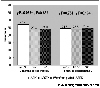
Table 2. There is no statistically significant difference of postoperative visual acuity and manifest refraction among three groups. The mean forward shift of the posterior corneal surface was 27.2±11.45 µm in LASIK eyes (n=53), 23.4±10.5 µm in LASEK eyes (n=75), 24.0±14.9 µm in wavefront-guided LASEK eyes (n=18) at 2 month postoperatively. On the other hand, it was 24.3±9.8 µm in LASIK (n=34), 23.6±10.6 µm in LASEK (n=72), 28.4±14.7 µm in wavefront-guided LASEK (n=16) at 4 month postoperatively. There were no statistically significant difference of posterior elevation among three groups at 2 and 4 months postoperatively (p=0.231, p=0.624, respectively, oneway ANOVA)(Fig. 1).
Time course of changes was also analyzed. There was no statistically significant change of posterior elevation in all three groups (p=0.176, oneway repeated-measured ANOVA) (Table 3).
The preoperative and postoperative HOA-RMS, coma, trefoil, and spherical aberration values were shown in Table 4. Preoperatively, HOA-RMS, coma, trefoil were statistically no difference between LASIK and LASEK (p=0.542, p=0.736, p=0.228, respectively, independent t-test), but significantly higher in wavefront-guided LASEK than LASEK (p=0.000, p=0.001, p=0.006, respectively). In case of spherical aberration, there was no statistically significant difference between LASEK and wavefront-guided LASEK (p=0.203).
Postoperatively, there were significant increases of HOA-RMS, coma, spherical aberration in LASIK (p=0.000, p=0.020, p=0.001, respectively, paired t-test) and LASEK group (p=0.000, p=0.000, p=0.000). Trefoil showed no significant increase in both two groups (p=0.188 in LASIK group, p=0.218 in LASEK group). In wavefront-guided LASEK, coma, spherical aberration and trefoil decreased postoperatively but there were no statistical significances (p=0.971, p=0.152, p=0.594, respectively). The mean changes of HOA-RMS, coma, spherical aberration from preoperative levels to the postoperative 2 month level showed significant difference among three groups (p=0.000, p=0.002, p=0.001, respectively, oneway-ANOVA test). The mean change of HOA-RMS was significantly smaller in wavefront-guided LASEK than LASIK or LASEK (p=0.000, p=0.000, respectively, Bonferroni-corrected). The mean changes of coma and spherical aberration were significantly smaller in wavefront-guided LASEK than LASEK (p=0.002, p=0.001, respectively, Bonferroni-corrected) (Table 5).
Discussion
LASIK & LASEK surgery are all widely accepted useful refractive surgical procedure for the correction of myopia.18,19 LASIK has advantage of reduced postoperative discomfort, improved immediate acuity and less corneal haziness.20 But visual outcomes, contrast sensitivity after LASEK may appear to be superior to those after LASIK in moderate myopia.21
Steepening of the posterior corneal surface and increases in the posterior corneal curvature resulting from anterior bulging of the cornea after photorefractive keratectomy and LASIK have been reported.4-6 Steepening of the posterior surface indicates an increase in the negative power of that surface, so it may reinforce the effect of surgery and be a cause of overcorrection.4 Because the anterior and posterior surfaces face different media - anterior surface contacts air, and posterior surface contacts aqueous humor, the anterior surface exerts far greater refractive changes. So anteroposterior corneal shift results in counteraction of refractive surgery for myopia. Forward shift of the cornea can be a causative factor of myopic regression.6 In the case of iatrogenic keratectasia, the precise causes are not known, but keratoconus that was not detected preoperatively, insufficient residual corneal thickness, subclinical keratoconus are presumed to be the causes. And it was thought that the residual corneal bed thickness was correlated with bulging of the posterior corneal surface.9,10
We used a difference map generated from the preoperative and postoperative elevation maps to evaluate posterior corneal surface. It was based on the scanning-slit corneal topography. The height data of the scanning-slit corneal topography are expressed as color-coded maps that are compared with best-fit sphere. Forward protrusion of the posterior surface induced steeper best-fit sphere, and there was a correlation between the amount of corneal forward shift and posterior best-fit sphere curvature, so forward protrusion of cornea can be masked if assessed on a single color-coded map of the postoperative elevation map alone.
In our study, all three groups showed some amount of anterior shift of the posterior corneal surface, and iatrogenic keratectasia was not noticed during follow-up period. In LASIK group, the posterior surface shifted anteriorly by 27.9 ±12.9 µm at 2 month and 24.7±9.52 µm at 4 month. It is a compatible or favorable result compared with previous studies : Wang Z. et al2 reported 17.2±7.2 µm to 41.0±22.1 µm after LASIK, Baek T et al3 reported 40.9±24.8 µm after LASIK.3 There were few reports about the forward shift of posterior corneal surface after LASEK or wavefront-guided LASEK. Comparing LASIK with LASEK or LASEK with wavefront-guided LASEK, there were no statistically significant differences.
From a technological point of view, surgical methods can affect the residual corneal thickness or mechanical strength of the cornea. LASIK has a thicker flap which consists of epithelium and stroma than LASEK. After lifting of flap, thin corneal bed was affected by intraocular pressure during laser ablation, and residual cornea was relatively thinner in LASIK than in LASEK if same amount of stroma was ablased. And wavefront-guided LASEK method has larger optical zone than standard method to smooth the edge area. So ablation amount may become greater than standard method. On the other hand, Kim et al.22 reported that posterior surface protruded more in LASEK than LASIK at an early time point postoperatively, in spite of the thicker residual corneal thickness. Direct damage of epithelium and Bowman's membrane was thought as a possible cause. Despite of such controversial points, our results showed LASIK, LASEK, wavefront-guided LASEK made similar and acceptable amount of forward shift of posterior surface in moderate myopia.
Time course of changes showed that the forwards shift of the posterior surface was not significantly increased from 2 month to 4 month after surgery in all groups. On the contrary, Miyata et al.7 reported that forward shifts of the cornea were induced after PRK, which were progressive up to 6 months postoperatively. But the preoperative refraction of that study ranged from -1.20 D to -10.00 D, they said the progressive forward shift did not occur in every case and progression was more prominent in those eyes with less preoperative corneal thickness and greater myopia requiring larger ablation. In our study, the posterior surface shifted early time after surgery, but the changes tend to be stable and not progressive in moderate myopia. Continued study with longer follow-up is needed.
In this study, we used the Orbscan topography to measure the forwards shift of the posterior surface. The previous results suggest that the scanning-slit topography possesses reasonable accuracy and reproducibility in the comparative assessment of posterior corneal surfaces.3,4,6 However, because it is based on optical analysis, Orbscan might be affected by a loss of transparency of the cornea. Prisant et al.23 reported that Orbscan pachymetric values might be underestimated and less accurate after LASIK and PRK. So it could be possible that haze after LASEK or interface of LASIK had an influence on the analysis of posterior elevation with Orbscan.
The most significant effort in refractive surgery today is improve what is commonly called the quality of vision as well as quantitative measures of visual acuity such as Snellen acuity. Previous studies reported that standard refractive surgeries- PRK, LASIK induce increase of higher-order aberration to result decrease of quality of vision.13,24 In our study, HOA-RMS, coma, and spherical aberrations were significantly increased 2 months after LASIK and LASEK surgery. The mean changes of HOA-RMS, coma and spherical aberration were larger in LASEK than LASIK, although did not reach significance. An effect of creating flap to higher-order aberrations is controversial. Pallikaris et al.25 reported that creating a LASIK flap increases HOA, especially coma and spherical aberration. On the contrary, Zadok D et al.26 reported that creating a corneal flap did not induce changes in HOA during the early postoperative period. Buzzonetti L et al.27 reported that spherical aberrations after LASEK and LASIK were significantly increased with 7.0 mm pupil, and the increase was greater in LASEK than in LASIK and this result was compatible with our results. They suggested the epithelial flap may induce aberration more than stromal flap because of different wound healing process. The minimal epithelial trauma that occurs with normal LASIK is believed to trigger a less intense cellular response.28 In LASEK, the ethanol may trigger necrosis of the anterior keratocytes, and may thus alter the overall wound healing response.
The effects of wavefront-guided refractive surgery have been discussed by previous studies. Phusitphoykai N et al.29 suggested that there was no statistically significant difference in postoperative higher-order aberration between conventional and wavefront-guided LASIK. But Mrochen M et al.30 reported that increase in spherical aberration and coma was significantly smaller than conventional PRK or LASIK. Aizawa D et al.31 reported that 3rd order aberrations decreased after wavefront-guided LASIK in low to moderate myopia group although increase of 4th or 5th aberrations. Our results showed that values of higher-order aberration were at least not increased after wavefront-guided LASEK. In wavefront-LASEK, postoperative RMS, coma and spherical aberration were significantly low compared with conventional LASEK. We performed wavefront-guided LASEK surgery on patients who had relatively high aberrations, and our results are very encouraging. There are previous studies that reported improved the higher-order aberrations might yield a superior quality of vision. Chalita MR et al.32 reported that a strong correlation between visual symptoms and ocular aberrations such as monocular diplopia with coma and starburst and glare with spherical aberration. Williams DR et al.17 also reported that great gains in correcting higher-order aberration are noted in improved contrast sensitivity especially under low light conditions. Our results suggested superior vision would be expected with wavefront-guided LASEK in patients with high value of HOAs.
In conclusion, the results of this study indicate that LASIK, LASEK, Wavefront-guided LASEK induced steepening of the posterior corneal surface but the changes are similar and not progressive regardless of surgical methods in moderate myopia. Changes of HOA were significantly smaller after wavefront-guided LASEK surgery than LASIK or LASEK. A long-term follow-up is needed.


 XML Download