

 PDF
PDF ePub
ePub Citation
 Print
Print


Antiphospholipid syndrome (APS) is an autoimmune disease in which patient are positive for lupus anticoagulant and anticardiolipin antibody with clinical symptoms of thrombosis, recurrent abortion, and thrombocytopenia. In the absence of clinical evidence of other autoimmune diseases the disease is referred to as primary APS.
APS is associated with a wide range of neurological manifestations. Among these, idiopathic intracranial hypertension (IIH) may occur through mechanisms unrelated to major venous thrombosis. IIH may be associated with ocular motility deficits to our knowledge there is one case of third cranial nerve palsy presenting secondary to IIH in primary APS.1 There are no reported cases of concurrent third, fourth, and sixth cranial nerve palsy presenting secondary to IIH as the initial ophthalmic manifestation of primary APS.
Case Report
A 16 year old female presented with a headache that had lasted for one week and diplopia that started two days previously. She did not have a history of any ocular or systemic diseases and her family history was also unremarkable. Her height was 163 cm and her body weight was 49 kg, and she was not obese. Her blood pressure was 120/80 mm Hg and a general medical examination revealed normal findings.
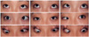
Ophthalmic examinations showed a visual acuity of 20/20 in both eyes, a mid dilated pupil of the left eye, and marked bilateral disc edema with peripapillary hemorrhage. She was unable to depress, adduct, and abduct the left eye and had limited abduction of the right eye (Fig. 1). Visual field examination showed peripheral constriction and enlargement of the blind spot in both eyes.
A complete neurological examination showed no abnormal findings except for limited ocular motion. Brain magnetic resonance (MR) imaging, MR angiography, and cerebral angiography with venous phase performed within 10 days of clinical onset were normal. Cerebrospinal fluid (CSF) examination revealed the following: pressure, 400 mm H2O; glucose, 61 mg/dl; protein, 17 mg/dl; red blood cells, 2/mm3; white blood cell, 1/mm3; and culture, negative.
Laboratory analysis showed that the platelet count was 19,000/mm3. Prothrombin time was normal, but the activated partial thromboplastin time measured by the (aPTT)-mixing test was prolonged at 55s (normal range 25-39s). The erythrocyte sedimentation rate was elevated at 26 mm/h. Urine analysis, an electrocardiograph and chest X-ray were normal. Values for C-reactive protein, fibrinogen, antithrombin III, protein C, and protein S were all normal. Immunologic tests was positive for IgG anticardiolipin antibody (36.7 GPLunit/ml: range <10 GPLunit/ml) and for lupus anticoagulant. Antinuclear antibody, anti-dsDNA antibody, antineutrophil cytoplasmic antibodies, antiplatelet antibody, thyroid autoantibodies and rheumatoid factor were negative. The serologic test for syphilis was negative. There was no other abnormal findings such as butterfly lash, discoid lash, photophobia, oral ulcer, arthritis, renal dysfunction, pleuritis or pericarditis. There was no clinical evidence of other autoimmune disease.
Six days later, the patient complained of central scotoma of the right eye and the visual acuity of the right eye had decreased to 40/200. Fundus examination of the right eye revealed new preretinal and subhyaloid hemorrhage. No evidence of retinal vascular occlusion was present in fluorescein angiographies performed at this time and two months later. No change was observed in the ocular motion examination. At this time, the platelet count was 20,000/mm3.
With an initial diagnosis of intracranial hypertension (IH), the platelet was treated with repeated lumbar puncture, acetazolamide, and diuretics. Since the CSF pressure did not decrease aspirin and steroid therapy was started and the CSF pressure was normalized after 28 days. Thirty days later, when the preretinal and subhyaloid hemorrhage of the right eye had decreased, heparin was administered and subsequently replaced with warfarin to maintain an international normalized ratio (INR) of 1.5-2.0.
A follow-up examination after two months revealed the disappearance of retinal hemorrhage in the right eye, visual acuity of 20/20 in both eyes, and normal ocular movement. Lupus anticoagulant and IgG anticardiolipin antibody remained positive.
Discussion
Based on the clinical and laboratory findings, the patient was diagnosed with bilateral sixth, and left third and fourth cranial nerve palsies secondary to IIH in primary APS.
Sixth cranial nerve palsy is known to occur in 10 to 40% of patients in most series of IIH,2-4 and third or fourth nerve palsy has been described occasionally.5,6 However, concurrent third, fourth, and sixth nerve palsies in one patient has not been reported previously. In addition previous reports provided no details regarding the incomitancy of the ocular motor deviation in various gaze positions. Other conditions associated with third cranial nerve palsy include diabetes mellitus, trauma, syphilis, aneurysm, tumor, or sinus infection; none of these was found in our patient.
The prevalence of IIH has not yet been reported for patients with primary APS although several cases have been described.7,8 The prevalence of anticardiolipin antibodies in patients with IIH was reported variously as 8.1%, 31% or 43%.8-10 However, the two highest results include patients with other risk factors for IIH and additional prothrombotic risk factors and are therefore considered to overestimate the prevalence with respect to APS. It is also important to take into account the fact that it may be possible to have circulating antiphospholipid antibodies without an increased risk for thromboembolism, and the prevalence of IIH may therefore be lower than predicted.
The term "idiopathic" requires the exclusion of intracranial venous sinus thrombosis. Our patient did not have overt sinus thrombosis although she may have partial dural sinus obstruction (venous sinus thrombosis involving the deep sinuses) that was missed by MR imaging or angiography. It is possible that non-occlusive thrombus lining the dural vessles could impede CSF absorption; however, cerebral vascular imaging was performed soon after the onset of symptoms while CSF pressure was still high and she recovered completely under therapy designed to lower the CSF pressure without anticoagulation. There is also a possibility that increased blood viscosity induced by anticardiolipin antibody without actual thrombosis may cause IH.11
Preretinal and subhyaloid hemorrhage of the right eye was considered to be the result of papilledema rather than a venous thrombus caused by by hypercoagulation or thrombocytopenia, as the hemorrhage was located near the disc and there was no evidence of retinal vascular occlusion present in fluorescein angiographies performed at this time and two months later. It is likely that an intraretinal hemorrhage caused by papilledema resulted in detachment of the internal limiting membrane, leading to subhyaloid hemorrhage.
This report describes the unusual presentation of a young female patient with bilateral sixth cranial nerve palsies, and left third and fourth cranial nerve palsies secondary to IIH with primary APS. Concurrent third, fourth and sixth cranial nerve palsies are a neurological complication that may be the initial ophthalmic manifestation of primary APS.
 XML Download