

 PDF
PDF ePub
ePub Citation
 Print
Print


Intramuscular hemangioma (IMH) is an uncommon type of tumor, accounting for less than 1% of the total number of hemangioma tumors diagnosed.1 IMHs are nonmetastasizing, benign hamartomatous congenital neoplasms that, after remaining unremarkable for long periods, may suddenly start to grow in the second and third decades of life.1-3 IMH in extraocular muscles are extremely rare; to the best of our knowledge, the cases of only three patients with such a neoplasm have been reported. These three cases, however, exhibited no abnormal ocular motility, their main symptoms being proptosis and lid swelling.4,5 Here, the case of a patient with large-angle hypertropia of an intramuscular hemangioma of the right superior rectus (SR) is presented in order to demonstrate the atypical clinical presentation which should alert surgeons to the possibility of such a lesion.
Case Report
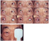
A 63-year-old man with progressive vertical deviation of the right eye presented to our strabismus department (Fig. 1a), noting that the ocular motility problem had been present for the past 6 months and was not associated with pain. The patient had no past medical history or systemic disease.
Physical examination indicated that corrected visual acuity and intraocular pressure were 20/20 and 18 mmHg, respectively, in both eyes. There was a 3mm-proptosis of the right eye compared with the left eye, and the upper and lower eyelids were slightly edematous, but with no chemosis or hyperemia; pulsation or bruit was not noted. He had 60PD of right hypertropia at both distance and near in primary gaze. He could fix objects with his right eye only when his left eye was occluded. (Fig. 1b) Significant limitation of his downgaze was noted. (Fig. 1a)
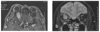
Orbital magnetic resonance imaging (MRI) studies revealed fusiform enlargement of the right superior rectus muscle, with prominent but irregular enhancement following gadolinium administration. (Fig. 2a, b) All other structures in the right orbit were normal except an incidental middle cranial artery (MCA) aneurysm in the left hemisphere, which was clipped by a neurosurgeon; the neurosurgeon indicated that the MCA aneurysm was not associated with the tumor.
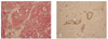
A biopsy through the infrabrow was performed under general anesthesia with a subperiosteal approach to the midsuperior orbit. Upon incision of the superior periorbita and levator muscles, the right SR appeared considerably thickened, and was dark purple and tense; the tendinous region was normal. An incisional biopsy was taken 15 mm behind the insertion of the muscle. The patient underwent a right superior rectus recession of approximately 15 mm by a hang-back technique and a 4-mm right inferior rectus resection with a conjuctival approach. The elasticity of the right SR was markedly reduced, and posterior portions were two or three times thicker than their normal size. Light microscopy of these sections revealed several blood-filled vessels of variable size surrounding muscle fibers and islands of fibrofatty deposits. (Fig. 3a) Capillary proliferation was not present, and there was no evidence of fibrotic intimal thickening or mitotic figures. An immunohistochemical study with antismooth muscle actin antigen (clone 1A4 at 1/25 titration, DAKO monoclonal, Denmark) demonstrated the presence of smooth muscle cells in the walls of blood vessels. (Fig. 3b) Biopsy results revealed an intramuscular hemangioma in the superior rectus muscle with cavernous-type vessels.
Systemic corticosteroids were utilized for four weeks postoperatively to ameliorate swelling, not to shrink the tumor. Three months after surgery there were no signs of tumor progression. Mild ptosis and 15 PD of right hypertropia has since been noted in the patient's primary gaze.
Discussion
Intramuscular hemangioma (IMH) classification is based on the size of the predominant vessel, as suggested by Allen and Enzinger.6 The frequency of IMHs are 50%, 29%, and 21% for capillary, cavernous, and mixed type, respectively.3
Christensen et al.4 reported the case of a 21-year-old woman with a 10-year history of proptosis, where exenteration showed four extraocular muscles affected by mixed capillary-cavernous hemangioma, all except the inferior rectus and inferior oblique muscles. Kiratli et al.5 described two cases of isolated primary IMHs of the extraocular muscles, one in the lateral rectus of a 3-year-old boy, and one in the medial rectus of a 40-year-old man. The three cases described above presented with gradual proptosis or lid swelling; abnormal ocular motility was apparently not present. The subject of this study likely represents the first reported case of an isolated primary IMH of pure carvenous type associated with large-angle strabismus. In our case, we assumed that the progressive fibrotic process of cavernous hemangioma caused the unusual hypertropia.
The most common cause of extraocular muscle (EOM) enlargement in adults is Graves' disease;7 both Graves' disease and myositis are associated with many inflammatory indicators. Orbital metastases of breast carcinoma and cutaneous melanoma have a predilection for EOMs and are a focal enhancement sign.8 Lymphoma may also cause significant enlargement of EOMs, and tends to frequently affect the superior rectus and levator palpebra complex; lymphoma usually shows minimal enhancement under contrast studies.9 Arteriovenous malformation consists of dilated muscularized blood vessel with fibrotic intimal thickening and displays both arterial and venous characteristics. Clinical symptoms and signs include loss of vision, chemosis, episcleral venous congestion, elevated intraocular pressure; it is associated with cranial nerve palsy.
Due to the rarity of ocular IMHs, we were unable to exclude all of the preceding diagnostic possibilities in our patient before biopsy results were obtained. However, the imaging characteristics of skeletal IMHs are well documented as a combination of large vessels with stagnant blood and nonvascular tissue within the tumors produce a typical serpiginous pattern, septated-striated high signal intensity, and curvilinear areas of low signal intensity.10 Similar features were observed in our patient. Preoperative clinical findings did not suggest inflammation or infectious processes.
The optimum management strategy appears to be total excision, whereby the mass must be excised beyond gross visible limits.2,3 In the present study, the patient complained only of hyperdeviation of the right eye because of cosmetic problems. He had minimal proptosis and lid swelling, normal visual acuity, and experienced no pain. Total excision was not considered a viable treatment option for the patient, as surgical results were unpredictable and may have created a potentially irreversible motility disturbance. Moreover, in cases of incomplete excision, there is an 18% risk of recurrence.3 In our case, establishing the diagnosis by biopsy and treating with strabismus surgery was at the very least potentially justified.
Systemic corticosteroids can decrease the bulk of the tumor, but IMHs of the extraocular muscles are insensitive to systemic corticosteroids because of encapsulation and the presence of cavernous elements which are resistant to corticosteroids.11
This case demonstrates that IMHs can occur in extraocular muscles, even causing ocular motility disturbances. Therefore, this type of tumor should be considered in the differential diagnosis of isolated extraocular muscle enlargement and unusual strabismus.
 XML Download