Abstract
Purpose
To determine the influence of clinical features and Indocyanine green (ICG) angiographic features on the visual outcome of patients with myopic sub-foveal choroidal neovascularization (CNV) who received photodynamic therapy (PDT).
Methods
Thirty-six consecutive patients (39 eyes) with myopic CNV who were followed up for more than one year after PDT were enrolled in this study. Clinical features included age, gender, refractive error, great linear dimension, and subretinal hemorrhage. ICG features included the lesion size, lacquer cracks, hypofluorescence surrounding the CNV (dark rim), peripapillary atrophy size, and visible prominent choroidal veins under the macula. Linear regression analysis was performed using the change in visual acuity (ΔlogMAR) as the dependent variable and the above factors as independent variables.
Pathologic myopia is a major cause of legal blindness in developed countries.1-2 The most common complication leading to decreased central vision in pathologic myopia is choroidal neovascularization (CNV), which affects 5% to 10% of patients with high myopia.3-5 Despite its importance as a cause of visual impairment in myopic patients, no effective treatments have been established for pathologic myopia except photodynamic therapy (PDT).6-7
Recently, Montero and Ruiz-Moreno8 showed that a patient's age at the onset of myopic CNV influenced the future prognosis of the disease. The younger patient group (≤55 years old) had a better final visual acuity at one-year follow-up after PDT than did the older group of patients (>55 years old). Their study included 31 eyes of 30 patients, and the mean age in the entire group was 54.9 「Standard Deviation (SD)=14.0」 years. Ergun and associates9 found in their 36-patient series that younger patients and patients with higher baseline visual acuities had better treatment outcomes at the two-year follow-up after PDT. The mean age of the patients was 59.7 (SD=15.1) years. Axer-Siegel and associates10 also demonstrated that the younger myopic CNV patient group (<60 years old) had a better final visual acuity and less visual loss after PDT. In their study of 29 patients, the mean age was 63.1 (SD=14.11) years with a mean follow-up period of 11.5 (SD=9.9) months. Based on their observations, they concluded that patient age was the most important prognostic factor of treatment outcome after PDT. The aforementioned studies have some limitations. They included a large number of elderly patients, which might cause neovascular degeneration of degenerative myopia to be confused with age-related macular degeneration.
In degenerative myopia, blood flow in the choroidal vessels is delayed and the choriocapillaries show severe circulatory disturbances.11-13 Since new vessels originate from the choroids, the hemodynamic characteristics of these vessels depend on those of the choroids. For example, the incidence of neovascularization may increase with the progression of degenerative changes in myopic eyes. However, the development of CNV requires the relative preservation of the choriocapillaries.14 It is also known that the fovea is almost entirely dependent on choroidal circulation for its nutrition. The visual prognosis of myopic CNV after PDT may not only be influenced by the characteristics of the CNV, but also by the state of the choroid and the choriocapillaries. Indocyanine green (ICG) angiography can better document choroidal vessels because of its better penetration of near-infrared light into the choroid. However, there have been no reports that have identified the ICG findings as a prognostic factor for the visual outcome of myopic CNV after PDT.
The 'dark rim' was first characterized by Scheider and associates.15 They observed the network of choroidal neovascularization within the choroidal hypofluorescent area in ICG angiograms using a scanning laser ophthalmoscope. There has also been a small number of reports describing a dark rim in idiopathic CNV and myopic CNV.15-18 However, no reports have investigated the relationship between the presence of a dark rim and the visual prognosis of idiopathic or myopic CNV.
The purpose of this study was to evaluate the visual improvement of myopic CNV when treated with PDT, and to establish a relationship between age and visual outcome. We also examined other clinical and ICG angiographic characteristics as prognostic factors for the visual outcome after PDT.
Thirty-eight consecutive patients (for a total of 41 eyes) with pathologic myopia and subfoveal CNV who were treated with verteporfin (Visudyne, NovartisAG, Basel, Switzerland) PDT at Yonsei University Medical Center between May 2002 and August 2004 were identified. Two patients were excluded because they were lost to follow-up, and the remaining 36 completed at least 12 months of follow-up. The study included 39 eyes of these 36 patients. Informed consent was obtained from all patients, and approval from the institution's ethics committee was not required for this retrospective study.
Pathologic myopia was defined as when an eye required a distance correction of at least -6.0 diopters (D=spherical equivalent). Patients with angioid streaks, evidence of histoplasmosis, choroiditis, idiopathic CNV, or other causes of CNV were excluded from the study. Subfovea was defined as an involvement of the center of the foveolar avascular zone.
PDT was administered by one surgeon (OW Kwon) according to the VIP and TAP study protocol6,7,19,20 with follow-ups at one week, one month, and three months after the therapy, and then every three months thereafter. If there were any signs of fluorescein leakage three months after PDT, then the PDT was repeated. Fluorescein angiography and ICG angiography were repeated for the initial examination and at every subsequent follow-up visit.
All patients received comprehensive ocular examinations (including dilated fundus photography, fluorescein angiography and ICG angiography) at the baseline investigation and again every three months thereafter. The best-corrected visual acuity was measured using a modified ETDRS chart.
The patient characteristics retrieved from the medical charts included age, gender, refractive errors, ETDRS visual acuity, and the number of treatments. The greatest linear dimension (GLD) (including the CNV, area of leakage and the area of blocked fluorescence in the lesion, according to the TAP study) was measured manually on the fluorescein angiograms.19
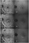
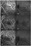
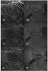
Indocyanine green (ICG) angiography was performed with a scanning laser ophthalmoscope (HRA, Heidelberg Engineering, Dossenheim, Germany). The ICG angiograms were reviewed by two examiners (SH Byeon and HJ Koh) to determine the lesion size on ICG angiography 「ratio of the total area of the lesion divided by the area of the optic disc (DA=Disc Area)」 and any associated findings, including hypofluorescence surrounding the CNV (dark rim), lacquer cracks, prominent large choroidal veins in the macula, and peripapillary choroidal atrophy size.21-24 The presence of an ICG angiographic dark rim (hypofluorescence around the CNV) was defined as a round-shaped background hypofluorescence within which a hyperfluorescent island appeared during the early phase (Fig. 1, 2).15-18 Lacquer cracks were identified using late-phase ICG angiography (Figures 1 and 3).16,22 Regarding the presence of visible large choroidal veins under the macula, the focal dilation of the choroidal veins in the posterior fundus during the early phase of ICG angiography was defined; some showed hyperfluorescence until the late-phase of the angiogram (Fig. 2).16,23 The area of peripapillary atrophy (islands of non-perfusion or reduced perfusion in the deep choroid in early or late hypoflurorescence) was defined as the ratio of the total area of peripapillary atrophy divided by the disc area on ICG angiography.22,24
To evaluate the relative impact of the baseline predictors (the clinical and ICG angiographic features) on the visual outcome, a linear regression analysis was performed. The main outcome measure was the change in the logMAR value during the follow-up period (ΔlogMAR), which was obtained by subtracting the baseline logMAR value from the final logMAR value 12 months after PDT. A linear regression analysis was performed using the change in visual acuity (ΔlogMAR) as the dependent variable and the above factors as independent variables. The potential prognostic effects were assessed using both univariate and multiple linear regression analyses. All reported p-values were the results of two-sided tests, and p-values ≤0.05 were considered statistically significant. SPSS 12.0 software for Windows (Chicago, Illinois, USA) was used for statistical calculations.
Thirty-nine eyes from 36 patients with subfoveal CNV secondary to pathologic myopia had completed 12 months of follow-up. All were Korean and 24 (66.7%) were females. The mean age of patients at presentation was 44.1 [standard deviation (SD)=15.0] years, with a range from 25 to 74 years. Refractive errors ranged from -6.00D to -23.00D, and the mean was -10.78 (SD=3.80) D. The average number of treatments during the 12 months of follow-up was 2.4 (SD=1.2). The mean Great linear dimension (GLD) of lesions on fluorescein angiography was 1775 (SD=976) µm (range= 400-4280) at presentation. Submacular hemorrhages visible on fundus photography and fluorescein angiography were found in 8 of 39 eyes. The mean area of lesions visible on ICG angiography was 0.71 (SD=0.61) DA (range=0.08-2.24). The lacquer cracks were found in 27 of 39 eyes. The mean area of peripapillary choroidal atrophy on ICG angiography was 2.0 (SD=2.4) DA. The hypofluorescence surrounding the CNV (dark rim) was found in 15 of 39 eyes, and prominent large choroidal veins were noted under the macula in 13 of 39 eyes.
The presenting best-corrected visual acuity measured with an ETDRS chart was greater than 20/40 in four (10.3%) eyes, from 20/40 to 20/200 in 26 (66.7%) eyes, and less than 20/200 in nine (23.0%) eyes. The baseline mean logMAR was 0.82 (SD=0.43). At the 12-month follow-up, the best-corrected visual acuity was greater than 20/40 in six (15.4%) eyes, from 20/40 to 20/200 in 25 (64.1%) eyes, and less than 20/200 in eight (20.5%) eyes. The mean logMAR had improved to 0.69 (SD=0.50).
The change in visual acuity from baseline at the 12-month follow-up was six lines or more of visual acuity improvement in seven eyes (18%), one to six lines of improvement in 14 eyes (36%), no change in nine eyes (23%), and a one-line or more decrease in visual acuity in nine eyes (23%).
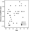
When clinical features including age, gender, refractive error, baseline visual acuity, baseline GLD, and submacular hemorrhage, and ICG features such as the size of CNV, lacquer cracks, area of peripapillary atrophy, dark rim, and a prominent large choroidal vein were examined as prognostic factors by univariate analysis, both age and the presence of a dark rim had a statistically significant effect on a change in logMAR(ΔlogMAR) at 12 months (Table 1). On a multivariate analysis, age and the presence of a dark rim still remained statistically significant at the 12-month follow-up (Table 1, Fig. 4).
More than 50% of the myopic CNV patients showed visual improvement 12 months after PDT. We found that patients older than 50 years rarely had visual improvement following PDT after 12 months of follow-up. Both the age at onset and the ICG angiographic feature (dark rim) were related to a change of visual acuity from the baseline. A younger age and the presence of a dark rim were good prognostic factors after PDT. Other factors such as gender, refractive error, baseline visual acuity, baseline GLD, submacular hemorrhage, and ICG features (including the size of the CNV, lacquer cracks, area of peripapillary atrophy, and a prominent large choroidal vein) were not predictive of outcome when the age and presence of a dark rim were accounted for. Our series contained a relatively large number of patients who had a visual improvement after PDT.6,7,8-10,25 This may be due to the younger age distribution in our series compared to other studies.
Among our participants, 24 eyes (62%) from 22 patients completed a 24-month follow-up. Their baseline characteristics were similar to those who had finished a 12-month follow-up (data not shown). Their baseline mean logMAR value was 0.89 (SD=0.45). The 24-month mean logMAR value improved to 0.80 (SD=0.57). In our 24-month follow-up group, a multivariate regression model also showed that a younger age (P=0.012) and a dark rim (P=0.031) were significantly correlated with an improvement of visual acuity at the 24-month follow-up.
Previous reports have suggested that patients who were younger at the onset of CNV might be able to retain good vision even without any treatment during follow-up. Because our study did not have a comparative group, it was not clear whether the better visual outcomes of young patients in our series were due to an effect of PDT or to the natural characteristics of myopic CNV in young patients. Reports on the natural history of CNV in high myopia patients vary, and most to date provide conflicting information.6,7,26-31 This may be because those studies enrolled a variety of eligible patients, represented variable lesion topographies, contained different patient age distributions, and had variable follow-up periods. Our study had similar eligibility criteria and age distributions to Yoshida's series,28 which found that younger, myopic CNV patients retained good visual acuity without any treatment. Japan and Korea have ethnic and geographic similarities as well as a prevalence of high myopia. Yoshida examined 63 consecutive patients (73 eyes) with myopic CNV, and then divided the patients into two groups according to their ages (≤40 and >40 years old). These patients were followed for more than three years. For comparison, we also divided our patients into two groups at the age of 40 (Table 2). In our study, the younger group (≤40) had significant improvement in visual acuity. In his natural history series, even though the younger patients retained relatively good visual acuity, they did not have significant visual improvement. We could hypothesize that PDT might increase the chance of visual improvement in myopic CNV patients, especially in the younger patient group. However, our study had a shorter follow-up period and our patients had a smaller mean refractive error.
In our 12-month multivariable regression model, the age of patients at onset significantly influenced their visual outcome, and a dark rim was another covariate significantly affecting the end results. We also found that this dark rim was more prominent as the CNV regressed with PDT during follow-up (Figure 1). We are the first group to study the dark rim as a predictive factor for visual outcome in CNV. Histologically, a dark rim corresponds to the multilayered, proliferated retinal pigment epithelium at the outer margin of the neovascular membrane.18,21 If large parts of the CNV remained unperfused for several weeks after PDT, the adjacent RPE cells may benefit from the interval required for the restoration of the CNV, and they could also migrate and proliferate around the vascular net.32 We postulated that patients with a dark rim had more potent regenerative potential of RPE and received a greater benefit from PDT. It was thought that the dark rim found on the baseline ICG angiography could be another indicator of the potent regenerative potential of RPE.
In age-related macular degeneration, the visual prognosis after PDT is related to the fluorescein angiographic subtype of CNV.19-20 The size of the CNV is also related to the prognosis. However, in myopic CNV, the relationship between the size of the CNV and visual outcome has not been fully determined.6-10,25 In our series, there was no significant relationship between size and visual outcome. New vessels originating from the choroids and the hemodynamic characteristics of new vessels must depend on those of the choroids. In eyes with severely decompressed choriocapillaries, the size and activity of neovascularization might be decreased. The visual prognosis could not only be influenced by the characters of the CNV but also by the state of the choroids and the choriocapillaries. This could obscure the relationship between the two.
Lacquer cracks are imaged using enhanced delineation with late phase ICG angiography.16,22 There were a few reports regarding the pathophysiological relationship between lacquer cracks, CNV and visual outcomes.22,33 However, in our results the presence of lacquer cracks had no significant effect on visual outcome after PDT.
There have been reports that an anatomic connection of the choroids with the arteries or veins is a factor influencing the natural course of CNV.16,23,34,35 We postulated that the visible prominent choroidal veins in the macula may reflect a thinned choroidal vasculature, which may in turn affect the visual prognosis of myopia. In our study, there were no significant relationships between the presence of a choroidal vein and the visual outcomes at the 12- and 24-month follow-ups (data not shown). In the univariate analysis of choroidal vessels on 24-month follow-up, the presence of a prominent choroidal vein in the macula correlated with a decrease in visual acuity, even though it was not significant (p=0.090). This relationship was the third-smallest p-value have an influence on the change in the logMAR value after 24 months, and it might be more prominent if it were to be examined over a longer period of time.
Our study, however, has its own limitations, including a small sample size, a short follow-up period and no comparative control group. Prevention of later chorioretinal atrophy development and further visual decrease over the long-term could not be assessed.36 Patients' ages at onset and ICG angiographic features (dark rim) were the significant prognostic indicators of the visual outcome after PDT in our study. In conclusion, younger patients and patients with a dark rim on ICG angiography had a higher chance of visual improvement after PDT in myopic CNV.
Figures and Tables
Fig. 1
Angiography of a 30-year-old man with myopic sub-foveal CNV in his left eye. At the initial examination, the patient's best-corrected visual acuity was 20/200. The refractive error was -9.0 diopters. (1A) An early-phase ICG angiogram revealed the hypofluorescence surrounding the CNV (dark rim). (1B) Late-phase ICG angiography showed lacquer cracks radiating from the lesion. At the 12-month follow-up, the patient's visual acuity had improved to 20/40. (1C, 1D) ICG angiography showed the decreased size of the lesion and a more prominent dark rim. At the 24-month follow-up, the patient's visual acuity remained at 20/40. (1E, 1F) ICG angiography also revealed the remaining smaller lesion.

Fig. 2
Angiography of the left eye of a 55-year-old man. At the initial examination, the patient's best-corrected visual acuity was 20/200. The refractive error was -9.0 diopters. (2A) An early-phase ICG angiography showed a dark rim surrounding the CNV, an adjacent small-blocked fluorescence due to hemorrhage and a bifurcate prominent choroidal vein. (2B) Late-phase ICG angiography revealed the visible hyperfluorscent choroidal vein on the macula, but no lacquer cracks were seen. (2C, 2D) At the 12-month follow-up, the patient's visual acuity had improved to 20/160. The lesion and the dark rim had become less prominent. Persistent prominent choroidal veins were noted. (2E, 2F) At the 24-month follow-up, the size of the CNV was enlarged even after receiving PDT five times. The patient's visual acuity was decreased to 20/200.

Fig. 3
ICG angiography of the right eye of a 58-year-old woman. At initial examination, the patient's best-corrected visual acuity was 20/63. The refractive error was -11.0 diopters. (3A) The mid-phase ICG angiography showed the lesion to be slightly more hyperfluorescent than the background. (3B) The late phase revealed dye leakage beyond the previous lesion with multiple lacquer cracks. (3C, 3D) At a 12-month follow-up, the patient's visual acuity had decreased to 20/200. The ICG angiography showed a similar appearance of the baseline ICG angiography despite four treatments of PDT during the previous 12 months. (3E, 3F) At the 24-month follow-up, the patient's visual acuity had returned to 20/125. During the 24-month follow-up, she had undergone PDT five times.

Fig. 4
Scattergram of the 12-month results of change in the logMAR (ΔlogMAR) after photodynamic therapy according to age and the presence of a dark rim.

References
1. Ghafour IM, Allan D, Foulds WS. Common causes of blindness and visual handicap in the west of Scotland. Br J Ophthalmol. 1986. 67:209–213.
2. Sperduto RD, Seigel D, Roberts J, et al. Prevalence of myopia in the Unites States. Arch Ophthalmol. 1983. 101:405–407.
3. Curtin BJ, Karlin DB. Axial length measurements and fundus changes in the myopic eye. Am J Ophthalmol. 1971. 71:42–50.
4. Grossniklaus HE, Green WR. Pathologic findings in pathologic myopia. Retina. 1992. 12:127–133.
5. Levy JH, Pollock HM, Curtin BJ. The Fuchs' spot: an opthalmoscopic and fluorescein angiographic study. Ann Ophthalmol. 1977. 9:1433–1443.
6. Verteporfin in Photodynamic Therapy (VIP) Study Group. Photodynamic therapy of subfoveal choroidal neovascularization in pathologic myopia with verteporfin: 1-year results of a randomized clinical trial - VIP report No. 1. Ophthalmology. 2001. 108:841–852.
7. Verteporfin in Photodynamic Therapy (VIP) Study Group. Photodynamic therapy of subfoveal choroidal neovascularization in pathologic myopia with verteporfin: 2-year results of a randomized clinical trial - VIP report No. 3. Ophthalmology. 2003. 110:667–673.
8. Montero JA, Ruiz-Moreno JM. Verteporfin photodynamic therapy in highly myopic subfoveal choroidal neovascularization. Br J Ophthalmol. 2003. 87:173–176.
9. Ergun E, Heinzl H, Stur M. Prognostic factors influencing visual outcome of photodynamic therapy for subfoveal choroidal neovascularization in Pathologic myopia. Am J Ophthalmol. 2004. 138:434–438.
10. Axer-Siegel R, Ehrlich R, Weinberger D, et al. Photodynamic therapy of subfoveal choroidal neovascularization in high myopia in a clinical setting: visual outcome in relation to age at treatment. Am J Ophthalmol. 2004. 138:602–607.
11. Avetisov ES, Salvitskaya NF. Some features of ocular microcirculation in myopia. Ann Ophthalmol. 1977. 9:1261–1264.
12. Curtin BJ. The pathogenesis of congenital myopia: a study of 66 cases. Arch Ophthalmol. 1963. 6:166–173.
13. Noble KG, Carr RE. Pathologic myopia. Ophthalmology. 1982. 89:1099–1100.
14. Steidl SM, Pruett RC. Macular complications associated with posterior staphyloma. Am J Ophthalmol. 1997. 123:181–187.
15. Scheider A, Kaboth A, Neuhauser L. Detection of subretinal neovascular membranes with Indocyanine green and an infrared scanning laser ophthalmoscope. Am J Ophthalmol. 1992. 113:45–51.
16. Iida T, Hagimura N, Kishi S, Shimizu K. Indocyanine green angiographic features of idiopathic submacular choroidal neovascularization. Am J Ophthalmol. 1998. 126:70–76.
17. Quaranta M, Arnold J, Coscas G, et al. Indocyanine green angiographic features of pathologic myopia. Am J Ophthalmol. 1996. 122:663–671.
18. Fukushima I, Takahashi K, Nishimura T, et al. Dark rim around choroidal neovascularization in indocyanine green angiography. Nippon Ganka Gakkai Zasshi. 1995. 99:1262–1270.
19. Treatment of Age-related Macular Degeneration with Photodynamic Therapy (TAP) Study Group. Photodynamic therapy of subfoveal choroidal neovascularization in age-related macular degeneration with verteporfin. One year results of 2 randomized clinical trials - TAP Report No. 1. Arch Opthalmol. 1999. 117:1329–1345.
20. Treatment of Age-related Macular Degeneration with Photodynamic Therapy (TAP) Study Group. Photodynamic therapy of subfoveal choroidal neovascularization in agerelated macular degeneration with verteporfin. Two year results of 2 randomized clinical trials - TAP Report No. 2. Arch Opthalmol. 2001. 119:198–207.
21. Parodi MB, Pozzo SD, Ravalico G. Angiographic features after photodynamic therapy for choroidal neovascularization in age related macular degeneration and pathologic myopia. Br J Ophthalmol. 2003. 87:177–183.
22. Axer-Siegel RD, Cotlear D, Priel E, et al. Indocyanine green angiography in high myopia. Ophthalmic Surg Lasers Imaging. 2004. 35:139–145.
23. Yannuzzi LA, Flower RW, Slakter JS. Indocyanine green angiography. 1997. 1st ed. St. Louis: Mosby;chap. 85.
24. Tokoro T, Mochizuki M, Yasuzumi K, et al. Peripapillary crescent enlargement in highly myopic eyes evaluated by fluorescein and indocyanine green angiography. Br J Ophthalmol. 2003. 87:1088–1090.
25. Lam DSC, Chan W-M, Liu DTL, et al. Photodynamic therapy with verteporfin for subfoveal choroidal neovascularization of pathologic myopia in Chinese eyes: a prospective series of 1 and 2 year follow up. Br J Ophthalmol. 2004. 88:1315–1319.
26. Fried M, Siebert A, Meyer-Schwickerath G. Natural history of Fuchs' spot: a long-term follow up study. Doc Ophthalmol. 1981. 28:215–221.
27. Avila MP, Weiter JJ, Jalkh AE, et al. Natural history of choroidal neovascularization in degenerative myopia. Ophthalmology. 1984. 91:1573–1581.
28. Yoshida T, Ohno-Matsui K, Ohtake Y, et al. Long-term visual prognosis of choroidal neovascularization in high myopia. Ophthalmology. 2002. 109:712–719.
29. Hayashi K, Ohno-Matsui K, Yoshida T, et al. Characteristics of patients with favorable natural course of myopic choroidal neovascularization. Graefes Arch Clin Exp Ophthalmol. 2005. 243:13–19.
30. Hotchkiss ML, Fine SL. Pathologic myopia and choroidal neovascularization. Am J Ophthalmol. 1981. 91:177–183.
31. Hampton GR, Kohen D, Bird AC. Visual prognosis of disciform degeneration in myopia. Ophthalmology. 1983. 90:923–926.
32. Schmidt-Erfurth U, Michels S, Barbazetto I, Laqua H. Photodynamic effects on choroidal neovascularization and physiologic choroid. Invest Ophthalmol Vis Sci. 2002. 43:830–841.
33. Ohno-Matsui K, Yoshida T, Futagami S, et al. Patchy atrophy and lacquer cracks predispose to the development of choroidal neovascularization in pathologic myopia. Br J Ophthalmol. 2003. 87:570–573.
34. Bischoff PM, Flower RW. High blood pressure in choroidal arteries as possible pathogenic mechanism in senile macular degeneration. Am J Ophthalmol. 1983. 96:398–399.
35. Dimitrova G, Tamaki Y, Kato S, Nagahara M. Retrobulbar circulation in myopic patients with or without myopic choroidal neovascularization. Br J Ophthalmol. 2002. 86:771–773.
36. Yoshida T, Ohno-Matsui K, Yasuzumi K, et al. Myopic choroidal neovascularization. Ophthalmology. 2003. 110:1297–1305.


 PDF
PDF ePub
ePub Citation
 Print
Print


 XML Download