

 PDF
PDF ePub
ePub Citation
 Print
Print


The abducens nerve passes through the deepest part of the cranial base. Duplication of the nerve has sometimes been discovered during surgery for petroclival tumors or at autopsy. During the embryological development of human embryos, the abducens nerve is in the form of two aberrant branches.1 The failure of these nerve branches to fuse could lead to the duplication of the abducens nerve.1 There has been no report of associated eye movement in a patient with duplicated abducens nerve. Here, we present magnetic resonance imaging (MRI) findings of duplicated abducens nerves and associated eye movement.
Case Report
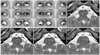
A 24-year-old woman was referred to us for the evaluation of eye movement from the otolaryngology department where she had undergone brain MRI for the evaluation of tinnitus. The 0.7-mm-thick MR images obtained with a T2-weighted 3D fast field echo technique in the axial plane at the level of the brainstem2 showed a duplicated left abducens nerve. One right abducens nerve and two left abducens nerves were observed, otherwise there was no abnormal findings in the brain (Fig. 1). She was born after an uneventful full-term pregnancy and delivery was normal. Data collection for this study conformed with Korean law.
On ophthalmologic evaluation, her best corrected visual acuities were 20/20 OU. She was orthotropic in 5 cardinal positions, and ductions and versions were full (Fig. 1). The anterior segment and fundi were normal.
Discussion
The abducens nerve can be reliably observed by MRI,2 thus MRI can be very helpful for accurate diagnosis as well as providing additional insight into the pathogenesis of various diseases related with congenitally anomalous abducens nerves, such as Duane's retraction syndrome,2-4 synergistic divergence,5 and congenital fibrosis syndrome.5
During the course of the abducens nerve between the brainstem and the lateral rectus muscle, the abducens nerve usually travels forward as a single trunk, but it is not uncommon for the nerve to split into two branches. Autopsy studies have described an incidence rate of the duplication of the abducens nerve from 5 to 28.6%,1,6 and MR images have also showed double rootlets of the abducens nerve in 25.2% of the study's subjects.7 However, details of associated eye movement have not previously been reported in the literature. In this case report, we present the findings of a duplicated abducens nerve with completely normal eye movement.
 XML Download