

 PDF
PDF ePub
ePub Citation
 Print
Print


Abstract
Purpose
We investigated hemodynamic changes in the ophthalmic artery (OA) using color Doppler imaging (CDI) after two horizontal rectus muscles surgery.
Methods
Eyes of the surgical group (n=18) underwent surgery on two horizontal rectus muscles, and the control group was the contralateral eyes. CDI of the OA was performed before operation and on postoperative days (POD) 1, 7 and 30. Peak systolic (Vmax), end diastolic (Vmin), and mean (Vmean) blood flow velocities were measured, and resistivity index (RI) and pulsatility index (PI) were calculated.
Results
Vmax, Vmin and Vmean were significantly higher, and RI and PI were significantly lower in the surgical group than in the control group on POD 1 (p<0.05). In the surgical group, Vmax, Vmin and Vmean were significantly higher, and RI and PI were significantly lower, on POD 1 than those mesured on other days (p<0.05).
Go to : 
REFERENCES
1. Erickson SJ, Hendrix LE, Massaro BM, et al. Color Doppler flow imaging of the normal and abnormal orbit. Radiology. 1989; 173:511–6.

2. Langham ME, Farrell RA, O'Brien V, et al. Blood flow in the human eye. Acta Ophthalmol. 1989; 191:9–13.
3. Guthoff RF, Berger RW, Winkler P, et al. Doppler ultrasonography of the ophthalmic and central retinal vessels. Arch Ophthalmol. 1991; 109:532–6.
4. Regillo CD, Sergott RC, Brown GC. Successful scleral buckling procedures decrease central retinal artery blood flow velocity. Ophthalmology. 1993; 100:1044–9.
5. Santos L, Carpeans C, Gonzalez F. Ocular blood flow velocity reduction after buckling surgery. Graefes Arch Clin Exp Ophthalmol. 1994; 232:666–9.
6. Trible JR, Sergott RC, Spaeth GL, et al. Trabeculectomy is associated with retrobulbar hemodynamic changes. A color Doppler analysis. Ophthalmology. 1994; 101:340–51.
7. Mittra RA, Sergott RC, Flaharty PM, et al. Optic nerve decompression improves hemodynamic parameters in papilledema. Ophthalmology. 1993; 100:987–97.
8. Lee JP, Olver JM. Anterior segment ischemia. Eye. 1990; 4:1–6.
9. Aburn NS, Sergott RC. Orbital colour Doppler imaging. Eye. 1993; 7:639–47.
10. Virdi PS, Heyreh SS. Anterior segment ischemia after recession of various recti. An experimental study. Ophthalmology. 1987; 94:1258–71.
11. Wilcox LM, Keough EM, Connolly RJ, Hotte CE. The contribution of blood flow by anterior ciliary arteries to the anterior segment in the primate eye. Exp Eye Res. 1980; 30:167–74.
12. Mckeown CA, Lambert HM, shore JW. Preservation of anterior ciliary vessels during extraocular muscle surgery. Ophthalmology. 1989; 96:498–506.
13. The Korean Strabismus and Pediatric Ophthalmological Society. Gross anatomy of the extraocular muscles. In: Current Concepts in Strabismus. 1st ed.Reston: Naewae Haksool;2004. p. 16–7.
14. Hayreh SS. Proceedings: Anatomy and pathophysiology of ocular circulation. Exp Eye Res. 1973; 17:387–8.
15. France TD, Simon JW. Anterior segment ischemia syndrome following muscle surgery; the AAPOS experience. J Pediatr Ophthalmol Strabismus. 1986; 23:87–91.
16. Elsas FJ, Witherspoon CD. Anterior segment ischemia after strabismus surgery in child. Am J Ophthalmol. 1987; 6:833–4.
17. Hiatt RL. Production of anterior segment ischemia. Trans Am Ophthalmol Soc. 1977; 75:87–102.
18. Saunders RA, Sandall GS. Anterior segment ischemia syndrome following rectus muscle transposition. Am J Ophthalmol. 1982; 93:34–8.
19. Simon JW, Price EC, Krohel GB, et al. Anterior segment ischemia following strabismus surgery. J Pediatr Ophthalmol Strabismus. 1984; 21:179–84.
20. Wagner RS, Nelson LB. Complications following strabismus surgery. Int Ophthalmol Clin. 1985; 25:171–8.
21. Hayreh SS. Scott WE. Fluorescein iris angiography. II. Disturbances of iris circulation following strabismus operation on the various recti. Arch Ophthalmol. 1978; 96:1390–400.
22. Hayreh SS. Scott WE. Fluorescein iris angiography. I. Normal pattern. Arch Ophthalmol. 1978; 96:1383–9.
23. Meyer PA, Watson PG. Low dose fluorescein angiography of the conjunctiva and episclera. Br J Ophthalmol. 1987; 71:2–10.
24. Ormerod LD, Fariza E, Hughes GW, et al. Anterior segment fluorescein videoangiography with a scanning angiographic microscope. Ophthalmology. 1990; 97:745–51.
25. Lieb WE, Cohen SM, Merton DA, et al. Color Doppler imaging of the eye and orbit: technique and normal vascular anatomy. Arch Ophthalmol. 1991; 109:527–31.
26. Saliba E, Laugier J. Circulation du nouveau-ne' et de l'enfant. Pourcelot L, editor. Dynamique Cardio-Vasculaire Foetale et Neonatale Echocardiographie-Doppler. 1st ed.Paris: Masson;2006. p. 139–61.
27. Freed KS, Brown CK, Carrol BA. The extracranial cerebral vessels. Rumack CM, Wilson SR, Charboneau JW, editors. Diagnostic Ultrasound. 1st ed.St Louis: Mosby;2006. p. 26–30.
28. Peter AN, Scott WS, Alon H. Color Doppler ultrasound measurements after topical and retrobulbar epinephrine in primate eyes. Invest Ophthalmol Vis Sci. 1997; 38:2655–61.
29. Pelit A, Barutcu O, Oto S, Aydin P. Investigation of hemodynamic changes after strabismus surgery using color Doppler imaging. J AAPOS. 2002; 6:224–7.
30. Bayramlar H, Totan Y, Cekic O, et al. Evaluation of hemodynamic changes in the ophthalmic artery with color Doppler ultrasonography after strabismus surgery. J Pediatr Ophthalmol Strabismus. 2000; 37:94–100.
31. Tane S, Hashimoto T. Estimation of blood flow in the carotid artery and intraorbital ophthalmic artery by color pulse Doppler ultra sonography. Acta Ophthalmol. 1992; 204:S62–5.
32. Han KH, Kim BJ, Youn JW, Lee HB. The measurement of ocular blood flow velocity using Doppler ultrasound in normal eyes. J Korean Ophthalmol Soc. 1994; 35:1685–90.
33. Hong JW, Kim YY, Lee TS. Measurement of blood flow velocity of ophthalmic and central retinal artery using color Doppler imaging. J Korean Ophthalmol Soc. 1996; 37:985–92.
34. Goebel W, Lieb W, Ho A, et al. Color Doppler Imaging: A new technique to assess orbital blood flow in patients with diabetic retinopathy. Invest Ophthalmol Vis Sci. 1995; 36:864–70.
35. Lizzi FL, Mortimer AJ. Bioeffects considerations for the safety of diagnostic ultrasound. J Ultrasound Med. 1988; 7:S1–38.
36. Lizzi FL, Packer AJ, Coleman DJ. Experimental cataract production by high frequency ultrasound. Ann Ophthalmol. 1978; 10:934–42.
37. Lizzi FL, Coleman DJ, Driller J, et al. Effects of pulsed ultrasound on ocular tissue. Ultrasound Med Biol. 1981; 7:245–52.
Go to :
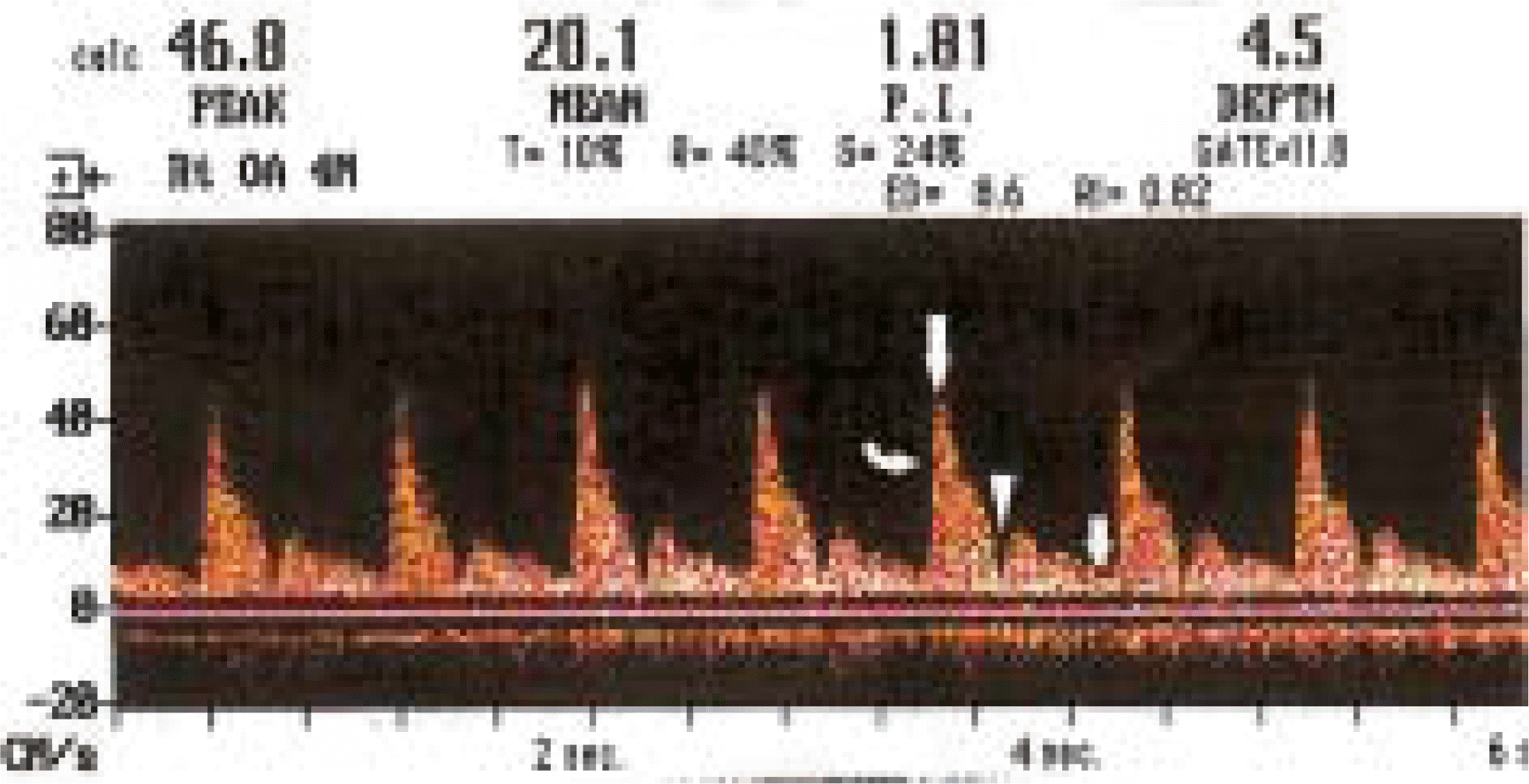 | Fig. 1.Color Doppler waveform of the ophthalmic artery showing initial steep systolic velocity rise (curved arrow)7 and notched “incisure” in middownslope (arrowhead). Only such waves were selected to measure the blood flow velocity parameters. The highest Value of the wave is the peak systolic blood flow velocity (long straight arrow) and the lowest value of the wave is the end diastolic blood flow velocity (short straight arrow). |
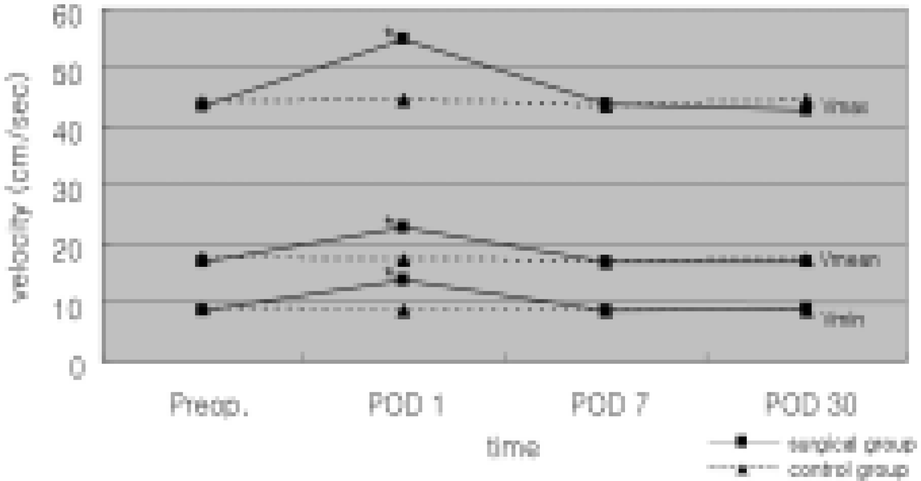 | Fig. 2.The Change of peak systolic (Vmax), end diastolic (Vmin) and mean (Vmean) blood flow in the ophthalmic artery in both groups with the lapse of time. ∗2 significantly different compared to the values measured at other examination times in the surgical group. |
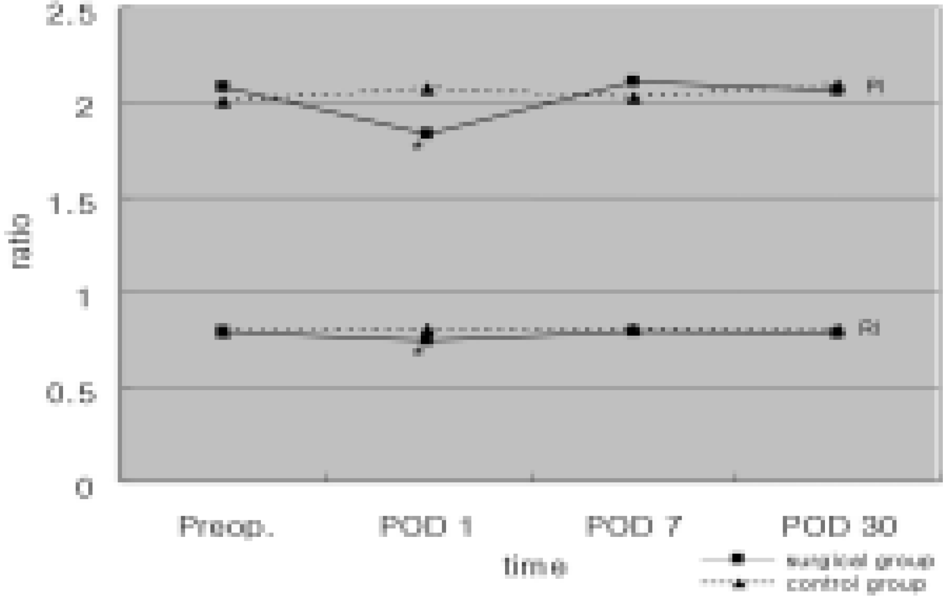 | Fig. 3.The change of resistivity index (RI) and pulsatility index (P1) in the ophthalmic artery in both groups with the lapse of time.∗: significantly different compared to the values calculated at other examination times in the surgical group. |
Table 1.
Demographics Of the subjects
| Surgical grout) | Control group | |
|---|---|---|
| Number of eves | 18 | |
| Male: Female | 10:8 | |
| Right: Left | 7:11 | 11:7 |
| Mean age (Years) | 25.22+165 | |
| Age range (Years) | 7~56 | |
Table 2.
Comparison of mean (뀁SD) preoperative and postoperative blood flow velocity parameter in the ophthalmic artery between surgical and control groups
| Parameter | Preoperation | POD 1 | POD 7 | POD 30 | |
|---|---|---|---|---|---|
| vmax | Surgical group | 43.33±7.16 | 54.91±6.49 | 43.78±6.79 | 42.84±9.44 |
| Control group | 44.01+8.76 | 44.52+9.23 | 43.35±8.91 | 44.62±8.19 | |
| p-Value | 0.72 | 0.00∗ | 0.74 | 0.42 | |
| Vmin | Surgical group | 8.63±1.16 | 13.74±1.43 | 8.61±1.18 | 8.91±1.59 |
| Control group | 17.50±3.25 | 22.61±3.18 | 16.86±3.11 | 16.83+3.77 | |
| p-Value | 0.52 | 0.00∗ | 0.74 | 0.96 | |
| Vmean | Surgical group | 16.76±3.86 | 22.61±3.18 | 16.86±3.11 | 16.83±3.77 |
| Control group | 17.50±325 | 17.18±2.82 | 17.06±353 | 17.14±3.25 | |
| group p-Value | 0.52 | 0.00∗ | 0.74 | 0.96 | |
| RI | Surgical group | 0.79±0.04 | 0.74±0.04 | 0.79±0.04 | 0.78±0.05 |
| Control group | 0.79±0.05 | 0.80±0.04 | 0.79±0.04 | 0.80±0.06 | |
| p-Value | 0.61 | 0.00∗ | 0.74 | 0.31 | |
| PI | Surgical group | 2.08±031 | 1.83±0.27 | 2.11±0.38 | 2.06±056 |
| Control group | 2.0±0.36 | 2.07±0.39 | 2.02±0.31 | 2.09±0.36 | |
| p-Value | 0.56 | 003∗ | 0.4 | 0.72 |
 XML Download