Abstract
This study was performed to demonstrate the ultrasound, biomicroscopic and dimensional changes of angle structure after laser iridotomy (LI) and primary trabeculectomy (PT) in primary angle-closure glaucoma (PACG). Angle-opening distance at a point 500(m from the scleral spur (AOD500), trabecular-iris angle (Θ1), trabecular ciliary process distance (TCPD), ciliary process-iris angle (CPI), iris thickness (ID1, ID3), length of iris-lens contact distance (ILCD) and anterior chamber depth (ACD) were assessed before and after each procedure. Thirteen patients with LI and 16 with PT were prospectively enrolled. There were statistically significant increases in AOD500, Θ1, and ILCD in both groups. CPI was decreased in both groups. ACD, TCPD, and iris thickness were not changed significantly. The changes in angle configuration after LI or PT may result more from alterations in aqueous pressure gradients across the iris and the changes of configuration were greater in the iris roots without rotation of ciliary body. However, we didn't find any significant differences in the changes of parameters between the two procedures.
References
1. Cho HJ, Woo JM, Yang KJ. Ultrasound biomicroscopic dimensions of the anterior chamber in angle-closure glaucoma patients. Korean J Ophthalmol. 2002; 16:20–25.

2. Garudadri CS, Chelerkar V, Nutheti R. An ultrasound biomicroscopic study of the anterior segment in Indian eyes with primary angle-closure glaucoma. J Glaucoma. 2002; 11:502–507.

3. Marchini G, Pagliarusco A, Toscano A, Tosi R, Brunelli C, Bonomi L. Ultrasound biomicroscopic and conventional ultrasonographic study of angle-closure glaucoma. Ophthalmology. 1998; 105:2091–2098.
4. Ishikawa H, Esaki K, Liebmann JM, Uji Y, Ritch R. Ultrasound biomicroscopy dark room provocative testing: a quantitative method for estimating anterior chamber angle width. Jpn J Ophthalmol. 1999; 43:526–534.

5. Salmon JF, Swanevelder SA, Donald MA. The dimension of eyes with chronic angle-closure glaucoma. J Glaucoma. 1994; 3:237–243.
6. Caronia RM, Liebmann JM, Stegman Z, Sokol J, Ritch R. Increase in iris-lens contact after laser iridotomy for pupillary block angle closure. Am J Ophthalmol. 1996; 122:53–57.

7. Jin JC, Anderson DR. The effect of iridotomy on iris contour. Am J Ophthalmol. 1990; 110:260–263.

8. Pavlin CJ, Harasiewicz K, Eng. P, Foster FS. Ultrasound biomicroscopy of anterior segment structures in normal and glaucomatous eyes. Am J Ophthalmol. 1992; 113:381–389.
9. Lee DA, Brubaker RF, Illstrup DM. Anterior chamber dimensions in patients with narrow angles and angle-closure glaucoma. Arch Ophthalmol. 1984; 102:46–50.

10. Ishikawa H, Esaki K, Liebmann J, Uji Y, Ritch R. Quantitative assessment of the anterior segment using ultrasound biomicroscopy. Curr Opin Ophthalmol. 2000; 11:133–139.

11. Markowitz SN, Morin JD. The ratio of lens thickness to axial length for biometric standardization in angle-closure glaucoma. Am J Ophthalmol. 1985; 99:400–402.

12. Sakuma T, Sawada A, Yamamoto T, Kitazawa Y. Appositional angle closure in eyes with narrow angles: an ultrasound biomicroscopic study. J Glaucoma. 1997; 6:165–169.
13. Quigley HA, Friedman DS, Cogdon NG. Possible mechanisms of primary angle-closure and malignant glaucoma. J Glaucoma. 2003; 12:167–180.

14. Jacobs IH, Krohn DL. Central anterior chamber depth after laser iridotomy. Am J Ophthalmol. 1980; 89:865–867.
15. Wishart PK, Atkinson PL. Extracapsular cataract extraction and posterior chamber lens implantation in patients with primary chronic angle-closure glaucoma: effect on intraocular pressure control. Eye. 1989; 3:706–712.

16. Gunning FP, Greve EL. Lens extraction for uncontrolled angle-closure glaucoma: longterm follow-up. J Cataract Refract Surg. 1998; 24:1347–1356.

17. Jacobi PC, Dietlein TS, Luke C, Engels B, Krieglstein GK. Primary phacoemulsification and intraocular lens implantation for acute angle-closure glaucoma. Ophthalmology. 2002; 109:1597–1603.
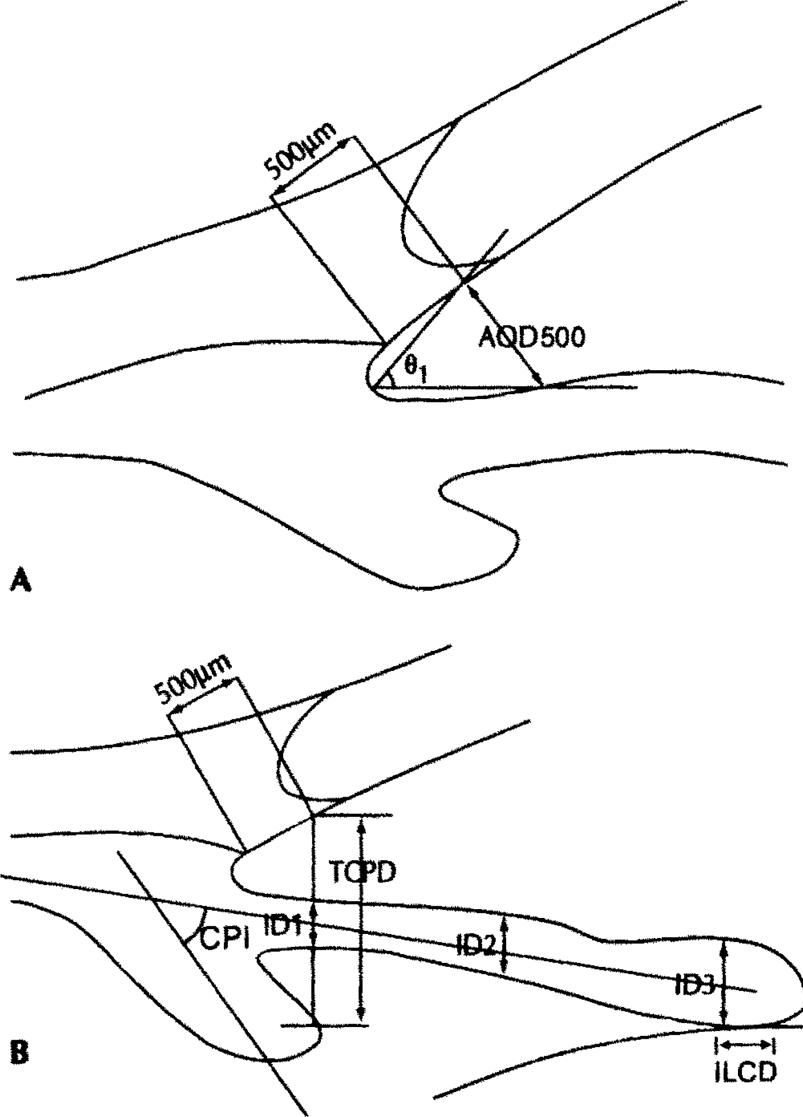
Fig. 1.
Ultrasound biomicroscopic measurement positions displayed on a diagrammatic representation of the anterior segment of the eye. (A) Angle-opening distance (AOD 500) was measured on the line perpendicular to the trabecular meshwork at a point 500 µm from the scleral spur. The trabecular-iris angle (Θ1) was measured with the apex in the iris recess and the arms of the angle passing through a point on the trabecular meshwork 500 µm from the scleral spur and through the point on the iris perpendicularly opposite. (B) Trabecular ciliary process distance (TCPD) was measured on the line extending from a point 500 µm from the scleral spur perpendicularly through the iris to the ciliary process. Iris thickness was measured 1mm from the iris root (ID1) and at its thickest point near the margins (ID3). Ciliary process-iris angle (CPI) and length of iris-lens contact distance (ILCD) are also indicated.

Table 1.
Patients demographics
| Angle-closure glaucoma patients | ||
|---|---|---|
| Laser iridotomy group | Trabeculectomy group | |
| Gender | ||
| (male/female, No. of patient) | 2/11 | 3/13 |
| Age (years) | ||
| Mean ± SD | 60.30 ± 8.17 | 65.83 ± 5.91 |
| (Range) | (47–70) | (59–74) |
Table 2.
Mean preoperative and postoperative intraocular pressures(mmHg) in the laser iridotomy and trabeculectomy groups.
| Laser iridotomy group | Trabeculectomy group | |
|---|---|---|
| Preoperative | 42.1 ± 15.9 | 43.7 ± 15.6 |
| Postoperative | ||
| 2 weeks | 14.8 ± 2.2 | 19.0 ± 2.4 |
| 2 months | 15.2 ± 2.0 | 14.5 ± 3.2 |
Table 3.
Mean preoperative and postoperative ultrasound biomicroscopy (UBM) parameters in the laser iridotomy group
| UBM parameter | Preoperative | Postoperative | P value |
|---|---|---|---|
| AOD∗500(µm) | 69.46 ± 25.39 | 187.15 ± 13.30 | .000 |
| θ1†(°) | 11.92 ± 3.40 | 27.31 ± 5.23 | .000 |
| ACD‡(µm) | 1917.61 ± 281.73 | 2050.00 ± 251.46 | .100 |
| TPCD§(µm) | 800.23 ± 106.52 | 872.54 ± 105.98 | .081 |
| CPIII(°) | 26.15 ± 7.89 | 4.39 ± 1.22 | .005 |
| ILCD#(µm) | 605.38 ± 100.38 | 1053.85 ± 106.34 | .000 |
| ID※1(µm) | 545.38 ± 108.68 | 505.38 ± 104.61 | .237 |
| ID※(µm) | 620.77 ± 108.28 | 569.23 ± 89.58 | .190 |
Table 4.
Mean preoperative and postoperative ultrasound biomicroscopy (UBM) parameters in the trabeculectomy group
| UBM parameter | Preoperative | Postoperative | P value |
|---|---|---|---|
| AOD∗500(µm) | 79.66 ± 31.12 | 230.00 ± 67.82 | .004 |
| θ1†(°) | 9.33 ± 3.88 | 25.30 ± 2.07 | .004 |
| ACD‡(µm) | 2036.66 ± 406.23 | 2186.66 ± 416.15 | .229 |
| TPCD§(µm) | 912.50 ± 127.03 | 1007.83 ± 140.02 | .109 |
| CPIII(°) | 24.17 ± 7.76 | 11.17 ± 6.91 | .020 |
| ILCD#(µm) | 588.33 ± 67.06 | 980.00 ± 128.99 | .004 |
| ID※(µm) | 581.67 ± 109.26 | 531.67 ± 85.42 | .335 |
| ID※(µm) | 648.33 ± 95.17 | 598.48 ± 96.3 | .199 |


 PDF
PDF ePub
ePub Citation
 Print
Print


 XML Download