Abstract
Primary lid tuberculosis after lid surgery is a very rare condition and is likely caused by the introduction of bacilli through epithelial injury. Secondary infection, due to direct hematogenous spread or contiguous spread from adjacent structures are more common presentations of lid tuberculosis. The authors experienced a case of primary lid tuberculosis occurring in a 19 year old female after blepharoplasty for making a eyelid crease. Her upper lid skin showed a reddish and non-tender mass lesion measured 3x1 cm, which was diagnosed as the tuberculosis through typical histopathological findings(caseous necrosis), acid-fast bacilli stain and PCR, and treated with anti-tuberculosis medications.
Tuberculosis is a infectious disease caused by mycobacteria species, which occasionally affects lung. However, the kidneys, lymph nodes, brain, bowels, skin, and eyes, can also be affected.1 Systemic tuberculosis arises in about 1-2% of human beings2 but primary lid tuberculosis is a very rare condition.
In most cases, lid infections are due to the contiguous spread from adjacent skin, conjunctival lesions or direct hematogenous spread.3 Primary lid infection caused by direct introduction of bacilli through lid injury or defect4 is less common condition than contiguous spread.
Eyelid tuberculosis begins with red-brownish papules in the early stage and can form nodules, plaque or tumor like lesions according to the disease progression. After all it leaves ulcerative lesions, skin fistulas and scars specific histopathologic examination sometimes demonstrates caseous necrosis with chronic granulomatous inflammation. Therefore if the diagnosis is delayed, there may be eyelid and tarsal plate destruction over large areas.5,6
The authors experienced a case of primary tuberculosis of the eye lid occurring in a 19 year old female after blepharoplasty for making an eyelid crease and would report this case.
The authors examined 19 year-old female complaining of a upper lid reddish mass on her right eye with one month duration. She had bilateral blepharoplasty for making an eyelid crease one month previously and the mass lesion developed after that operation. She was treated for several days in local eye clinics but no improvement was observed.
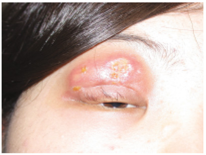
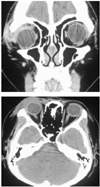
In the initial examination, there was a non-tender mass present on the right upper lid. The lesion measured 3 cm × 1 cm on the upper lid space (Fig. 1) and there weren't any other systemic infection signs or lymphadenopathies. Her corrected visual acuity was 1.25 in both eyes and other ocular examinations and routine biochemical investigations were normal. Computer tomography of the orbit revealed a diffuse homogenous enhancement of the soft tissue density in the right upper lid (Fig. 2).
At first time, she was prescribed 30 mg of prednisolone (Delta-Cortef)® once a day, and the lesion size appeared to be decreased. So the dosage was reduced to 20 mg/day. But the mass size appeared to be increased and exacerbated, we increased the dosage to 60 mg for 5 days. After drinking alcohol, the mass size were increased further more and the patient were received 250 mg of methylprednisolone (Solumedrol®) four times a day for 3 days. While undergoing intravenous steroid therapy, an excisional biopsy was performed through the previous lid operation scar.
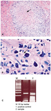
Upon operation, it was found that the mass was firmly attached to the upper orbital rim and a subtotal excision was performed. In histopathologic examination, only lymphocytic infiltration were showed on hematoxylin-eosin stain (Fig. 3). We thought it was just only a non-specific inflammatory reaction and radiation therapy was performed for a month without further systemic evaluations for detection of primary focus lesion. The total dose were 3600 cGy and it was applied to the patient divided with 20 times. After radiation therapy, lid swelling around the mass appeared to be subsided but the mass itself showed an ulcerative change associated with purulent discharge. So we started antibiotic therapy.
After that, clinical symptoms was seemed to be improved. However a newly developed nodular mass lesion was detected after two week radiation therapy (Fig. 4). We sent her to another hospital for further evaluations.
After transfer, the excision biopsy was done. The mass size was gradually increased and more eyelid swelling and associated skin defect were developed (Fig. 5). The result of the histopathologic examination were caseous necrosis with chronic granulomatous inflammation and bacilli were detected through acid-fast bacilli stain. Finally, Mycobaterium tuberculosis was demonstrated through PCR (polymerase chain reaction) (Fig. 6).
The authors made definitive diagnosis as upper eyelid tuberculosis and treated her with anti-tuberculosis medications with 9 month schedule. After 9 months of INH and rifampin therapy, the inflammation was reduced but a large skin defect was left on the upper lid and we directly repaired lid defect (Fig. 7).
M. tuberculosis was first discovered by Koch7 in 1882. It affects one-third of the global population and 3 million people lose their lives due to this bacterial infection in every year.
M. tuberculosis is a gram positive aerobic bacilli 10 µm long and 1 µm wide. It grows well between 35-44℃ and needs an oxygen rich environment for maximal growth. In the Ziehl-Neelsen stain, the organism shows acid fastness because of the mycolic acid in the cell wall that is not decolorized in acid alcohol.8 Mycobacteria can be classified as M. tuberculosis, M. africanum, M. bovis, and M. leprae and atypical mycobacteria is the organism which influences in immunosuppressive environment.9,10
Ocular tuberculosis was firstly reported in 1771 by Maitrejan.11 After this, Gueneau de Mussy12 reported choroidal tuberculoma in miliary tuberculosis in 1830. The most common manifestation of the ocular tuberculosis is anterior uveitis or choroiditis13 caused by hematogenous infection or a hypersensitivity after another organ infections.
Primary lid tuberculosis is a very rare condition and likely to be caused by introduction of bacilli through epithelial injury.4 Secondary infection due to direct hematogenous spread or contiguous spread from adjacent structures are more common presentations of lid tuberculosis.3
Eyelid tuberculosis begins with red-brownish papules in early stages and with progression, it forms an indurated nodule, plaque or tumor and later becoming an ulcerative lesion and skin fistula with scar. When diagnosis is delayed, there may be eyelid and tarsal plate destruction in large areas and abscess, skin fistula and cicatrical ectropion can also occur.6
Diagnosis is established through specific histopathologic examinations, tuberculin skin testing and the stain and culture of acid-fast bacilli. It is also done indirectly through the response to anti-tuberculosis medications.14 To detect bacilli, Gas chromatography15 or PCR(polymerase chain reaction)16 technique is also currently used.
Tuberculin skin tests provides the evidence for recent or past infection by the hypersensitivity reaction to tuberculin. It is a very useful diagnostic and epidemiologic tool. Tuberculin, originally known as old tuberculin (OT), was a rough extract and a more refined tuberculin, called as purified protein derivative (PPD), was prepared by precipitation with 50% saturated ammonium sulfate several times. Tuberculin hypersensitivity can be evaluated by intradermal injection 0.1 ml (0.0001 mg (5TU)) of an appropriate dilution of standardized PPD into the superficial layer of the skin on the forearm. The average diameter of induration at the injected site is measured at the 48 hour period and reactions of 10mm or more are generally considered to be positive. This result means the subject has been exposed to the organisms, and the test can be considered negative in immunocompetent patients though they have excess antigen. This negative response can also be caused by severe malnutrition, chronic renal failure, lymphoma, advanced age, acute stress or the administration of steroids.14
Histologically, the bacillus exhibits specific characteristics. When bacilli are infected, the macrophages and leukocytes around them initially undergo a dramatic modification of polymorphonuclearization and granulomatous lesions are formed when the individual becomes hypersensitive to tuberculoprotein. The macrophages then become concentrically arranged in the form of elongated epitheloid cells to form the tubercules, characteristic lesion of this disease. Some of these cells in the center of tubercles may be fused to form one or more giant cells with dozens of nuclei arranged at their periphery and viable bacilli often visible in their cytoplasm. Outside the multiple layers of epithelioid cells is a mantle of lymphocytes and proliferating fibroblasts and it is eventually developed an extensive fibrosis and the subsequent caseous necrosis.9
Incipient primary ocular tuberculosis is rare. Ahn et al5 reported primary ocular tuberculosis that was misdiagnosed as chalazion and Joseph17 reported 13 cases of primary eyelid tuberculosis after blepharoplasty.
In our case, initially the lesion exhibited just only a nonspecific inflammatory findings and there weren't any other systemic infectious signs. So we had difficulties in diagnosis. The primary tuberculosis focus is commonly pulmonary, but extrapulmonary sites, such as cervical lymphadenopathy or abdominal disease, may be present. The disease reaches the intraorbital space or periorbital region by contiguity from the paranasal sinuses or through the blood stream.18 There has usually been evidence of disseminated tuberculosis in previously reported cases. Although our patient was not investigated extensively for primary tuberculosis of other organs, she did not have evidence of any systemic evaluation and signs or lymphadenopathy. We could not ascertain the origin of the tuberculosis in the present case. Furthermore, we considered the lesion as nonspecific inflammation because of only lymphocytic infiltration was showed in the histopathological findings. As a result, our diagnosis was delayed. After transfer, we could proved the typical caseous necrosis with chronic inflammation through excision biopsy, and bacilli was detected through AFB stain and eventually M. tuberculosis was demonstrated through PCR and we could make the definitive diagnosis as primary lid tuberculosis.
Currently the recommended therapy of lid tuberculosis is a combination of INH and rifampin therapy for 9 months. We used this treatment regimen as prescribed.
Lid tuberculosis is a rare disease and when diagnosis is delayed, there may be eyelid and tarsal plate destruction over large areas with abscess, skin fistula and cicatrical ectropion can be also occurred as another complications.6 Therefore when the patient does not respond to conventional anti-inflammatory and antibiotic therapy, it is important that practitioners should ascertain whether primary or secondary tuberculosis is the cause and perform the further evaluations to detect them. Thereby we can avoid misdiagnosis and reduce the chance for the occurrence of the associated serious complications.
Figures and Tables
Fig. 3
Histopathologic examination demonstrating lymphocytic infiltration into perivascular area (arrow) (×200, H&E).

Fig. 5
After biopsy, the mass size gradually increased, and showed more lid swelling and even skin defect had developed.

References
1. Isselbacher KJ, Braunwald E, Wilson JD, Martin JB, Fauci AS, Kasper DL. Harrions's Principles of Internal Medicine. 1994. 13th ed. New York: McGrawHill;705–718.
2. Jean D, Christopher S, Cha SB. William T, Edward AJ, editors. Tuberculosis & atypical mycobacteria. Duane's Clinical Ophthalmology. 1991. Philadelphia: Lippincott Company;1–8.
3. Sasikala P, Timothy J. Orbital Tuberculosis. Ophthal Plast Reconstr Surg. 1995. 11:27–31.
4. Dinning WJ, Marston S. Cutaneous and Ocular Tuberculosis. J Roy Soc Med. 1985. 78:576–581.
5. Mehta DK, Sahnikamal , Ashok P. Bilateral tubercular abscess. Indian J Ophthalmol. 1989. 37:98.
6. Salas D, Murthy S, Champ C, Hawksworth N. Primary tuberculosis of the conjunctiva. Eye. 2001. 15:674–676.
7. Liesegang TJ, Cameron JD. Mycobacterium bovis infection of the conjunctiva. Arch Ophthalmol. 1980. 98:1764–1766.
8. Schluger NW. The diagnosis of tuberculosis: what's old, what's new. Semin Respir Infect. 2003. 18:241–248.
9. John S, Franz VL. Ramzi SC, Vinay K, Stanley L, editors. Infectious disease. Robbins Pathologic Basis of Disease. 1994. 5th ed. Saunders;324–327.
10. Myrvic QN, Weiser RS. Fundamentals of Medical Bacteriology and Mycology. 1988. 2nd ed. Philadeiphia: Lea & Febiger;373–397.
11. Craig JH, Gary NH. Ocular tuberculosis. Surv Ophthalmol. 1993. 38:299–256.
12. Sharma PM, Singh RP, Kumar A, Prakash G, Mathur MB, Malik P. Choroidal tuberculoma in miliary tuberculosis. Retina. 2003. 23:101–104.
13. Rosen PH, Spalton DJ, Graham EM. Intraocular tuberculosis. Eye. 1990. 4:486–492.
14. Morimura Y, Okada AA, Kawahara S, Miyamoto Y, Kawai S, Hirakata A, Hida T. Tuberculin skin testing in uveitis patients and treatment of presumed intraocular tuberculosis in Japan. Ophthalmology. 2002. 109:851–857.
15. French GL. Diagnosis of pulmonary tuberculosis by detection of tuberculostearic acid in sputum by using gas chromatographymass spectrometry with selected monitoring. J Infect Dis. 1987. 156:356.
16. Biswas J, Kumar SK, Rupauliha P, Misra S, Bharadwaj I, Therese L. Detection of mycobacterium tuberculosis by nested polymerase chain reaction in a case of subconjunctival tuberculosis. Cornea. 2002. 21:123–125.
17. Mauriello JA Jr. Atypical bacterial infection of the periocular region after periocular and facial surgery. Ophthal Plast Reconstr Surg. 2003. 19:182–188.
18. Deepak A, Ashish S, Ashok KM. Orbital tuberculosis with abscess. J Neuroophthalmol. 2002. 22:208–210.


 PDF
PDF ePub
ePub Citation
 Print
Print


 XML Download