

 PDF
PDF ePub
ePub Citation
 Print
Print


INTRODUCTION
Diabetes mellitus can lead to various ocular complications such as diabetic retinopathy (DR), cataract, glaucoma, keratopathy, refractive changes, palsy of the oculomotor nerve, and chronic inflammation of the lids. Among these, keratopathy associated with diabetes mellitus comprises superficial punctate keratopathy, recurrent corneal erosion, persistent epithelial defect, and corneal endothelial damage. In addition, many diabetic patients complain of typical dry eye syndrome.1-4 Diabetic keratoepitheliopathy is sometimes hard to cure and can induce quantitative and qualitative abnormalities in tear secretion, decreased corneal sensitivity, and poor adhesion of regenerating epithelial cells.5-8
Changes of tear function parameters in diabetes have been studied, but the results remain controversial. In addition, there has been a lack of research related to the changes of ocular surface in diabetic patients to clinical parameters of diabetes.9 In the present study, we investigated the changes of tear film and ocular surface in diabetic patients by assessing the keratoepitheliopathy score, corneal sensitivity test, tear secretion test, and impression cytology, and by comparing the results with those in a normal control group. We also investigated ocular and systemic factors related to diabetic keratoepitheliopathy.
MATERIALS AND METHODS
We studied 94 eyes of 47 patients with noninsulin-dependent diabetes mellitus (26 men and 21 women) and 60 eyes of 30 normal control subjects (16 men and 14 women) from June to November 2003. The mean age of the diabetic patients was 55.7 years (range, 35-75 years) and that of the nomal subjects was 53.5 years (range, 37-75 years).
Diabetes was diagnosed by endocrinologic examination and criteria in the department of internal medicine. We investigated the duration of diabetes, fasting blood glucose level, hemoglobin A1c, presence of diabetic neuropathy, and DR stage. According to the duration of diabetes, we classified the patients into 2 groups: less than 10 years and more than 10 years. Patients with a fasting blood glucose level of less than 140 mg/dl and a glycosylated hemoglobin level of less than 7.8% were regarded as having good metabolic control. Diabetic neuropathy was diagnosed on the basis of symptoms and signs of neuropathy and abnormal nerve conduction velocity. The DR stage was classified on the basis of Early Treatment of Diabetic Retinopathy Study criteria as either nonproliferative diabetic retinopathy (NPDR) (mild, moderate, severe) or proliferative diabetic retinopathy (PDR) (early and high-risk).
Individuals who had a history of drug abuse, contact lens wear, topical medication, ocular surgery within the previous 3 months, abnormalities in the cornea, conjunctiva, or eyelid, and secondary ocular and systemic disease were excluded from this study. Informed consent was obtained from each subject enrolled in this study, and the study was performed in accordance with the guidelines of the declaration of Helsinki.
We performed the evaluation of keratoepitheliopathy, corneal sensitivity test, tear film break-up time (BUT), Schirmer test, and conjunctival impression cytology, and compared the parameters in the diabetic group with those in the control group. We also investigated ocular and systemic factors, such as age, sex, duration of diabetes, metabolic control, diabetic neuropathy, and DR stage, related to ocular surface changes.
Keratoepitheliopathy was evaluated by staining the cornea with fluorescein and scoring the area and density of staining.8,10 The severity of keratoepitheliopathy was scored by multiplying the area score by the density score, and this product was used as an index of corneal surface damage. The staining area was graded on a numerical scale of 0 to 3, with 0 representing no punctate staining, 1 representing less than one third, 2 representing one third to two thirds, and 3 representing more than two thirds staining. The staining density was also graded on a numerical scale of 0 to 3, with 0 representing no punctate staining, 1 representing sparse density, 2 representing moderate density, and 3 representing high density with overlapping lesions.
Corneal sensitivity was measured using a Cochet-Bonnet esthesiometer. The tip of the fully extended nylon filament was applied perpendicular to the surface of the central cornea and advanced steadily. When the subject felt its presence, the length of the filament was recorded in millimeters. A measurement of less than 45 mm was considered as low corneal sensitivity.
Tear film BUT, Schirmer test without topical anesthesia (total tear secretion test), and Schirmer test with topical anesthesia (basal tear secretion test) were measured as previously described.9,11 A BUT value of less than 10 sec and a tear secretion value of less than 5 mm were regarded as abnormal.
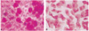
Impression cytology was performed as follows.12 After topical anesthesia with 0.5% proparacaine hydrochloride (Alcaine®, Alcon, USA), strips of cellulose acetate filter paper (MFS membrane filters, Advantec MFS, USA) (6.2 mm diameter) were applied, dull side down, to the lower nasal bulbar conjunctiva adjacent to the corneal limbus. The filter strips were pressed gently with blunt, smoothtipped forceps for 2-3 seconds. They were then gently removed in a peeling motion, avoiding shearing. A solution containing three parts acetone and one part of a mixture of 1/4 95% methanol and 3/4 95% ethanol was freshly prepared. Immediately after pressing the filter strips onto the slides, the slides were placed horizontally in a glass Petri dish for 3-4 hours in the above solution. The slides were then fixed in absolute alcohol, stained with periodic acid-Schiff (PAS) and mounted. Photographs were taken using a light microscope fitted with a calibrated grid at a magnification of ×400 and the degree of squamous metaplasia of conjunctival epithelial cells and goblet cell density were then evaluated. The degree of squamous metaplasia was graded from 0 to 3 according to Nelson's13 grading scheme and the goblet cell density was represented as the number of cells per square millimeter. Grade 2 or greater was regarded as abnormal.
Data were expressed as mean ± SD. Parameters between groups were analyzed by the Student t-test and analysis of variance with SPSS software. The Mann-Whitney U test and Kruskal-Wallis test were used for the analysis of nonparametric values such as keratoepithelial score and grade of conjunctival squamous metaplasia. A P value of less than 0.05 was considered statistically significant.
RESULTS
There were no significant differences in age or sex between the diabetic and normal control groups. In the diabetic group (47 patients), the duration of diabetes was more than 10 years in 31 patients (65.9%). The metabolic control was poor in 20 patients (42.6%) and 16 patients (34.1%) had diabetic neuropathy. Thirty eyes (31.9%) had no DR, 28 eyes (29.8%) had NPDR, and 36 eyes (38.3%) had PDR.
In the diabetic group, 30 eyes (31.9%) had low corneal sensitivity, 61 eyes (64.9%) had an abnormal BUT value, and 22 (23.4%) and 33 (35.1%) eyes had abnormal total and basal secretion test values, respectively. Thirty-four eyes (36.2%) had abnormal impression cytologic findings.
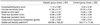
Comparison of tear film and ocular surface parameters between the two groups is shown in Table 1. The keratoepitheliopathy score was significantly higher in the diabetic group (1.14 ± 0.89) than in the control group (0.34 ± 0.48) (P < 0.001). Corneal sensitivity was significantly lower in the diabetic group (51.06 ± 6.17 mm) than in the control group (57.84 ± 2.50 mm) (P < 0.001). Tear film BUT was significantly shorter in the diabetic group (7.82 ± 2.12 sec) than in the control group (10.95 ± 1.56 sec) (P < 0.001). Total and basal tear secretions were 12.88 ± 5.06 mm and 6.95 ± 3.76 mm in the diabetic group, and 19.26 ± 3.06 mm and 11.22 ± 2.10 mm in the control group, respectively. The differences between the two groups were statistically significant (P < 0.001). In impression cytologic analysis, the average grade of conjunctival squamous metaplasia was significantly higher in the diabetic group (1.25 ± 0.63) than in the control group (0.65 ± 0.57), and goblet cell density was significantly lower in the diabetic group (429.68 ± 108.35 cell/mm2) than in the control group (545.17 ± 77.56 cell/mm2) (P < 0.001) (Fig. 1).
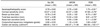
The relation of tear film and ocular surface parameters in the diabetic patients to the DR stage is shown in Table 2. In the no DR group, the keratoepitheliopathy score, corneal sensitivity, BUT, total tear secretion, basal tear secretion, grade of conjunctival squamous metaplasia, and goblet cell density were 0.79 ± 0.63, 53.57 ± 5.42 mm, 8.79 ± 2.45 sec, 15.57 ± 4.99 mm, 8.46 ± 4.00 mm, 0.92 ± 0.53, and 488.93 ± 104.11 cell/mm2, respectively. The respective parameters in the NPDR group were 0.75 ± 0.65, 53.57 ± 4.88 mm, 8.25 ± 1.82 sec, 13.04 ± 3.93 mm, 6.93 ± 2.89 mm, 1.10 ± 0.49, and 456.07 ± 71.66 cell/mm2, and in PDR group, 1.75 ± 0.91, 47.08 ± 5.78 mm, 6.75 ± 1.65 sec, 9.81 ± 2.70 mm, 5.11 ± 2.26 mm, 1.63 ± 0.63, and 360.56 ± 102.17 cell/mm2. There were no significant differences of parameters between the no DR and the NPDR groups, except for total tear secretion. However, there were statistically significant differences of parameters between the no DR and the PDR groups, and between the NPDR and the PDR groups (P < 0.05).
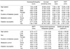
Patient age, sex, and duration of diabetes were not significantly correlated with the tear film and ocular surface parameters, but poor metabolic control and presence of diabetic neuropathy were significantly correlated with the parameters (Table 3).
DISCUSSION
In the present study, we assessed keratoepitheliopathy scoring, corneal sensitivity test, tear film BUT, total and basal tear secretion tests, and conjunctival impression cytology in noninsulin-dependent diabetic patients and compared the results with those in normal subjects. The degree of keratoepitheliopathy was severe, and both corneal sensitivity and tear film parameters were significantly reduced in the diabetic patients. The conjunctival impression cytologic results also supported these changes. These results indicate that dry eye is a significant feature of the diabetic ocular surface disease. To evaluate the ocular and systemic factors related to diabetic keratopathy, we also assessed the relationship of tear film and ocular surface changes with age, sex, duration of diabetes, metabolic control, diabetic neuropathy, and DR stage.
The corneal changes associated with diabetes mellitus suggest that diabetic patients have clinical or subclinical abnormalities in the corneal epithelium. The overall occurrence and degree of keratoepitheliopathy are higher in diabetic patients and correlate with the DR severity.8,14 Recent study using anterior fluorophotometer demonstrated that the corneal epithelial function was impaired in diabetic patients.15 Diabetic patients with higher serum hemoglobin A1c levels are more predisposed to impaired barrier function in the corneal epithelium. In our results, keratoepitheliopathy was related to poor metabolic control, presence of diabetic neuropathy, and advanced DR stage.
Several studies have reported decreased corneal sensitivity in diabetic patients, but the mechanism is unclear.8,9,16 Abnormal glucose metabolism may induce the functional disorder of corneal nerve fiber through activated polyol pathway.17,18 Another hypothesis is that loss of corneal sensation is a manifestation of diabetic polyneuropathy.16 Dogru et al9 reported that corneal sensitivity was significantly lower in diabetes with poor metabolic control and peripheral neuropathy, but it was not related to the duration of diabetes or the stage of retinopathy. On the other hand, Rogell19 and Saito et al16 insisted that the decrease in corneal sensitivity was correlated with the DR stage. In our results, decreased corneal sensitivity in diabetic patients was related to poor metabolic control, presence of diabetic neuropathy, and advanced DR stage.
Changes of tear function parameters in diabetes have been studied, but the results remain controversial. In some studies, total and reflex tear secretions were significantly reduced, but basal tear secretion and tear film BUT did not change.16,20 However, other studies reported a decrease in basal tear secretion and BUT.9,21,22 Dogru et al9 reported that BUT and basal tear secretion were decreased, especially in diabetes with poor metabolic control and peripheral neuropathy, but they were not related to the duration of diabetes or the stage of retinopathy, suggesting a neuropathy involving the innervation of the lacrimal gland. Saito et al16 also reported that neither total or reflex tear secretion was correlated with the DR stage. On the other hand, Nepp et al14 and Ozemir et al22 reported that abnormal tear function tests were associated with poorer metabolic glucose control, panretinal argon laser photocoagulation, and PDR. In our results, all tear function parameters, including BUT, and total and basal tear secretions, were lower in the diabetic group, and these abnormalities were related to poor metabolic control, presence of diabetic neuropathy, and advanced DR stage. Only total tear secretion was different between the no DR and NPDR groups. This result suggests that the decrease in total or reflex tear secretion may be the first change of tear film when DR progresses. Decreased reflex tearing in diabetes may be the result of a diminished corneal sensitivity, and decreased tear production and abnormal tear composition may result in superficial ocular lesion.
Our impression cytologic analysis showed a higher grade of squamous metaplasia and lower goblet cell density in the diabetic patients. Epithelial cells were larger and more polygonal, and the nucleocytoplasmic ratio was increased. In severe cases, multinucleated, variable staining cytoplasms and small nuclei, even pyknotic or absent nuclei, were found. The mechanisms of these ocular surface changes in diabetes are still not clear, but several reasons may be responsible for these findings. The decrease in tear secretion and disturbed trophic function of tear film such as vitamin A and epithelial growth factors may induce chronic damage of the conjunctival surface, resulting in conjunctival metaplsia.20 The loss of neurotrophic effects evidenced by corneal hypesthesia, fluctuation in glucose level and insufficiency of metabolic control may induce conjunctival squamous metaplasia.9 Also, the ocular surface changes found in diabetics may at least partially be the result of a primary surface disease independent of tear film abnormalities.20
In conclusion, our study indicates that tear film and ocular surface changes in patients with noninsulin-dependent diabetes mellitus include keratoepitheliopathy, decreased corneal sensitivity, decreased tear stability and secretion, squamous metaplasia, and low goblet cell density. Our results also suggest that poor metabolic control, presence of diabetic neuropathy, and advanced DR stage are risk factors for tear film and ocular surface disorder in diabetes mellitus. Therefore, diabetic patients with these conditions should be examined for tear film and ocular surface changes.
 XML Download