

 PDF
PDF ePub
ePub Citation
 Print
Print


INTRODUCTION
Dystonia, one of the most prevalent forms of movement disorder, is defined as sustained or intermittent muscle contractions usually producing twisting and repetitive movement or abnormal posture (12). Oral medications and botulinum toxin injections have been the mainstays of treatment for a time, but are not sufficiently effective in many patients. Internal globus pallidus (GPi) deep brain stimulation (DBS) has been widely accepted as an effective treatment modality of medically refractory dystonia (34). However, few studies have been reported regarding the safety issue of pregnancy and childbirth and its long term outcome of the GPi DBS during the pregnancies. This report describes a female patient with generalized dystonia implanted with GPi DBS who delivered a baby during 84-month follow-up period after DBS surgery.
CASE DESCRIPTION
History
A 33-year-old female patient, a mother of one child, developed a left upper limb dystonia after being asphyxiated under blankets at the age of 4 weeks. The dystonia in both lower limbs progressively developed into a generalized form, accompanied by a left facial spasm. She had undergone orthopedic surgery because of right ankle eversion and lateral deviation of the right great toe 2 years ago. The most painful symptom to her was a dystonia of left upper limb accompanied by tremor occurred in both resting and exercise. The symptoms were not improved by medical treatment including nortriptyline, levodopa, clonazepam, baclofen, and biperiden. There was no family history of movement disorders. The patient was referred to our institution for evaluation of the DBS (November 14, 2006).
Examination
She had alert consciousness and normal intellectual ability. Motor power grades were V in all extremities, and sensory was also intact. After orthopedic surgery, the right ankle was nearly corrected but the right great toe was still deviated laterally. Her gait was mildly ataxic. The left arm was adducted in the shoulder and extended in the elbow. The left wrist was hyperextended, with continuing rhythmic clenching and opening of the fist (Fig. 1A). The dystonia severity was measured using Burke-Fahn-Marsden Dystonia Rating Scale (BFMDRS); her preoperative movement score was 33 points, and functional disability score was 10 points. Mini-mental state examination (MMSE) was 29 points, and Beck’s depression inventory (BDI) scale was 3 points. DYT-1 gene was not detected. On magnetic resonance imaging (MRI), there were no definite focal lesions in the brain parenchyma.
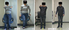
Fig. 1
Movie frames obtained from preoperative and postoperative video. (A) Movie frames obtained from a preoperative video showing the patient lifting both arms and walking. (B) Movie frame obtained from a postoperative video showing the same patient 7 years after deep brain stimulation (DBS) surgery.
Operation
In November 2006, she underwent bilateral GPi implantation. A Leksell stereotactic frame was secured to the patient’s head after application of a local anesthetic and she was transferred to the MR suite. The posteroventral portion of the GPi was targeted by means of axial, sagittal, and coronal MRI images (5). The pallidal target was 12.4 mm anterior to the midcommisural point, 20 mm lateral to the midline, and 3 mm below the intercommisural line in both side. The procedure was performed under general anesthesia, with the assistance of microelectrode recording (MER). A set of four microelectrodes (Differential microTargeting® Electrodes; FHC, Chemnitz, Germany; 1.5 MΩ impedance) were sequentially inserted toward the anatomical target within the GPi, which was vertically on the axial slice at the level of anterior commissure and horizontally at the junction between the two posterior quarters of the GPi (6). Permanent DBS electrodes (DBS 3387; Medtronic, Minneapolis, MN, USA) placements were determined without interpreting vessels, ventricles, and sulci. The electrode of the left side was inserted earlier than that of the right side, to minimize the error of the dominant side by brain shifting after cerebrospinal leakage. The electrodes were fixed to the burr hole and connected to pulse generators (IPG, Soletra 7426; Medtronic) implanted on both subclavicular pouch. There were no adverse events. One day after surgery, stimulation was begun using an N’vision programmer (Medtronic). The initial setting was as follows: monopolar stimulation by using Contact 1 as the negative and the internal pulse generator case as the positive pole with an amplitude of 3.72 V, pulse width 60 μsec, and frequency 130 Hz. Six months later, we performed a repeat computed tomography (CT) scan and fused it to the preoperative MRI to confirm the locations of the leads (Fig. 2).
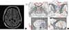
Fig. 2
Postoperative imaging showing the location of the electrodes. (A) Postoperative magnetic resonance imaging (MRI) scans demonstrating the bilateral deep brain stimulation electrodes in the posteroventral internal globus pallidus (GPi) Axial fluid-attenuated inversion recovery (Axial FLAIR). (B) Postoperative assessment of implanted electrodes by image fusion of a postoperative computed tomography (CT) scan with the corresponding preoperative inversion-recovery image. The bilateral electrodes located in the external globus pallidus (GPe).
Postoperative course
The movement and disability scores and the stimulation parameters were recorded, as described in Tables 1 and 2. After DBS implantation, considerable improvement was noted. Three months later, she showed improvement in dystonic dyskinesia, but facial dyskinesia on left eyelid was still noted. Subjectively, she handled the cleaning supplies more easily. Her movement score was decreased to 29 points and disability score to 5 points, but Karnofsky performance scale (KPS) score was still checked as 80 points at 6-month follow-up. The stimulation parameters were adjusted based on the patient’s symptom. The left-sided dystonic posture and walking had kept improving (Fig. 1B). She found it easier to clean, but still had difficulty in washing. At 12-month follow-up, the movement score decreased to 28 points (improvement rates, 15%) and disability score to 5 points (improvement rates, 50%) were checked, and she had sustained such improved state. She could do housework including cleaning and doing the laundry without difficulty. Four years later, the obstetricians consulted us about her 38 weeks pregnancy state. The maternal serum triple test and amniocentesis were performed because of her old age, and revealed low risk of congenital anomaly. A scheduled caesarian section was carried out under general anesthesia. After induction using thiopental and succinylcholine, intubation was done quickly, followed by DBS turn off. The surgery lasted for one hour, and the blood pressure was maintained within the normal range during the surgery. For hemostasis, only bipolar electrocautery was used. Before awakening from the anesthesia, DBS was turned on as the same parameters previously adjusted. After delivery, she could feed her baby by herself, because the dystonia of left upper extremity and hand was improved. Six years after DBS surgery, her general condition was more improved, and she started to play ping-pong and billiards. Until now, she has been showing continual improvement and being good at housework, carrying for children, with no trouble in daily life based on her KPS score of 90 points and MMSE score of 30 points. She even obtained a driver’s license in April 2014 and now drives a car by herself.
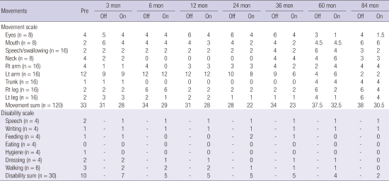
Table 1
The movement and disability scores during the follow periods

Table 2
The stimulation parameters during the follow up periods

DISCUSSION
This report is one of the rare reports on GPi DBS for dystonia with long-term follow up over 7 years. Dystonia can be classified according to the involved body distribution: focal, segmental, multifocal, generalized, and hemidystonia, or according to the etiology: inherited dystonia of proven genetic origin, acquired dystonia with a known specific cause (e.g., perinatal brain injury, infection, drugs, toxicity, vascular, neoplastic, or brain injury), and idiopathic dystonia of unknown cause. Previous reports have shown promising results of GPi DBS especially in the patients diagnosed as primary generalized dystonia (PGD) with DYT-1 positive, focal, and tardive dystonia (78910).
The patient was diagnosed as a patient with acquired dystonia that has been known to show less response to GPi DBS (111213). However, Vercueil et al. (14) reported a few cases with secondary dystonia showing successful response. Speelman et al. (15) also reported that GPi DBS was useful in some secondary dystonia patients, but many patients diagnosed as tardive dystonia and Hallervorden-Spatz disease (HSD) were included into their secondary dystonia group. Since March 2005, the author experienced 12 cases of acquired dystonia including this patient. The improvement rate of the patients diagnosed as acquired dystonia was about 29% at 12-month follow up according to our unpublished study. Although the improvement rate was not high as much as those of PGD with or without DYT-1 positive, the patients diagnosed as acquired dystonia also gained benefit from GPi DBS. The reason for favorable outcome in this patient might be an absence of the structural abnormality on preoperative MRI.
MER facilitates the selection of the final target in DBS in our experience, although some authors reported no benefit from MER (16). All procedures of GPi DBS for dystonia were performed under general anesthesia because of the patients’ abnormal posture and muscle contractions. General anesthesia did not interfere with the MER signals from the GPi. The typical bursting pattern could be identified, whereas amplitude was decreased and bursting pattern was emphasized more than in awaken surgery.
One notable finding was that this patient revealed improved outcome despite the bilateral electrodes located in the external globus pallidus (GPe), as shown in Fig. 2B. A possible explanation is that GPi might receive near impact from GPe stimulation. Another assumption is that the gamma-aminobutyric acid-ergic (GABAergic) pathway from striatum to GPi could be stimulated.
Few authors reported several cases about pregnancy and delivery in patients who underwent DBS surgery. Paluzzi et al. (17) reported three women who were pregnant and had babies by vaginal delivery after bilateral GPi DBS surgery. These women showed no worsening of dystonic symptoms during the pregnancy and labor periods, and had no problems in breast feeding. There is no evidence of that the pregnancy and delivery should not be allowed to the patients who underwent DBS implantation. Gwinn-Hardy et al. (18) tried to figure out the correlation between hormones and dystonia in 279 female patients, but they found no clear-cut relationship between pregnancy, menopause, postmenopausal hormone replacement therapy, and worsening of dystonic symptoms.
DBS is also not a contraindication for general or regional anesthesia. If the surgeon is careful not to use short wave (around 2,727 MHz), microwave (2.45 GHz) diathermy, or therapeutic ultrasound (1–5 MHz) diathermy, delivery can be performed safely. The bipolar electrocautery should be used at least 15 cm away from the IPG device, extension cable, and lead (17). As the indication for DBS increases, the patients’ distribution will be more diverse. A standardized protocol for DBS implanted patients who undergo pregnancy, delivery, and surgery irrelevant to neurosurgery would be required. In conclusion, GPi DBS is a safe and effective therapeutic method for treatment of dystonia. Favorable outcomes could be expected even for the patients with not only PGD, but also acquired dystonia. The patients who underwent DBS could safely be pregnant and deliver a baby.
 XML Download