

 PDF
PDF Citation
 Print
Print


INTRODUCTION
In the current medical practice, radiological imaging studies are of critical importance in every aspect of patient management, and thus, they have been dramatically expanded in recent years. Most commonly used radiological imaging methods are planar X-ray and computed tomography (CT), which cause radiation exposure of patients (1). Although the benefit from medical imaging far surpasses the potential risk of radiation, medical doctors need to properly understand the risk and benefit of radiation exposure in decision making of imaging studies.
18F-fluorodeoxyglucose (FDG) positron emission tomography (PET) is a molecular imaging method that visualizes glucose metabolism in vivo. Currently, a hybrid imaging of FDG PET/CT is widely used in clinical practice for diverse diseases such as cancers, inflammatory diseases, neurological disorders, and myocardial metabolic disorders. Due to its usefulness, FDG PET/CT has been rapidly expanded; in Korea, more than 500,000 examinations were performed in 2014 (2). With the increase of FDG PET/CT examinations, a concern has been raised with regard to the radiation exposure by PET/CT, because it causes both internal and external radiation from radiopharmaceutical administration and CT acquisition.
The radiation dose of FDG PET/CT depends on both injected activity of FDG and CT protocol. Notably, radiation dose may be reduced with recent PET/CT scanners, which enable sensitive gamma ray detection for PET and dose-reduction algorithms for CT. Thus, radiation dose from FDG PET/CT should be estimated separately in each society that has different conditions regarding scanner equipment and cultural background of medical imaging. However, there have been scarce data on radiation dose of FDG PET/CT based on a real world survey in Korea.
In this study, a nation-wide survey was conducted in Korea to estimate the average radiation dose of FDG PET/CT examinations. Additionally, correlations were investigated between the radiation dose and possible affecting factors.
MATERIALS AND METHODS
Questionnaire survey
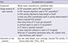
The study design was exempted from the ethical review by the decision of the Institutional Review Board of Seoul National University Hospital (E-1511-003-713). This survey aimed to include all working PET/CT scanners in Korea, which were known to be 154 scanners in 117 institutions according to a survey in 2013 (3). In July 2015, a survey questionnaire was e-mailed to the persons in charge of PET/CT examinations in all institutions where PET/CT was in operation. The questionnaire was designed for dosimetry-related information in usual FDG PET/CT examinations covering torso (from the skull base to the upper thigh) area. The questionnaire was composed of 3 parts (Table 1); the first part was related to the equipment information such as manufacturer, model name, and installation date; the second part was related to the examination protocol in terms of FDG injection and PET/CT acquisition, including image-enhancing methods such as time-of-flight (TOF) acquisition and point spread function (PSF)-recovery algorithms. In the third part, patient dosimetry data of PET/CT in real practice were requested, for the most recent 10 patient results per each scanner, including age, sex, body weight, scan-covered area, scan length, injected activity of FDG, CT parameters of volume CT dose index (CTDIvol) and dose-length product (DLP).
Table 1
Surveyed items and questions in the questionnaire
![]()
Estimation of radiation dose
Effective dose from FDG PET was calculated from the injected FDG activity using the conversion factor presented by the International Commission on Radiological Protection (4). Effective dose from CT was calculated from CT parameters using the CT-Expo method (version 2.4, Institut fűr Diagnostische und Interventionelle Radiologie, Hannover, Germany) with tissue weighting factors defined in the publication 60 of the ICRP (56). When additional contrast-enhanced CT scans were obtained after conventional FDG PET/CT scan, only the CT scan for attenuation correction and lesion localization was included in the analysis.
Statistical analysis
Radiation dose of FDG PET/CT was calculated from the injected FDG activity and DLP in real practice. Additionally, the influence of equipment characteristics on radiation dose was assessed in terms of equipment age (installation year) and use of dose-reduction software, TOF acquisition, and PSF-recovery algorithm. All values were expressed as mean ± standard deviation. In comparison of continuous variables, χ2 test and one-way ANOVA test with Bonferroni’s post-hoc correction were used and P values less than 0.05 were regarded significant. All statistical analyses were conducted using a commercial statistical software package (SPSS version 22, IBM SPSS statistics, Chicago, IL, USA).
RESULTS
Collection of questionnaires
The questionnaires were returned from 73 institutions and information of 105 PET/CT scanners was collected. The response rate was 62.4% on institution-basis and 68.2% on scanner-basis. Regional distribution of the institutions that responded in this nation-wide survey is shown in Fig. 1. The response included PET/CT results of 1,041 adults (M:F = 633:408, age 60 ± 13 years, body weight 61.4 ± 11.4 kg) and 3 children. One responder returned only FDG PET results without CT information, and only the PET results were included in the analysis. Characteristics of the enrolled PET/CT scanners are summarized in Table 2.
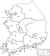
Table 2
Characteristics of the dedicated PET/CT scanners included in the survey
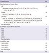
BGO, bismuth germanium oxide; CT, computed tomography; GSO, cerium-doped gadolinium oxyorthosilicate; LBS, lutetium based scintillators (not otherwise specified); LSO, cerium-doped lutetium oxyorthosilicate; LYSO, cerium-doped lutetium-yttrium oxyorthosilicate; PET, positron emission tomography.
![]()
Injected FDG activity and radiation dose
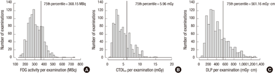
The distributions of FDG activity and CTDIvol are shown in Fig. 2. In adults, mean injected activity was 310 ± 77 MBq (range 126–729 MBq). In 90.2% of responding institutions, the injected activity was determined primarily based on body weight of a patient; mean value of injected activity per body weight was 5.11 ± 1.19 MBq/kg (range, 2.56–11.40 MBq/kg) (Fig. 2A). When radiation dose was calculated from the injected activity of all real practice data, 75th percentile of injected activity was 368 MBq (Fig. 3A) and mean effective dose from FDG was estimated to be 5.89 ± 1.46 mSv (range 2.39–13.85 mSv).
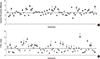 | Fig. 2Distribution of injected FDG activity and CTDIvol values of CT examinations in each institution. (A) Median value of injected FDG activity is 5.00 MBq/kg (25th–75th percentile, 4.24–5.62 MBq/kg; gray box). (B) Median value of CTDIvol is 4.10 mGy (25th–75th percentile, 2.80–5.96 mGy; gray box).
|
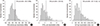 | Fig. 3Distribution of injected FDG activity (A), CTDIvol (B), and DLP (C) of each PET/CT examination.
|
In children, the injected activity was determined primarily based on body weight of a patient in 75.5% of the surveyed institutions. Mean value of injected activity per body weight was 4.47 ± 1.20 MBq/kg, which was slightly lower than that of adults (P = 0.01). Among the collected data of real examinations, 3 were results of children, in which the injected dose was 4.37, 4.81, and 4.93 MBq/kg.
With regard to CT scan, mean CTDIvol was 4.60 ± 2.47 mGy (range 0.97–15.19 mGy) in adults. Mean CTDIvol of the surveyed 73 institutions are shown in Fig. 2B and 75th percentile was 5.96 mGy (Fig. 3B). Mean DLP was 429.2 ± 227.6 mGy∙cm (range, 99.0–1274.0 mGy∙cm) and 75th percentile was 561 mGy∙cm (Fig. 3C). Based on the results, radiation dose from CT component in adult patients was estimated to be 6.26 ± 3.06 mSv. In 9 institutions (12.3%), additional CT scan was routinely performed with contrast-enhancement or breath-holding, for which radiation dose was not evaluated.
Factors affecting radiation dose of FDG PET/CT
Among the surveyed PET/CT scanners, 73 were equipped with software for CT dose reduction. Mean DLP was not significantly different between scanners equipped with the software and those without the software (436.1 ± 217.1 mGy∙cm vs. 412.9 ± 250.4 mGy∙cm, P = 0.14).
When PET/CT scanners were classified into 3 groups according to installation year, 42 were less than 5 years old, 50 were 5–10 years old, and 13 were more than 10 years old. Injected FDG activity was significantly reduced in more recently installed scanner groups (P < 0.001, Table 3). In addition, radiation dose from CT was also significantly lower in more recently installed scanner groups (P < 0.001 for both CTDIvol and DLP).
Table 3
Radiation dose of FDG PET/CT according to installation year
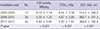
CTDIvol, volume computed tomography dose index; DLP, dose-length product; FDG, F-18 fluorodeoxyglucose.
![]()
In PET acquisition, TOF technology was available in 45 PET scanners. Mean injected FDG activity for the TOF-available scanners was lower than that for TOF-unavailable scanners (4.76 ± 0.96 MBq/kg vs. 5.37 ± 1.28 MBq/kg, P < 0.001). PSF-recovery algorithm was equipped in 36 PET scanners. Mean injected FDG activity for these scanners was also lower than that for PSF recovery-unavailable scanners (4.64 ± 0.85 MBq/kg vs. 5.36 ± 1.27 MBq/kg, P < 0.001).
DISCUSSION
In this nation-wide survey, which covered approximately 55% of the total PET/CT scanners in operation in Korea, the average radiation dose from FDG PET/CT was estimated to be 12.2 mSv; 5.89 mSv from FDG PET and 6.26 mSv from CT. It was also demonstrated that more recent PET/CT scanners equipped with certain image-enhancing methods are related to lower radiation dose.
With the recent expansion of radiological imaging procedures, medical radiation exposure has been rapidly increased during the last 3 decades; in the United States, annual per capita medical radiation exposure has been increased from 0.53 mSv in 1980 to 3.0 mSv in 2006, the largest source of which was CT (7). The proportion of CT examination has become more considerable in medical radiation exposure because the amount of CT examination is related to economic development (8). However, a concern recently has been raised that FDG PET/CT would be another large source of medical radiation exposure because it has been increased rapidly over the last 10 years. In Korea, a total of 308,663 PET/CT examinations were performed in 2009 (3), and approximately 513,000 FDG PET/CT examinations, in 2014 (9). Additionally, FDG PET/CT is a source of both internal and external radiations; internal radiation from intravenously injected FDG, and external radiation from CT imaging. On the other hand, a single examination of FDG PET/CT may substitute several CT scans and nuclear imaging studies because it covers whole body in a single scan. Thus, medical doctors need to understand the radiation dose from FDG PET/CT and to make a decision for diagnostic imaging based on appropriate risk-benefit assessment.
There have been nation-wide surveys of radiation dose from FDG PET/CT and its quality control in some European countries (10,11,12). However, radiation dose can vary widely according to imaging protocols and scanner models. Additionally, because recent PET/CT scanners are equipped with highly sensitive detectors and dose reduction algorithms, FDG PET/CT can be performed with lower radiation dose than before. In Korea, many PET/CT scanners have been installed in recent years with expansion of its use. Thus, a real world survey is required to estimate overall radiation dose of FDG PET/CT.
The average radiation dose demonstrated in this study is grossly similar to the previously reported results. In a nation-wide survey in France, mean radiation dose from FDG PET/CT was estimated to be 14.3 mSv; 5.6 mSv from FDG PET and 8.7 mSv from CT (10). In our study, the dose from FDG PET was slightly higher whereas the dose from CT was lower than that in the French survey. The injected FDG activity recommended by the European Association of Nuclear Medicine (EANM) is 2.5–5.0 MBq/kg (13), which is slightly lower than the mean injected FDG activity surveyed in our study (mean 5.11 MBq/kg). It is speculated to be caused by different imaging protocol. In the EANM guideline, the injected activity is based on a protocol using a fixed scan time of 5 minutes/bed; however, in Korea, scan time is usually less than 2–3 minutes/bed, chiefly for patients’ convenience and high throughput. In a recent guideline, injected activity is determined considering scan time; 7–14 MBq⋅min⋅bed-1⋅kg-1 (14). Although the current FDG activity is within a reasonable range, more efforts should be made in the future based on the balance of risk and benefit.
An intriguing point of this study is the relationship between equipment characteristics and radiation dose. Both the radiation doses from FDG and CT were significantly reduced by using more recently installed scanners equipped with image-enhancing methods. TOF acquisition algorithms can reduce background signal noise and cause an increase in sensitivity (15). As PSF-recovery algorithms can enhance image resolution and overall image quality (16), TOF technology combined with PSF-recovery algorithm would be a main cause of the reduced injected activity. The optimal injected FDG activity is determined in each institution by considering image quality and patients’ radiation safety. The present study demonstrated in a real world study that injected FDG activity is reduced by using more recent scanners equipped with these algorithms based on the improved image quality. Additionally, the radiation dose from CT was also lower in more recent scanners, although mean DLP was not significantly different between scanners with and without dose reduction software. It is speculated that hardware factors such as multi-detectors are more important than software factors. Additionally, the influence of specific CT protocol in each institution should be investigated in further studies. Considering the results of the current study, use of obsolete scanners should be discouraged by health insurance reimbursement system or healthcare policy, for patients’ radiation safety.
Quality control programs of imaging equipment and protocol are also important for maintaining the performance of diagnostic tests and reducing unnecessary radiation exposure. In a quality control program, various steps of image acquisition and reconstructions are checked up and authoritative recommendations for standardized quality control protocols have been reported regarding daily procedures, calibration of PET/CT scanners and image quality evaluation (1314). Quality control and standardization of imaging procedures are necessary not only for radiation safety but also for comparing image results between different institutions in case of multicenter clinical trials (1718).
The International Commission on Radiological Protection recommended constitution of the national diagnostic reference levels (DRL) to achieve evidence-based medical radiation protection (19). DRLs are defined as dose levels in medical radiological diagnostic practices or typical levels of radiopharmaceutical activity for groups of standard-sized patients or standard phantom (20). In terms of radiation protection and standard procedure, DRLs are recommended to be implemented for medical radiation diagnostic procedures. The present study is expected to be a basis for establishing the national DRLs for FDG PET/CT scan; the DRL for CT component of whole body FDG PET/CT may be suggested as 560 mGy∙cm (75th percentile of DLP in Fig. 3C), which is lower than the value of 750 mGy∙cm proposed in a French survey (10). DRL for FDG activity may be suggested as 370 MBq (75th percentile of injected activity in Fig. 3A), which is similar to proposed values of 350–385 MBq in other countries (10,21,22).
The present study has a limitation that it is based on a questionnaire survey without actual evaluation of radiation dose in each scanner. However, this is the first study that conducted a nation-wide survey on radiation dose of FDG PET/CT in Korea, and more than 50% of scanners were included in this study. Further studies are required regarding actual measurement of radiation dose.
In conclusion, the average radiation dose from FDG PET/CT is estimated to be 12.2 mSv from this nation-wide survey in Korea. The radiation dose is reduced with more recent scanners equipped with image-enhancing algorithms. The results are expected to be a basis for establishing the national DRLs for FDG PET/CT scan.
 XML Download