

 PDF
PDF ePub
ePub Citation
 Print
Print


INTRODUCTION
In 2011, several young women with no known previous medical conditions were admitted with severe respiratory distress to the intensive care unit (ICU) of a tertiary care hospital in Seoul (1). These women exhibited severe respiratory failure that was ultimately fatal despite the provision of aggressive supportive care and broad-spectrum antimicrobial treatment. The concerning factors were that the patients were mostly at the peripartum stage, the respiratory failure was refractory to all supportive measures available, and an extensive laboratory investigation yielded no plausible explanations. Therefore, the Korea Centers for Disease Control and Prevention (KCDC) planned to investigate these unforeseen cases of respiratory failure and created a multidisciplinary team comprising many healthcare professionals. Using a case-control study and a disinfectant inhalation animal study, we confirmed that the inhalation of humidifier disinfectants was the cause of respiratory failure in those cases. Thereafter, monitoring for humidifier disinfectant-associated lung injury was initiated. Here we describe the clinical characteristics of 84 adult patients who were identified as having humidifier-associated lung injury through a national survey.
SUBJECTS
Based on the index cases, the conferences at Asan Medical Center focused on a working definition of the disease. For a diagnosis of this condition, the following three characteristics had to be present: bilateral, centrilobular, or diffuse ground-glass opacity on high-resolution computed tomography (HRCT); symptoms or signs not consistent with other clinical diagnoses; and age 20–54 years (young adults) (2). Several multidisciplinary conferences led to the suspicion that this outbreak may be associated with an inhalation injury. An age- and sex-matched hospital-based case-control study was performed to examine the inhalation exposure to various agents. As the epidemiologic study suggested a high prevalence of exposure to humidifier disinfectants in the patient group, the humidifier disinfectants exposure experiment in an animal inhalation model was performed. Based on the pathologic, radiologic, and clinical features of the index cases and the lung histopathology of the exposed experimental animals, the diagnostic criteria for humidifier disinfectant-associated lung injury (Table 1) were refined in 2013 (3).
Table 1
Clinical criteria for humidifier disinfectant-associated lung injury

CLINICAL COURSE
Clinical characteristics
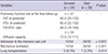
A total of 88 cases of humidifier disinfectant-associated lung injury leading to severe illness or death were identified. Table 2 presents the demographic and clinical characteristics of the 84 patients who consented to the monitoring. The mean patient age was 35 years (range, 26–70 years), and most were young women in the peripartum period. The presenting symptoms were nonspecific. Most of the patients complained of cough and dyspnea, whereas fever was infrequently noted. The clinical course was subacute, and some patients progressed rapidly and developed symptoms resembling severe acute respiratory distress syndrome (ARDS).
Table 2
Initial clinical characteristics of the 84 study patients with humidifier disinfectant-associated lung injury
The time of symptom onset was commonly in late winter or early spring. There were no notable features in terms of geographical location, occupational history, or drug history, although all of the patients reported using a humidifier and humidifier disinfectants. The humidifier disinfectant products used by the patients contained various concentrations of antimicrobial chemicals including oligo(2-(2-ethoxy)-ethoxyethyl guanidinium chloride, polyhexamethyleneguanidine phosphate, methylisothiazolinone, and chloromethylisothiazolinone. Although the estimated concentration of humidifier disinfectant or exposure time, which was recorded based on the patients’ or families’ memory, may not be accurate, it was clear that all of the patients were exposed to the humidifier disinfectant at night. The patients with mild hypoxemia who were admitted to the general ward responded to supportive care such as oxygen therapy, antibiotic treatment, and corticosteroid administration. However, those patients with refractory hypoxemia exhibited rapid progression leading to death despite the administration of medications such as corticosteroids, cyclosporine, cyclophosphamide, antibiotics, antiviral agents, anti-fungal agents, and immunoglobulin as well as ventilator care and extracorporeal membrane oxygenation.
A total of 39 patients who underwent a pulmonary function test at diagnosis exhibited a restrictive lung disease pattern and decreased diffusion capacity.
Pathologic findings of the reported cases
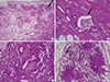
A fibroinflammatory process predominantly involving the bronchioles and centrilobular lung parenchyma, without notable granulomas, was observed (1). Similar pathologic findings were noted in cases of constrictive bronchiolitis and bronchiolitis obliterans (BO), which differ from the present cases in that no predisposing conditions were present (4). Most of the patients in the present study exhibited bronchiolar destruction consistent with the inhalation of toxic material. The bronchiolar lesions were characterized by epithelial sloughing and replacement with flattened regenerating cells, mild to severe subepithelial fibroblastic proliferation resulting in bronchiolar obliteration, and varying degrees of peribronchiolar fibrosis. The parenchymal lesions showed alveolar damage and a disease spectrum ranging from an early exudative/inflammatory phase to an extensive fibroproliferative/fibrosing phase. Furthermore, the subpleural and paraseptal airspaces were relatively well-preserved, even in an end-stage explanted lung (Fig. 1).
Fig. 1
Lung histology in a typical case.
The main pathology included a fibroinflammatory process predominantly involving bronchioles and centrilobular lung parenchyma without notable granuloma (arrow, panel A). Bronchiolar lesions were characterized by epithelial sloughing and replacement by flatten regeneration cells (arrow, panel B), and subepithelial fibroblastic proliferation resulting in bronchiolar obliteration (panel B). Parenchymal lesions showed histological patterns from the early exudative phase to the extensive fibrosing phase (panel C). Moreover, subpleural and paraseptal airspaces were relatively preserved, even in the end-stage explanted lung (arrow, panel D).
Radiologic findings of the reported cases
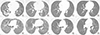
Chest radiography and HRCT were performed in all patients. The radiologic findings were unique and characterized by multifocal, patchy areas of consolidation at the lower lungs with relative subpleural sparing at the initial stage. Diffuse centrilobular ground-glass opacity involving the entire lung was observed without any zonal predominance in the late stage (Fig. 2). Pneumomediastinum or pneumothorax was observed in 11 patients.
Fig. 2
Typical lung radiographs at the early and late stage.
(A-D) A 30-year-old woman who was not admitted to the intensive care unit. (A) An axial computed tomography image showing irregular patchy consolidation and ill-defined centrilobular nodules approximately 1 week after the onset of respiratory symptoms including cough, sputum, and dyspnea. (B) After 2 months, the consolidation decreased and progressed to diffuse centrilobular nodules and ground-glass opacity; moreover, focal pneumomediastinum was noted. (C) The attenuation of the centrilobular nodules gradually diminished at the early chronic stage approximately 6 months after symptom onset. (D) The diffuse centrilobular nodule attenuation had decreased, but it persisted in the lungs even after 5 years. (E-H) A 35-year-old woman was admitted to the intensive care unit. (E, F) Computed tomography images showed diffuse dense centrilobular nodules with a ground-glass opacity, spontaneous pulmonary interstitial emphysema, and pneumomediastinum at 2 months after severe dyspnea. (G, H) The attenuation of the diffuse ill-defined centrilobular nodules and ground-glass opacity had decreased, but it persisted even after 8 years. This patient died after 9 years due to recurrent infection and deteriorated pulmonary function.
A total of 180 HRCT scans from 76 patients were examined, and the chronological changes in the HRCT findings are presented in Fig. 3. The HRCT scans for all the patients were categorized on the basis of the interval between the HRCT scan date and the initial respiratory symptom presentation: within 1 month, 2 months, 3 months, 6 months, 1 year, or annually. Two radiologists (K.D. and E.J.C.) reviewed all HRCT images at the lung (window width, 1,500 Hounsfield unit [HU]; level, −700 HU) and mediastinal window (width, 450 HU; level, 50 HU) settings (5). The consolidation extent, ground-glass opacity, and centrilobular nodules were categorized in 10th-percentile intervals (Fig. 3A). The presence of pleural effusion, pneumomediastinum, pneumothorax, pulmonary interstitial emphysema, fibrosis, and bronchiectasis are presented as percentages of patients (numbers of patients with the symptoms of all examined patients) (Fig. 3B).
Fig. 3
Chronological changes in the computed tomography findings. (A) Proportion of extent by duration. (B) Number (%) of patients by duration.
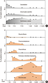
The initial HRCT images at admission, which were obtained within 1 week of the presentation of respiratory symptoms, primarily showed irregular patchy consolidation, ground-glass opacity, and centrilobular nodules with a subpleural sparing pattern. Within 1–3 months of the initial symptoms, spontaneous pulmonary interstitial emphysema, pneumomediastinum, and pneumothorax developed, all of which were unrelated to the intubation attempts. The diffuse extensive ill-defined centrilobular nodules had covered the entire lung, and although the attenuation of the nodules had gradually diminished, it persisted even after 5 years. Fibrosis and bronchiectasis had gradually increased after 2–3 months and had persisted.
Differential diagnosis
Diagnosing this condition was challenging. Its clinical manifestation was similar to that of ARDS, acute interstitial pneumonitis (AIP), or hypersensitivity pneumonitis (HP). However, the condition differed from ARDS, AIP, and HP in that the hyaline membrane and granuloma were not dominant pathologic findings (6).
The radiologic features observed also differed from the known diffuse lung diseases in several aspects. The multifocal patchy consolidation observed in the early phase is also noted in patients with cryptogenic organizing pneumonia or ARDS. However, features such as the sparing of the subpleural zones and evolution into diffuse centrilobular ground-glass opacity are not observed in those conditions. The most likely radiological diagnosis, based on the presence of diffuse centrilobular ground-glass opacity of the later phase, was subacute HP. However, the rapid fibrotic progression of ground-glass opacity despite corticosteroid treatment was inconsistent with the features of subacute HP. The acute exacerbation of unclassified interstitial pneumonia or AIP could explain the patients’ rapid deterioration despite the administration of conventional treatment; however, the presence of airway-centered inflammation, reflected by the presence of centrilobular ground-glass opacity on HRCT, is inconsistent with such a diagnosis.
BO, which is characterized by the disruption of the epithelium and obliteration of the lumen of the small airway, is a subset of chronic lung transplant rejection and has similar pathologic findings to those of humidifier disinfectant-associated lung injury. The important feature of BO is the limitation of airflow with a decrease in the forced expiratory volume in one second (FEV1) and the FEV1/forced vital capacity (FVC) ratio; the associated radiologic changes include hyperinflation and bronchiectasis in the advanced stage. However, the important feature of humidifier disinfectant-associated lung injury was the restrictive pattern, with a decrease in FEV1 and FVC, since the disease causes both bronchiolar destruction and alveolar damage from the early stage.
PROGNOSIS
The overall mortality rate was 36.0%. A total of 46.4% of patients were admitted to the ICU; of those requiring mechanical ventilation, 74.4% died. Moreover, six patients required lung transplantation due to progressive lung fibrosis and refractory hypoxemia.
During the follow-up period, the lung function recovered to normal in 54.0% of patients, whereas persistently severe restrictive lung function was seen in 13.5% (Table 3).
Table 3
Clinical outcomes of 84 patients with humidifier disinfectant-associated lung injury during follow-up
DISCUSSION
Although indoor and outdoor pollutants are reportedly the major causes of environmental hazards, many other seemingly innocuous inhalants can endanger human health (78). First, we reported the outbreak of lung injury associated with the inhalation of aerosolized disinfectants that are commonly used at home (12). The association between inhaled humidifier disinfectants and lung injury has been proven in epidemiologic studies (239). In the first hospital-based case-control study, the prior use of humidifier detergents was significantly associated with lung injury (crude odds ratio [OR], 47.3; 95% confidence interval [CI], 6.05–369.70) (2). Moreover, investigators reviewed cases of children with severe acute interstitial lung disease of unknown cause that showed similar clinical, radiologic, and pathologic patterns as those noted in adult patients. In the present study, we found that humidifier disinfectant inhalation causes an idiopathic type of children’s interstitial lung disease (chILD), which is characterized by rapid progression, spontaneous air leaks, high mortality, lack of treatment response, spring-season predominance, and a familial tendency (9). In our study, 138 children were diagnosed with this type of chILD, and the overall mortality was 58%. In a retrospective, 1:3 matched case-control study of chILD, the prior use of a humidifier disinfectant (OR, 2.73; 95% CI, 1.41–5.90; P = 0.00) was independently associated with an increased risk of lung injury (10).
In an animal model, the inhalation of humidifier disinfectants induced severe respiratory injury that was characterized by the same histological features as those observed in humans (11). Considering the accumulating evidence of this public health hazard, the Korean government ordered an immediate recall of all humidifier disinfectant products from the market in winter 2011; no new cases were reported during the subsequent 2 years.
In 2013, the KCDC conducted a nationwide study of humidifier disinfectant-associated lung injury to assess the dose-response relationships. The dose-response analysis indicated that the development of humidifier disinfectant-associated lung injury and death were strongly associated with recurrent, intense, acute exposures without sufficient recovery time between exposures; this association was stronger than that for long-term cumulative exposure. These findings could explain the occurrence of reversible or clinically unapparent cases in some co-exposed family members, particularly among children aged < 6 years and pregnant women (23).
Ultrasonic humidifiers effectively aerosolize water particles and other particulates that may be present in the water tank. Humidifier disinfectants were added to the water tank in humidifiers to prevent microbial contamination. After aerosolizing, these humidifier disinfectants were transformed into nanoparticles (peak mass median aerodynamic diameter, –100 nm), which could be deposited deep in the human lung and accumulated in the organ in cases of long-term exposure. Since 1994, more than 10 humidifier disinfectant products were marketed in Korea without any toxicological testing for the inhalation health effects. Home-dwellers in particular would be the most affected due to prolonged exposure to the potential lung irritants.
Although we are not currently aware of the entire spectrum of diseases induced by these humidifier disinfectants, this tragic outbreak event prompted many changes. First, the KCDC performed an epidemiologic investigation of the non-infectious cause of the condition. Second, this event indicated the importance of an approach involving a multidisciplinary team. Finally, this event increased the attention and awareness of the public regarding the chemicals used at home. Thus, we should enforce certain safety regulations to protect the health of the public against household chemical products that have toxic potential.
 XML Download