Journal List > J Korean Med Sci > v.31(10) > 1023113

Park, Chung, Kang, Ryu, Cho, and Lee: The Change of Stretched Penile Length and Anthropometric Data in Korean Children Aged 0–14 Years: Comparative Study of Last 25 Years

Abstract

There has been a great improvement in height and weight of Korean children owing to economic development over the last 25 years. This study aimed to evaluate the penile length of Korean children today and to compare it with a previous Korean study reported in 1987. The cross-sectional study was conducted with 909 Korean boys aged 0–14 years who had been brought to outpatient clinics of five tertiary hospitals (Busan, Ulsan, and Changwon) between September 2013 and May 2015. The stretched penile length (SPL) was measured and the testicular size was measured using orchidometry (mL). Student’s t-test or Mann-Whitney U test was used to compare the result of our study and the study reported in 1987. SPL of Korean children gradually increased from 4.1 ± 0.8 cm at 0-1 year old to 9.6 ± 3.0 cm at 13-14 years old, the most rapidly during the age of 13. While body weight and testicular size significantly increased from 1987 in most of age groups, there were no significant changes in SPL although there was in some age groups. Height decreased in the infants < 1 year old and increased in the children > 6 years old. With the great economic development over the last quarter century in Korea, height, body weight, and testicular size of children significantly increased but there was no significant change in SPL except penile growth pattern.

INTRODUCTION

Early diagnosis of abnormal penile size is important both medically and psychologically (123), and the exact measurement of penile size and current references of penile size are most important in diagnosing penile problems such as micropenis. Studies on the penile length of children have been very rarely conducted, especially in Korea (45). In 1987, a study on the penile and testicular size was conducted with 1,071 Korean children (6). Since then, only a few studies have been conducted with small numbers of children.
The authors previously evaluated the mean penile length of Korean boys and neonates in order to establish the reference of the current Korean children’ penile length (78). However, the preliminary study included a relatively small number of boys (n = 233) and neonates (n = 33) and limited to only Ulsan area in Korea. Therefore, the current study was conducted with more boys (n = 909) in Busan, Changwon, and Ulsan area. This study also compared the penile length and anthropometric measures such as height, body weight, and the testicular size of the current study and the previous Korean study conducted in 1987 to evaluate the association between them.

MATERIALS AND METHODS

This cross-sectional study was conducted in the outpatient clinic of 5 hospitals between September 2013 and May 2015. The subjects of this study were 909 Korean boys aged 0–14 years. Exclusion criteria were penile diseases including hypospadias and concealed penis, cryptorchidism, varicocele, and other growth problems such as chronic renal failure and endocrinologic disorders. Therefore, most children who were brought for general periodic examination, urinary tract infection, enuresis and lower urinary tract symptoms were included in the current study (7).
For all boys, penile length and the testicular size were measured by a pediatric urologist of each hospital. The stretched penile length (SPL) was measured and the testicular size was evaluated using orchidometry (mL) with the presence of their parents in a warm room. SPL was measured with a stiff ruler by compressing the fat tissue with one end of the ruler through the pubic ramus; then, the penis was fully stretched and the distance to the glans of the stretched penis was plotted (78). Foreskins of the uncircumcised children were not involved in the measurement.

Statistical analysis

Data analysis was performed using the software package SPSS 17.0 K for Windows. Continuous variables were expressed as the mean ± SD. Student’s t-test was used to compare SPL, height, body weight and the testicular size between the study in 1987 and the current study and P value < 0.05 was accepted to be statistically significant.

Ethics statement

This study was approved by the institutional review board of Ulsan University Hospital (IRB No. 2014-09-020). Clinical data all the participants were collected by retrospectively reviewing their medical records. Therefore, our institutional review board permitted this study without acquisition of informed consent.

RESULTS

SPL gradually increased until the age of 13 and the most rapidly increased during 13 years old (Table 1). As compared with those reported in 1987, there was no significant change in SPL in most age groups although there was in some age groups (Fig. 1). The result showed that the age of rapid growth of SPL has been 1–2 years younger.
Table 1

Age-related changes of stretched penile length and comparison with a previous study (1987)

jkms-31-1631-i001
Age, yr Current study (Mean ± SD, cm) 1987 year (Mean ± SD, cm) No. of patients (N = 909) P value*
0-1 4.1 ± 0.8 3.5 ± 0.6 86 < 0.001
1-2 4.4 ± 0.8 4.1 ± 0.7 165 0.012
2-3 4.6 ± 0.9 4.4 ± 0.7 143 0.118
3-4 4.6 ± 0.8 4.4 ± 1.0 90 0.178
4-5 4.9 ± 0.9 4.9 ± 0.9 83 0.938
5-6 5.2 ± 1.0 5.1 ± 1.0 67 0.482
6-7 5.4 ± 0.9 5.2 ± 0.8 76 0.118
7-8 5.7 ± 1.0 5.2 ± 0.9 33 0.005
8-9 5.4 ± 1.1 5.4 ± 0.9 24 0.987
9-10 5.8 ± 1.0 5.6 ± 0.8 33 0.330
10-11 6 ± 1.1 5.7 ± 0.7 42 0.156
11-12 6.5 ± 1.5 6.1 ± 0.8 28 0.138
12-13 7.1 ± 1.6 6.5 ± 1.1 19 0.115
13-14 9.6 ± 3.0 7.5 ± 1.0 20 0.016
SD, standard deviation.
*Student’ t-test or Mann-Whitney U test.
Fig. 1
Age-related changes of stretched penile length and comparison with a previous study (1987).
jkms-31-1631-g001
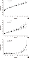
Fig. 2A showed that the height had a continuously increased pattern and as compared with those as of 1987, the increased change was more significant in children aged over 6 years old although the height of infant aged below 1 year old decreased.
Fig. 2
Age-related changes of height (A), body weight (B) and testicular sizes (C) and comparison with a previous study (1987).
jkms-31-1631-g002
Body weight and testicular size (Table 2) significantly increased in most age groups as compared with those reported in 1987. Although there were no significant differences in body weight of infants aged below 2 years old, it significantly increased afterwards, compared with data in 1987 (Fig. 2B). There were no significant changes in testicular size of children aged 0–9 years but it increased afterward and more rapidly during age of 12–14 years (Fig. 2C).
Table 2

Age-related testicular size (mL)

jkms-31-1631-i002
Age, yr Current study (Mean ± SD, mL) No. of patients (N = 909)
0–1 2.0 ± 0.5 86
1–2 2.0 ± 0.4 165
2–3 2.2 ± 0.9 143
3–4 2.3 ± 1.4 90
4–5 2.1 ± 0.5 83
5–6 2.2 ± 0.5 67
6–7 2.3 ± 0.6 76
7–8 2.5 ± 1.1 33
8–9 2.5 ± 0.8 24
9–10 3.9 ± 2.3 33
10–11 4.0 ± 2.1 42
11–12 5.2 ± 3.5 28
12–13 8.6 ± 4.4 19
13–14 13.2 ± 5.7 20
SD, standard deviation.
The changes of the testicular and penile sizes have a similar pattern. There was a rapid growth at 12 or 13 years old and the significant change of the age has become 1–2 years younger, compared with data reported in 1987. Nevertheless, in the current study, there were no changes in the testicular size of children aged 0–9 years while the penile size continuously increased.

DISCUSSION

The accurate measurement of the penile length is very important in children with abnormal genital development such as micropenis, buried penis and hypospadias and is essential to find out the sign of underlying severe endocrine and chromosome disorders (9). Another important thing is age specific standard data to compare the accurate measurement of SPL. However, studies on SPL in boys have been very rarely conducted, especially in a wide range of age groups. Therefore, the authors previously evaluated the mean SPL of Korean boys in order to establish the reference of the current Korean children’ penile length and compare with that of reported in 1987 (7). The study showed increases of SPL, height, body weight and testis size comparing with those reported in 1987, however, the preliminary study included a relatively small number of 233 boys. Therefore, current study was conducted with a larger sample including 909 boys and it revealed that there was no significant change in SPL except in some age group as compared with those reported in 1987 (Fig. 1) in the contrary to the previous preliminary study results. Unfortunately, the current study do not provide the data regarding the penile length of neonates so, it cannot be compared with other studies.
The reasons for the inconsistent result are as follows. First, the sample sizes were different. The preliminary study included 30 children in each age group while the current study included more subjects in order to bring more creditability to the statistics. Second, there is a regional difference. The current study was conducted in Busan, Ulsan and Changwon, Korea while the preliminary study was conducted in only Ulsan, Korea. However, the cohorts included in the preliminary and current study were all Korean people and it is thought to be a minor factor. Third, penile measurement was checked by different physicians in each hospital and in each study. Although SPL was measured in the same way in both studies, the results may differ depending on the examiners because the measurement process of SPL can be affected by the strength of examiners (10). Although the differences may significant, it is accounted to be only a few millimeters. A previous Korean study reported that there was no difference in SPL between two observers (3.3 ± 0.2 cm and 3.2 ± 0.3 cm, P = 0.165) in newborn infants with normal weight while there was a significant difference between two observers in newborn infants with low weight (2.9 ± 0.4 cm and 2.7 ± 0.4 cm, P = 0.001) (8). In a recent study, no significant fixed or proportional bias could be found for inter-observer variation (11). The reliability of the measurements of SPL remains a subject of debate (1213). Nevertheless, it is suggested that SPL is one of the most reliable methods in children to compare the penile size (1415).
In the current study, SPL gradually increased until 13 years old and the most rapidly increased during the age of 13 years (Fig. 1). It is thought that penile size had little change in boys aged 4–14 years since 1987 (6). The previous study reported that penile size increased slowly till 4 years old followed by a steady phase. It then increased rapidly during the puberty. Those patterns of penile growth are similar to the penile growth reported in previous Turkey or Brazil studies (1617). However, in current study, penile size increased continuously till 13 years old. Gradual penile growth curve before the puberty was also revealed in previous studies conducted in other countries such as Turkey, India and Japan (11181920).
In a recent Japanese study, SPL increases continuously during the prepubertal period, and the most rapidly increases during the first 4 months of life (11). This was explained by Boas et al. (21). They showed that penile growth rate during the first 3 months of life was positively correlated with serum testosterone levels. In the current study, the testicular size also increased after 9 years old and more rapidly in boys aged 12–14 years. This means that the age of rapid SPL growth has become 1–2 years younger as the age of puberty has become younger over the last quarter of century.
Fig. 2A showed that the height had a continuously increased pattern and as compared with those as of 1987, the increased change was more significant in children aged over 6 years old although the height of infant aged below 1 year old decreased. It may be suggested that Korean mothers today intend to keep fetal weight under control for safe and problem-free delivery. The exact causes of decreased height of infants are unknown and have to be evaluated later.
With the great economic development over the last quarter century in Korea, height, body weight and testicular size of children significantly increased but there was no significant change in SPL except penile growth pattern. The results of the current study may be used as the new reference values for boys aged 0 to 14 years. However, these results should be readjusted by the further epidemiologic studies with more children in a community-based population.

Notes

DISCLOSURE The authors have no potential conflicts of interest to disclose.

AUTHOR CONTRIBUTION Conception and design: Park S, Lee SD. Acquisition of data: Park S, Chung JM, Kang DI, Ryu DS, Cho WY, Lee SD. Analysis and interpretation of data: Park S, Chung JM, Lee SD. Writing or revision of the manuscript: Park S. Study supervision: Lee SD.

References

1. Reilly JM, Woodhouse CR. Small penis and the male sexual role. J Urol. 1989; 142:569–571.
2. Wiygul J, Palmer LS. Micropenis. ScientificWorldJournal. 2011; 11:1462–1469.
3. Tsang S. When size matters: a clinical review of pathological micropenis. J Pediatr Health Care. 2010; 24:231–240.
4. Kim KS, Han J. Reconstructive surgery for intersex and micropenis. J Korean Soc Pediatr Endocrinol. 2009; 14:1–10.
5. Phillip M, De Boer C, Pilpel D, Karplus M, Sofer S. Clitoral and penile sizes of full term newborns in two different ethnic groups. J Pediatr Endocrinol Metab. 1996; 9:175–179.
6. Chung KH, Choi H, Kim SW. Penile and testicular sizes of Korean children. Korean J Urol. 1987; 28:255–258.
7. Lee JH, Ji YH, Lee SK, Hwang HH, Ryu DS, Kim KS, Choo HS, Park S, Moon KH, Cheon SH, et al. Change in penile length in children: preliminary study. Korean J Urol. 2012; 53:870–874.
8. Park JY, Lim G, Oh KW, Ryu DS, Park S, Jeon JC, Cheon SH, Moon KH, Park S, Park S. Penile length, digit length, and anogenital distance according to birth weight in newborn male infants. Korean J Urol. 2015; 56:248–253.
9. Lee PA, Danish RK, Mazur T, Migeon CJ, Micropenis II. Primary hypogonadism, partial androgen insensitivity syndrome, and idiopathic disorders. Johns Hopkins Med J. 1980; 147:175–181.
10. Khan S, Somani B, Lam W, Donat R. Establishing a reference range for penile length in Caucasian British men: a prospective study of 609 men. BJU Int. 2012; 109:740–744.
11. Ishii T, Matsuo N, Inokuchi M, Hasegawa T. A cross-sectional growth reference and chart of stretched penile length for Japanese boys aged 0-7 years. Horm Res Paediatr. 2014; 82:388–393.
12. Matsuo N, Ishii T, Takayama JI, Miwa M, Hasegawa T. Reference standard of penile size and prevalence of buried penis in Japanese newborn male infants. Endocr J. 2014; 61:849–853.
13. Habous M, Muir G, Tealab A, Williamson B, Elkhouly M, Elhadek W, Mahmoud S, Laban O, Binsaleh S, Abdelwahab O, et al. Analysis of the interobserver variability in penile length assessment. J Sex Med. 2015; 12:2031–2035.
14. Guo LL, Tang DX. Measurement of penile length in children and its significance. Zhonghua Nan Ke Xue. 2013; 19:835–840.
15. Hatipoğlu N, Kurtoğlu S. Micropenis: etiology, diagnosis and treatment approaches. J Clin Res Pediatr Endocrinol. 2013; 5:217–223.
16. Tomova A, Deepinder F, Robeva R, Lalabonova H, Kumanov P, Agarwal A. Growth and development of male external genitalia: a cross-sectional study of 6200 males aged 0 to 19 years. Arch Pediatr Adolesc Med. 2010; 164:1152–1157.
17. Gabrich PN, Vasconcelos JS, Damião R, Silva EA. Penile anthropometry in Brazilian children and adolescents. J Pediatr (Rio J). 2007; 83:441–446.
18. Camurdan AD, Oz MO, Ilhan MN, Camurdan OM, Sahin F, Beyazova U. Current stretched penile length: cross-sectional study of 1040 healthy Turkish children aged 0 to 5 years. Urology. 2007; 70:572–575.
19. Teckchandani N, Bajpai M. Penile length nomogram for Asian Indian prepubertal boys. J Pediatr Urol. 2014; 10:352–354.
20. Cinaz P, Yeşilkaya E, Onganlar YH, Boyraz M, Bideci A, Camurdan O, Karaoğlu AB. Penile anthropometry of normal prepubertal boys in Turkey. Acta Paediatr. 2012; 101:e33–6.
21. Boas M, Boisen KA, Virtanen HE, Kaleva M, Suomi AM, Schmidt IM, Damgaard IN, Kai CM, Chellakooty M, Skakkebaek NE, et al. Postnatal penile length and growth rate correlate to serum testosterone levels: a longitudinal study of 1962 normal boys. Eur J Endocrinol. 2006; 154:125–129.
TOOLS
ORCID iDs

Sungchan Park
https://orcid.org/http://orcid.org/0000-0002-2337-983X

Jae Min Chung
https://orcid.org/http://orcid.org/0000-0003-3593-9643

Dong Il Kang
https://orcid.org/http://orcid.org/0000-0003-2283-4041

Dong Soo Ryu
https://orcid.org/http://orcid.org/0000-0002-7557-0820

Won Yeol Cho
https://orcid.org/http://orcid.org/0000-0001-6640-7872

Sang Don Lee
https://orcid.org/http://orcid.org/0000-0001-9459-3887

Similar articles