

 PDF
PDF ePub
ePub Citation
 Print
Print


INTRODUCTION
Rotator cuff lesions are the most common causes of painful shoulder in the elderly, accounting for up to 70% of cases (1). Although magnetic resonance (MR) arthrography is reported to be the most sensitive and specific technique for diagnosing rotator cuff tears, ultrasound (US) is widely used since it is a cost-effective and easily assessable tool, providing dynamic imaging studies (234). Ultrasound is reported to be accurate, with a sensitivity range from 92.4% to 96% and a specificity range from 93% to 94.4% for diagnosing full thickness tear and a sensitivity range from 66.7% to 84% and a specificity range from 89% to 93.5% for partial thickness tear (25). Measuring the tendon thickness by ultrasound is proven to be a valid method in diagnosing enthesopathy of plantar fascia, distal Achilles tendon, patellar ligament, distal quadriceps and brachial triceps tendon in spondyloarthropathy patients (6). In addition, ultrasound dimension of Achilles tendon thickness can be used as one of the diagnostic criteria in Achilles tendon pathology (7). In diagnosing rotator cuff lesions, tendon thickness should be compared with normal dimensions of the rotator cuff. There have been researches suggesting diagnostic cut off values of rotator cuff tear or supraspinatus tendinopathy (58). However, there is no definite reference providing normal ultrasound dimensions of the shoulder with a wide range of age groups, especially in the Korean population. In this study, the ultrasound dimensions of the rotator cuff, long head of biceps tendon, deltoid muscle, subacromial subdeltoid bursa and acromioclavicular joint in healthy Korean adults were measured and the possible variability among different sex, dominant hands, and ages were assessed to provide accurate references for rotator cuff measurements.
MATERIALS AND METHODS
Subjects
A total of 100 subjects were recruited in the study via open invitation. All of the subjects enrolled in the study were recruited from volunteers who visited our institution, from April 2014 to February 2015.
Healthy adults aged between 20 and 70 years who had no shoulder problems were included. Exclusion criteria were the following: Subjects with 1) shoulder pain, 2) history of shoulder instability or dislocation, 3) shoulder pathology such as rotator cuff injury, impingement syndrome, biceps tendinopathy, adhesive capsulitis, subacromial bursitis, or acromioclavicular joint injury, 4) history of surgery involving rotator cuff injury, 5) shoulder weakness due to underlying pathology such as suprascapular neuropathy, brachial plexopathy, cervical root disorder, cervical myelopathy and stroke 6) diabetes mellitus, 7) rheumatic disorders or systemic diseases (renal, hepatic, cardiac, etc.). To exclude preexisting shoulder pathology, physical examinations including shoulder range of motion and palpation were performed. In addition, provocative tests to evaluate glenohumeral instability, labral pathology, rotator cuff injury, impingement, acromioclavicular joint pathology, bicep tendon injury were performed. Patients who have pain, tenderness, limited range of motion, or positive findings in at least one of those physical examinations were excluded.
Data collection
Demographic details including age, sex, and hand dominance were collected. Ultrasound examination was performed by a single physiatrist, who is a formal member of Korean Academy of Rehabilitation Medicine, with experience in musculoskeletal ultrasound scanning for more than 10 years. Both shoulders were evaluated in each individual. Acuson Sequoia 512 (Siemens, Germany) ultrasound scanner with an 8-15 MHz linear array probe was used. The axial spatial resolution for this probe was 0.280 mm. Ultrasonographic scanning was performed according to the protocol recommended by the European Society of Musculoskeletal Radiology.
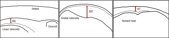
The measurements of following structures were evaluated (Fig. 1).
 | Fig. 1Schematic figures of measuring thickness of subscapularis, supraspinatus, and infraspinatus tendons. SSc, subscapularis; SST, supraspinatus; IST, infraspinatus.
|
The thickness of long head of biceps tendon
The thickness of subscapularis tendon
The thickness of supraspinatus tendon
The thickness of subacromial subdeltoid (SASD) bursa
The interval of acromioclavicular joint (AC joint)
The thickness of infraspinatus tendon
The thickness of deltoid muscle
The tendon of long head of biceps was measured in the transverse view at the highest point of the groove with the subject having neutral shoulder and elbow flexed 90' with palm up position. The probe was tilted to be positioned perpendicular to the direction of the tendon. Then, with the probe fixed in the same position, the subject was instructed to externally rotate the arm, fixing the elbow on the lateral chest. When the subscapularis tendon emerged inferior to the coracoid process, the probe was slightly moved to find the site of insertion to the lesser tuberosity and the thickness of subscapularis tendon was measured at just medial to the insertion site (Fig. 2A). The thickness of supraspinatus tendon was measured on the coronal view at the sulcus located between greater tuberosity and articular cartilage with the Modified Crass position (Fig. 2B). The Modified Crass position means placing the subjects' arm posteriorly and the palmar side of the hand on the superior aspect of the iliac wing with the elbow flexed, directed posteriorly. With this position, the probe was positioned more parallel to the supraspinatus tendon at the insertion site. The reason why we chose the Modified Crass position over the Crass position is that the majority of patients with rotator cuff pathology experience less pain and are able to position closer to the instruction in the former than the latter. The probe was moved anteriorly and posteriorly to precisely observe the insertion of supraspinatus tendon located anteriorly to the running of the biceps tendon. The thickness of the subacromial subdeltoid bursa and supraspinatus tendon were measured on the coronal view in the same plane. Acromioclavicular joint interval was measured over the top of the shoulder in a coronal plane. Infraspinatus tendon thickness was measured at the level of the posterior border of the acromion with the hand placing on the opposite shoulder (Fig. 2C). Deltoid muscle thickness was measured at the anterolateral edge of acromion with the same position as the infraspinatus tendon thickness.
 | Fig. 2Ultrasound dimensions of subscapularis, supraspinatus and infraspinatus tendon. (A) The thickness of subscapularis tendon was measured at just medial to the site of insertion to the lesser tuberosity. (B) The thickness of supraspinatus tendon was measured on the coronal view at the sulcus located between greater tuberosity and articular cartilage. (C) The thickness of infraspinatus tendon was measured at the level of the posterior border of the acromion.
|
Considering previous researches of measuring ultrasound dimensions in asymptomatic adults, our sample size was decided (89). In our study, we assumed ultrasonographic dimensions of shoulders would be values of standard deviation of 0.7 and a difference of 0.5 mm in thickness of rotator cuff between males and females would be significant statistically with P value of 5%. The sample size of 43 in each sex group was assumed to be adequate to compare with each other with power of 80%. The rate of falling out was assumed to be 15%, we planned to enroll 55 patients for each group.
Statistical analysis
Unpaired t-test (or Wilcoxon rank sum test for nonparametric test) was used to determine differences in measurements between males and females. Paired t-test (or Wilcoxon signed rank test for nonparametric test) was used to compare measurements of dominant and non-dominant shoulders. Differences in values according to the age were determined by analysis of variance (ANOVA or Kruskal-Wallis for nonparametric tests). Data analyses were performed with SPSS ver. 21.0 for Windows (SPSS Inc., Chicago, IL, USA).
RESULTS
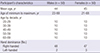
Out of 109 volunteers, 9 subjects were excluded because of positive findings in physical examinations and experiences of shoulder injection to relieve pain. A total of 200 shoulders from 100 subjects (50 males and 50 females, 95 right handed and 5 left handed, age 21 to 69 years) were scanned. Baseline demographic features of the participants are presented on Table 1.
Table 1
Study participant characteristics
![]()
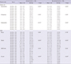
In male subjects, the mean thickness of supraspinatus, infraspinatus and subscapularis tendon was 5.1 ± 0.8 mm, 4.7 ± 0.6 mm, 4.6 ± 0.8 mm in dominant arm and 5.0 ± 0.7 mm, 4.8 ± 0.7 mm, 4.7 ± 0.7 mm in non-dominant arm, respectively. In female subjects, the mean thickness of supraspinatus, infraspinatus and subscapularis tendon was 4.6 ± 0.9 mm, 4.0 ± 0.7 mm, 4.1 ± 0.7 mm in dominant arm and 4.4 ± 0.8 mm, 4.1 ± 0.6 mm, 4.2 ± 0.6 mm in non-dominant arm, respectively (Table 2). The supraspinatus, infraspinatus, subscapularis tendon and deltoid muscle thickness were significantly different between males and females for dominant and non-dominant arms. The measurements of SASD bursa thickness were significantly different between males and females for non-dominant arms.
Table 2
Differences between males and females in dominant side and non-dominant side

Data are presented as the mean ± standard deviation. Minimal and maximal values are shown in parenthesis. All data are presented in millimeters.
*
P < 0.05; †unpaired t-test; ‡Wilcoxon rank sum test.
![]()
The differences in measurements of rotator cuff tendons between the dominant and non-dominant arm among males and females showed no statistical significance (Table 3). Only deltoid muscle thickness and AC joint interval in males and deltoid muscle thickness in females were significantly different between dominant and non-dominant arms.
Table 3
Differences between dominant side and non-dominant side in males and females

Data are presented as the mean ± standard deviation. Minimal and maximal values are shown in parenthesis. All data are presented in millimeters.
*P < 0.05; †unpaired t-test; ‡Wilcoxon rank sum test.
![]()
When subjects were stratified by the age groups, divided by ten years, the measurements of supraspinatus tendon thickness showed tendency of increase with the age, whereas the AC joint interval showed decreasing tendency (Table 4). In the other measurements, no significant difference among age groups was found.
Table 4
Differences among different ages by decades
All data are presented in millimeters.
SD, Standard deviation; Min, minimum; Max, maximum.
*P < 0.05; †ANOVA; ‡Kruskal-Wallis.
![]()
DISCUSSION
This study suggests normative reference data of rotator cuff tendon thickness and acromioclavicular joint interval among Korean population. To make ultrasonographic diagnosis of rotator cuff pathology, especially rotator cuff tear or tendinopathy, the measurements should be compared with reference values to make objective and accurate diagnoses. Although there has been a report suggesting diagnostic criteria of supraspinatus tendinopathy demonstrating maximal thickness of supraspinatus tendon based on comparison between symptomatic patients and asymptomatic controls, there is no report suggesting normal reference values of rotator cuff dimension (10). This study is in great value for the fact that it is the first report providing normative ultrasound dimensions of the rotator cuff in healthy Korean adults with varying age.
The results of our study possess certain degree of validity since the results show the correlation with prior studies (911). In our study, the supraspinatus, infraspinatus, subscapularis tendon and deltoid muscle thickness were significantly different between males and females for dominant and non-dominant arms. The increasing tendency of rotator cuff thickness in male subjects in our research is assumed to be related to larger strength of the shoulder in males than females. There has been research demonstrating significant correlations between supraspinatus thickness and external rotation strength, infraspinatus thickness and internal rotation thickness, subscapularis thickness and internal rotation strength (8). In addition, in other prior research on the rotator cuff dimensions of young adults (aged 18-40 years), the rotator cuff dimensions between males and females were significantly different (9).
The dimensions between dominant and non-dominant arms were not significantly different in all of the thickness of rotator cuff tendon, biceps tendon and subacromial subdeltoid bursa with the exception of AC joint interval and the thickness of deltoid muscle. Our study showed similar results with previous researches, which means the asymptomatic contralateral shoulder can be used to estimate the expected dimension (912). However, in the male group, the AC joint interval of dominant arms was significantly lower than non-dominant arms. This result could be associated with arthritic change as a consequence of more usage of dominant arm in males. Further researches regarding association of activities of upper extremities with AC joint interval are needed.
When the measurements were stratified by age, the measurements of supraspinatus tendon thickness revealed increasing tendency with increasing age groups. This tendency could be related to asymptomatic rotator cuff tendinopathy which shows frequent incidence rate with aging. The prevalence of rotator cuff pathology is reported to increase by natural aging process (13). In a research of ultrasonographic findings in asymptomatic shoulders, supraspinatus tendon was significantly thicker and demonstrated a lower echogenicity ratio in elderly patients aged more than 60 years and the thickness showed positive correlation with age. This study suggested the increase in thickness of the supraspinatus tendon might be due to chronic tendinopathy by age-related degeneration. In a research study of ultrasonographic findings of asymptomatic shoulders, supraspinatus tendinosis was the third most common abnormal finding, accounting for 39%, followed by subacromial subdeltoid bursal thickening and acromioclavicular joint osteoarthritis (14). Further studies for clarifying correlations between sonographic findings and pathology are needed.
In our data, acromioclavicular joint interval showed the tendency of decrease with increasing age. This tendency may be the result of natural degeneration by aging process and similar results are documented in the other studies. Stein et al. (15) reported more advanced arthritic changes in acromioclavicular joint were detected in MRI in the over 30 age group. Nicholson et al. (16) reported significant increase in degenerative changes at the acromial facet of the acromioclavicular joint occurred with advancing age.
This study has some limitations to be taken into consideration. First, the ultrasound scanning was performed once by a single physiatrist, which means intraobserver and interobserver agreement was not assessed. The previous study regarding rotator cuff dimension in young healthy adults showed good intraobserver and interobserver agreement (9). In the study of sonographic evaluation of the painful shoulder, the examiners were in very good agreement for full-thickness rotator cuff tears, supraspinatus tendinosis, abnormalities of the long head of biceps tendon, subacromial bursa abnormalities, acromioclavicular osteoarthritis and moderate agreement for partial thickness tear and intratendinous tears (17). The other study revealed good interobserver reliability in grading fatty degeneration of rotator cuff muscles (18). Although operator dependence has been considered a limitation of ultrasonography, good interobserver and intraobserver reliability was reported by previous studies. Although our study could not assess the intraobserver and interobserver reliability, ultrasonography measurements were performed on both arms for each participant and the results of each measurement showed congruence, indirectly increasing the reliability of the single physiatrist's measurement. Second, the number of subjects was not large enough to objectively assess differences among age. However, this is the first report that evaluated the normal dimensions of rotator cuff tendons where at least 10 subjects were recruited within each subgroup of stratified age, having a range of 20 to 70 years. Third, all of the subjects were chosen from one institution and there could be selection bias. However, participants in our study possess diversity in age and gender, which could represent general Korean population. Fourth, subjects' anthropometric factors that can possibly affect the measurements such as height, weight and body mass index (BMI) were not assessed. As the previous study showed there were no significant correlation between the height or weight of the subjects and the rotator cuff tendon measurements, we assumed weight and height could be omitted in measuring normal rotator cuff tendon (9). However, because there has been a study demonstrating obesity is related to increased risk for rotator cuff tendinitis and rotator cuff related surgery, further studies assessing relationship between body mass index and cuff tendon thickness under adjustment of age and gender are needed (19). Fifth, echogenicity that could be useful in diagnosing rotator cuff tendinopathy was not measured in our research. Further studies assessing echogenicity as well as rotator cuff tendon thickness in healthy adults would be needed to suggest more accurate normal reference data.
This study has suggested normative reference values of rotator cuff dimensions of Korean adults. Further studies with larger groups of subjects assessing normal rotator cuff dimensions and defining influencing factors including a wide range of age would be needed to further validate our normative reference values. Furthermore, studies regarding comparison between measurements of normal healthy adults and patients with rotator cuff lesion and suggesting the cut-off value of rotator cuff lesions in Korea would be needed.
 XML Download