

 PDF
PDF ePub
ePub Citation
 Print
Print


INTRODUCTION
Zika virus is a mosquito-borne flavivirus related to dengue virus, yellow fever virus, and West Nile virus. The virus was first isolated from a sentinel rhesus monkey stationed in the Uganda’s Zika Forest in 1947, during the epidemiological research of yellow fever (1). Human infection by Zika virus was first recognized in 3 patients in Nigeria in 1953 (2). Subsequently, only 14 sporadic cases have been reported until the first outbreak of Zika virus in Yap Islands of Micronesia in 2007 (3). Most cases during the Yap Islands outbreak were mild cases. Six years later, however, another outbreak occurred in French Polynesia, and neurological and autoimmune complications were reported for the first time (45).
The Zika virus outbreak in the Americas was first recognized in March 2015, when an epidemic of an illness characterized by fever, rash, arthralgia, myalgia, and conjunctivitis occurred in Bahia, Brazil (6). By September 2015, reports of an increase in the number of congenital microcephaly in Zika virus-affected areas began to emerge (7). Because of the cluster of microcephaly cases and other neurological disorders reported in Brazil and French Polynesia, the World Health Organization declared a Public Health Emergency of International Concern on February 1, 2016. As of May 18, 2016, 46 countries are experiencing a first outbreak of Zika virus transmitted by mosquitos, and 10 countries have reported evidence of person-to-person transmission of Zika virus, probably via a sexual route (8).
In order to prevent an outbreak of Zika virus in Korea, early detection and isolation of returning travelers with Zika virus infection from countries with ongoing outbreak is of paramount importance. Here, we report the first imported case of Zika virus infection into Korea.
CASE DESCRIPTION
A 43-year-old Korean man visited Chonnam National University Hospital due to fever and rash. The patient had history of staying and mosquito bites at Cumbuco, Ceara, Brazil for 3 weeks from 17 Feb 2016 to 9 Mar 2016, and returned to Republic of Korea on 11 Mar 2016. He had fever, chill, myalgia, and eyeball pain on 6 days after return from Brazil. Three days later, rash also developed. The patient visited nearby clinic and the blood was sampled for Zika virus reverse-transcriptase polymerase chain reaction (RT-PCR) on the 6th day of illness. The RT-PCR result was reported to be positive by the Korea Center for Disease Control and Prevention. The patient was admitted to Chonnam National University Hospital for further evaluation and management on the 7th day of illness.
Upon admission, he had a blood pressure of 110/70 mmHg, pulse rate of 80 beats/min, respiratory rate of 20/min, and a body temperature of 36.3°C. Painless multiple erythematous maculopapular rash with itching was observed in trunk and both upper and lower extremity (Fig. 1A). Hyperemia was present in both eyes and the patient complained of feeling of dryness in both eyes. Enlarged lymph node was not observed. Neurologic symptoms or signs including headache, vomiting, decrease in motor power, or abnormal sensation were absent. Initial laboratory findings performed on the day of admission were as follows: white blood cell count 4,900/µL (neutrophils 55%, lymphocytes 29%, monocytes 15%), hemoglobin level 16.5 g/dL, platelet count 221,000/µL, erythrocyte sedimentation rate 2 mm/hr, serum C-reactive protein 0.47 mg/dL, procalcitonin 0.05 ng/mL, serum neutrophil gelatinase-associated lipocalin 89.4 ng/mL, blood urea nitrogen 12.8 mg/dL, total protein 7.4 g/dL, albumin 4.6 g/dL. Serum aspartate aminotransferase, alanine transaminase, and lactate dehydrogenase were 61 U/L, 92 U/L, and 459 U/L, respectively. Serum level of ferritin was 417 ng/mL and serum adenosine deaminase was 37 IU/L.
Fig. 1
Clinical manifestation and virus shedding. (A) Maculopapular rash on the trunk and palm. (B) Time course of symptom, sign and the results of Zika virus RT-PCR.
+, positive; −, negative.

He was only medicated with cetirizine 10 mg/day per oral. Intravenous fluid or other medication was not prescribed during the hospital stay. The patient no longer felt myalgia or febrile sense during the hospital stay. Conjunctival hyperemia continued but maculopapular rashes were fading out. He was discharged from the hospital on 8th day of illness, and then was followed up weekly for 6 weeks. During the 6 weeks of follow-up, he did not have symptoms or signs of neurologic abnormality. Zika virus RT-PCR was positive from saliva and urine for 2 weeks after the symptom onset but became negative after 3 weeks (Fig. 1B).
Culture isolate of Zika virus was obtained by inoculating monolayers of Vero cells with his semen sample at the 7th day of the illness and culturing the cells at 37°C in a 5% carbon dioxide atmosphere. In the culture of first passage, the serial change of cycle threshold value by RT-PCR using RealStar Zika virus RT-PCR kit 1.0 (Altona Diagnostics, Hamburg, Germany) was shown in Fig. 2A.
Fig. 2
Isolation of Zika virus from semen sample. (A) Temporal change of cycle threshold (Ct) value for Zika virus RT-PCR in the culture of first passage. (B) Transmission electron microscopy image of Vero cells infected with Zika virus. White arrows denote virus particles. Black scale bar indicates 200 nm. (C, D) Immunofluorescence assay shows that Zika virus-infected Vero cells reacted with human convalescent anti-Zika virus IgG-positive serum (C) and did not with control serum (D). White scale bar denotes 100 μm.
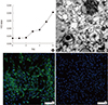
For electron microscopic observation, Vero cell monolayer inoculated with culture supernatant of second passage was fixed as previously described (9). It was cut on ultramicrotome (RMC MT-XL) at 65 nm. Ultrathin sections were stained with saturated 4% uranyl acetate and 4% lead citrate before examination with a transmission electron microscope (JEM-1400; JEOL Inc., Tokyo, Japan) at 80 kV. Virus particles were observed within the cytoplasm (Fig. 2B).
Immunofluorescence staining was performed by previously described method (1011). Briefly, Zika virus-infected and mock-infected cells were fixed with 4% paraformaldehyde in PBS for 1h at room temperature. Slides were blocked and then incubated with the 3 weeks convalescent serum of patient (1:40 dilution) and serum of healthy control (1:40 dilution) at -4°C overnight. Cells were washed and incubated for 1 hour at room temperature with fluorescein isothiocyanate-conjugated anti-human IgG. 4',6-Diamidino-2-phenylindole dihydrochloride was used to stain the nucleus. Preparations were examined with a confocal microscope (Leica, Buffalo Grove, IL, USA). Immunofluorescence was observed in Zika virus-infected Vero cells applied with convalescent serum rather than control serum (Fig. 2C and 2D).
DISCUSSION
In the present report, we describe clinical findings of the first imported patient (43 years old man) of Zika virus infection in Korea, from whom the virus was isolated. The incubation period between the mosquito bite and the onset of clinical manifestations of the current case was estimated to be 1 to 4 weeks, as his symptoms began 6 days after returning from Brazil, where he had stayed for 3 weeks. Clinical spectrum of Zika virus infection includes acute febrile illness, neurologic complications, and adverse fetal outcomes (12). Most cases with Zika virus infections are mild or asymptomatic; only 19% of cases had symptoms, and no patients were hospitalized during the Yap outbreak (3). Most common symptoms were macular or papular rash, fever, arthralgia, non-purulent conjunctivitis, myalgia, headache, and retro-orbital pain (313). A Brazilian study suggested that clinical features of pruritic rash, conjunctival injection, and lymphadenopathy should raise the suspicion of Zika virus infection (13). In our case, conjunctival injection was present and it lasted longer than rash. Neurologic complications of Zika virus infection include Guillain-Barré syndrome (14), myelitis, and meningoencephalitis (15). None of these neurologic complications were observed in our case. There is no antiviral treatment for Zika virus yet. Management consists of symptomatic treatment. The patient recovered spontaneously.
The diagnosis of Zika virus infection is made by detection of Zika viral RNA or serology. Zika viral RNA can be detected in blood, saliva, urine, and semen specimens by RT-PCR. Urine is the preferred sample for RT-PCR (16), as it yields higher detection rates than serum, even 5 days after symptom onset, when all serum specimens were negative by RT-PCR (17). For patients presenting > 7 days after symptom onset, serologic tests should also be included. However, serologic test results should be interpreted with caution, because Zika virus antibody can react extensively with other flavivirus antibodies (18). In our case, initial diagnosis was made by RT-PCR using blood sample. After 2 weeks, Zika virus RNA was detected only in saliva and urine but not in blood samples. Still, findings this case emphasize the sensitivity of urine and saliva than serum in the diagnosis of Zika virus infection, especially, in patients with time interval more than one week between symptom onset and specimen collection.
As for semen sample, which was obtains on day 7 of illness was positive by Zika virus RT-PCR and by virus culture. One of the possible modes of transmission of Zika virus infection is sexual transmission. Reports of the virus detected in semen as long as 62 days after onset of the illness (19), and the high viral load in semen compared to that of blood or urine (20) has brought great concern, as prolonged presence of virus in semen may increase the possibility of sexual transmission to pregnant woman. Zika virus infection during pregnancy can cause microcephaly and ocular abnormalities of the fetus (1214). These findings emphasize the importance of early detection of Zika virus infected patients and the application of preventive measures including abstain from unprotected sex. Further study is needed on the duration of detection of viable Zika virus in semen, to determine the period of sexual precaution for infected cases. In this case, the duration of viral excretion in semen is still under investigation. Besides sexual contact, there are two more possible route of transmission of Zika virus from this patient to other individuals in Korea, i.e. mosquito-borne and blood transfusion. However, we think the possibilities are negligible because he had no mosquito bite after returning home and during the febrile period. Fortunately, the period was in March when active mosquitoes are absent in Korea. He also did not donate blood.
In conclusions, in order to prevent an outbreak of Zika virus in Korea, early detection and isolation of returning travelers with Zika virus infection is important. Zika virus infection should be suspected in patients with acute febrile illness who visit endemic area. Saliva and urine samples are useful for diagnosis of Zika virus using RT-PCR.
 XML Download