Journal List > J Korean Med Sci > v.30(5) > 1022902

Kim, Jin, Cho, Kwon, Kee, and Park: Deficiencies of Circulating Mucosal-associated Invariant T Cells and Natural Killer T Cells in Patients with Acute Cholecystitis

Abstract

Mucosal-associated invariant T (MAIT) cells and natural killer T (NKT) cells are known to play crucial roles in a variety of diseases, including autoimmunity, infectious diseases, and cancers. However, little is known about the roles of these invariant T cells in acute cholecystitis. The purposes of this study were to examine the levels of MAIT cells and NKT cells in patients with acute cholecystitis and to investigate potential relationships between clinical parameters and these cell levels. Thirty patients with pathologically proven acute cholecystitis and 47 age- and sex-matched healthy controls were enrolled. Disease grades were classified according to the revised Tokyo guidelines (TG13) for the severity assessment for acute cholecystitis. Levels of MAIT and NKT cells in peripheral blood were measured by flow cytometry. Circulating MAIT and NKT cell numbers were significantly lower in acute cholecystitis patients than in healthy controls, and these deficiencies in MAIT cells and NKT cell numbers were associated with aging in acute cholecystitis patients. Notably, a reduction in NKT cell numbers was found to be associated with severe TG13 grade, death, and high blood urea nitrogen levels. The study shows numerical deficiencies of circulating MAIT and NKT cells and age-related decline of these invariant T cells. In addition, NKT cell deficiency was associated with acute cholecystitis severity and outcome. These findings provide an information regarding the monitoring of these changes in circulating MAIT and NKT cell numbers during the course of acute cholecystitis and predicting prognosis.

INTRODUCTION

Acute cholecystitis is an acute inflammatory disease of the gallbladder (1). Gallstones are etiologically involved in 90%-95% of cases, whereas acute acalculous cholecystitis accounts for 5%-10% of cases, in which severe systemic conditions, such as surgery, trauma, long-term intensive care unit stay, infection, burns, and parenteral nutrition, are commonly involved (1). The majority of patients with acute cholecystitis follow a mild clinical course, but about 6% of patients progress to accompanying organ dysfunction (grade III) according to the Tokyo guidelines 2007 (TG07) for severity assessment (2). Recently, it was reported that older patients with acute acalculous cholecystitis are more likely to have more severe complications and to succumb to the disease (3). With the exception of one study, which showed a decline in T cell numbers and functional activity in elderly patients (4), little is known of the status of the immune system before intervention in patients with acute cholecystitis.
Human mucosal-associated invariant T (MAIT) cells and natural killer T (NKT) cells are two distinct subsets of innate-like T cells that express an invariant TCR Vα7.2-Jα33 chain paired with a limited repertoire of Vβ chains or an invariant TCR pair of Vα24-Jα18/Vβ11 chains together with the Vβ chain, respectively (5, 6). Using these unique pairs of TCR chains, MAIT cells recognize bacteria-derived vitamin B2 metabolite antigens presented by major histocompatibility complex (MHC) class Ib-like related protein MR1 (7), whereas NKT cells react with endogenous and exogenous glycolipid antigens, such as isoglobotrihexosylceramide (iGb3) and α-galactosylceramide (α-GalCer), presented by MHC class I-like protein CD1d (8). Following antigen recognition, MAIT cells rapidly produce Th1/Th17 cytokines, such as interferon-γ (IFN-γ) and interleukin (IL)-17 (9), whereas NKT cells rapidly produce large amounts of Th1 and Th2 cytokines, such as IFN-γ and IL-4, in an innate-like manner (8). Accordingly, MAIT cells play important roles in mucosal immunity and gut homeostasis (9, 10), and NKT cells act as regulatory and/or effector cells and are implicated in the control of autoimmune diseases, cancer, and infectious diseases (11). Furthermore, these two invariant T cells are known to play protective roles in host immune responses against a variety of infectious pathogens, including certain enterobacterial and mycobacterial species, viruses, fungi, and parasites (12, 13). In humans, MAIT and NKT cells are more abundant in liver as compared with peripheral blood, suggesting that effector functions of MAIT and NKT cells might overlap and lead to protect the liver from infectious agents and intestinal floral products that reach the intrahepatic blood circulation (14, 15).
Recently, it was reported that patients with severe cholecystitis have high microorganism culture rates in bile and blood (2). Of the microorganisms cultured in bile or blood, Escherichia coli was most prevalent followed by other enterobacterial species. However, little is known about the relevances of MAIT and NKT cells in acute cholecystitis. Thus, the aims of the present study were to measure MAIT and NKT cell numbers in the peripheral blood of patients with acute cholecystitis and to investigate potential relationships between these cell numbers and clinical parameters.

MATERIALS AND METHODS

Study population

The study cohort was composed of 30 patients with a diagnosis of acute cholecystitis (7 women and 23 men; mean±SD age 66.7±10.8 yr) according to the revised Tokyo guidelines (TG13) regarding diagnostic criteria for acute cholecystitis (16), and 47 age- and sex-matched healthy controls (11 women and 36 men; mean±SD age 65.3±8.3 yr). None of the controls had a documented history of autoimmune disease, pregnancy, infectious disease, malignancy, chronic liver or renal disease, or diabetes mellitus, or had ever received immunosuppressive therapy or experienced fever during the 72 hr prior to enrollment.

Monoclonal antibodies (mAbs) and flow cytometry

The following mAbs and reagents were used in this study: fluorescein isothiocyanate (FITC)-conjugated anti-CD3, phycoerythrin (PE)-Cy5-conjugated anti-CD161, FITC-conjugated anti-TCR γδ and PE-conjugated 6B11 (all from Becton Dickinson, San Diego, CA, USA), allophycocyanin (APC)-conjugated anti-TCR Vα7.2 (BioLegend, San Diego, CA, USA) and APC-Alexa Fluor 750-conjugated anti-CD3 (Beckman Coulter, Marseille, France). Cells were stained with combinations of appropriate mAbs for 20 min at 4℃. Stained cells were analyzed on a Navios flow cytometer using Kaluza software (Beckman Coulter, Brea, CA, USA).

Isolation of peripheral blood mononuclear cells (PBMCs) and the identification of MAIT and NKT cells

Peripheral venous blood samples were collected in heparin-containing tubes, and PBMCs were isolated by density-gradient centrifugation using Ficoll-Paque Plus solution (Amersham Biosciences, Uppsala, Sweden). MAIT and NKT cells were identified phenotypically as CD3+TCRγδ-Vα7.2+CD161high and CD3+6B11+ cells, respectively, by flow cytometry, as previously described (17, 18, 19, 20, 21).

Statistical analysis

Percentages and absolute numbers of MAIT and NKT cells were compared using the Mann-Whitney U test. Linear regression analysis was used to examine potential relationships between MAIT/NKT cell numbers and clinical or laboratory parameters. P values of less than 0.05 were considered statistically significant. The statistical analysis was performed using SPSS version 18.0 (SPSS, Chicago, IL, USA).

Ethics statement

The study protocol was approved by the institutional review board of Chonnam National University Hospital (IRB No. CNUH-2012-093), and written informed consent was obtained from all participants in accordance with the Declaration of Helsinki.

RESULTS

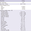
The clinical and laboratory characteristics of the acute cholecystitis patients are summarized in Table 1. Thirty patients with acute cholecystitis treated during a 6-month period were included in this study. Of these patients, 24 (80%) and 6 (20%) patients had moderate and severe acute cholecystitis, respectively, according to the Tokyo guidelines (TG13) (16).
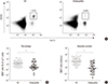
The percentages and absolute numbers of MAIT cells in the peripheral blood samples of the 30 patients and the 47 age- and sex-matched healthy controls (HCs) were determined by flow cytometry. MAIT cells were defined as CD3+TCRγδ- cells expressing TCR Vα7.2 and CD161high (Fig. 1A). Percentages of circulating MAIT cells were significantly lower in patients than in HCs (median 0.34% vs. 1.37% [P<0.05]) (Fig. 1B). Absolute numbers of MAIT cells were calculated by multiplying MAIT cell fractions by CD3+γδ- T cell fractions and total lymphocyte numbers (per microliter of peripheral blood). Acute cholecystitis patients had significantly lower absolute numbers of MAIT cells than HCs (median 0.75 cells/µL vs. 8.51 cells/µL [P<0.001]) (Fig. 1C).
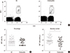
The percentages and absolute numbers of NKT cells in the peripheral blood samples of the 30 patients and the 47 age- and sex-matched HCs were determined by flow cytometry. NKT cells were defined as CD3+6B11+ cells (Fig. 2A). No significant difference was observed between the NKT cell percentages of patients and HCs (Fig. 2B). Absolute NKT cell numbers were calculated by multiplying NKT cell fractions by total lymphocyte numbers (per microliter of peripheral blood). Acute cholecystitis patients had significantly lower absolute NKT cell numbers than HCs (median 0.47 cells/µL vs. 0.78 cells/µL [P<0.05]) (Fig. 2C).
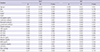
To investigate the clinical relevances of MAIT and NKT cell levels in patients, we explored relationships between the absolute numbers of MAIT cells and NKT cells in peripheral blood with clinical and laboratory parameters using regression analysis (Table 2). Because distributions were skewed, the absolute numbers of MAIT cells and NKT cells were log-transformed for the analysis. Linear regression analysis showed that log-transformed MAIT cell numbers were significantly correlated with age and lymphocyte count (P=0.039 and P=0.025, respectively). Log-transformed NKT cell numbers were found to be significantly correlated with age, TG13 grade, death, lymphocyte count, and BUN level (P=0.011, P=0.024, P=0.002, P=0.007, and P=0.044, respectively) (Table 2).

DISCUSSION

To the best of our knowledge, this is the first study to measure levels of circulating MAIT cells and NKT cells in acute cholecystitis and to examine the clinical relevances of these two distinct invariant T cell levels. The present study showed that percentages and numbers of circulating MAIT cells were lower in patients than in healthy controls. Recently, MAIT cells were reported to express high levels of CCR6 and CXCR6 and intermediate levels of CCR9 (12, 14), and these chemokine receptors are known to be involved in cell trafficking to intestines and liver (22, 23, 24). Thus, a loss of circulating MAIT cells in acute cholecystitis might be due to the transmigration of these cells from blood to sites of inflammation. This notion is supported by several lines of evidence that MAIT cells number are diminished in blood but obviously elevated in the intestines of patients with inflammatory bowel disease and in the ascetic fluids of patients with ovarian cancer or active tuberculosis (12, 25, 26). In the present study, no significant differences were observed between NKT cell percentages in the peripheral blood of patients and HCs. However, circulating NKT cell numbers were found to be significantly reduced in patients. This decline in NKT cell numbers might be due to lower lymphocyte counts in patients (data not shown), as a similar finding was reported in patients with head and neck squamous cell carcinoma. In this previous study, circulating NKT cell numbers and total lymphocyte counts were significantly reduced in patients, but NKT cells per million T cells in patients and HCs were not significantly different (27).
In addition, our data also revealed that numbers of MAIT and NKT cells were negatively correlated with age in acute cholecystitis patients, which is reminiscent of a previous report that demonstrated an age-dependent decrease in circulating NKT cell numbers in cancer patients and HCs (28). Similarly, we previously reported circulating MAIT and NKT cell numbers are affected negatively by age in healthy subjects (21, 29). These findings show that an age-related decline in these invariant T cell numbers can be expected regardless of patients or healthy subjects.
We also found that reductions in NKT cell numbers are associated with TG13 grade and death (i.e., disease severity and outcome). However, correlations between NKT cell numbers and grade or death might be obtained because all of these parameters are age-dependent. Thus, in the present study, age was included as a control variable in a partial correlation analysis and correlations were reanalyzed. It was found that death (r=-0.485, P=0.008), but not TG13 grade (r=-0.265, P=0.165), retained its association with NKT cell deficiency after this adjustment, which suggests that screening for NKT cell levels in blood samples could be used to predict prognosis in acute cholecystitis. Interestingly, this prognostic value of circulating NKT cell levels was also addressed in a previous study on head and neck cancer (27).
The present study has some limitations that require consideration. The study was performed on small populations with unbalanced sex ratios and narrow age ranges (41-83 yr). Furthermore, selection bias may have been caused by the inclusion of patients with pathologically proven acute cholecystitis in a tertiary-care regional referral center. Thus, patients with mild cholecystitis were not included in this study. Moreover, male (76.7%) and elderly (mean age, 66.7 yr) patients accounted for the majority of the patients, and several studies have reported patients with more severe cholecystitis tend to be male and that elderly patients have more severe grades (i.e., grade III) (2, 3). In addition, levels and functions of circulating immune cells are known to be affected by several factors, such as drugs and invasive procedures (30, 31). To exclude the possibility that MAIT and NKT cell levels were affected by these factors, blood samples were obtained before surgery and study subjects with a history of drug exposure (e.g., steroid or immunosuppressant use) were excluded. Nonetheless, follow-up analyses of MAIT and NKT cell levels before and after treatment are required to evaluate changes in these cell levels according to disease activity. Further investigations for large populations with balanced sex ratios and broad age ranges are needed and in particular functional studies are required to assess MAIT and NKT cell functions and to elucidate the roles these invariant cells play in immune responses to acute cholecystitis.
Summarizing, the present study describes numerical deficiencies of circulating MAIT cells and NKT cells in acute cholecystitis and age-related declines in these invariant T cells. In addition, NKT cell deficiency was found to be associated with disease severity or outcome. These findings provide important information regarding the monitoring of changes in circulating MAIT and NKT cell levels during disease course and predicting the prognosis of acute cholecystitis.

Figures and Tables

Fig. 1

Reduced circulating MAIT cell numbers in the peripheral blood of acute cholecystitis patients. (A) Representative MAIT cell percentages as determined by flow cytometry. (B) MAIT cell percentages among peripheral blood αβ T cells. (C) Absolute MAIT cell numbers (per microliter of blood). *P < 0.05; P < 0.001. HC, healthy control.

jkms-30-606-g001
Fig. 2

Reduced circulating NKT cell numbers in the peripheral blood of acute cholecystitis patients. (A) Representative NKT cell percentages as determined by flow cytometry. (B) NKT cell percentages among peripheral blood lymphocytes. (C) Absolute NKT cell numbers (per microliter of peripheral blood). *P < 0.05. HC, healthy control.

jkms-30-606-g002
Table 1

Clinical and laboratory characteristics of the 30 patients with acute cholecystitis

jkms-30-606-i001
Parameters Findings
No. male/female 23/7
Age, mean±SD (yr) 66.7±10.8
Clinical variables, No. (%)
 Fever 12 (40.0)
 Grade III* 6 (20.0)
 Death 4 (13.3)
Laboratory variables, mean±SD
 Hemoglobin (g/dL) 12.4±2.2
 Leukocyte (cells/µL) 13,290±3,987
 Neutrophil (cells/µL) 11,352±3,659
 Lymphocyte (cells/µL) 1,022±558
 Platelet (103 cells/µL) 172±64
 AST (U/L) 70±127
 ALT (U/L) 68±99
 ALP (U/L) 143±70
 Albumin (g/dL) 3.4±0.5
 Bilirubin (mg/dL) 1.7±1.3
 BUN (mg/dL) 17.8±7.4
 Creatinine (mg/dL) 0.8±0.3
 CRP (mg/dL) 15.8±9.5
 LDH (U/L) 528±172
 PaO2 (mmHg) 85.5±14.5
 Prothrombin time (INR) 1.20±0.13

*Indicates grade according to the 2013 Tokyo guidelines (TG13) for the severity assessment of acute cholecystitis. ALP, alkaline phosphatase; ALT, alanine aminotransferase; AST, aspartate aminotransferase; BUN, blood urea nitrogen; CRP, C-reactive protein; INR, international normalized ratio; LDH, lactate dehydrogenase; PaO2, partial pressure of oxygen in arterial blood; SD, standard deviation.

Table 2

Regression coefficients for log-transformed MAIT and NKT cell numbers with respect to clinical and laboratory findings in acute cholecystitis patients

jkms-30-606-i002
Variables MAIT NKT
β SE P value β SE P value
Age (yr) -0.026 0.012 0.039 -0.029 0.011 0.011
Sex -0.106 0.320 0.743 0.085 0.297 0.776
Fever 0.310 0.272 0.264 0.196 0.254 0.447
Grade* -0.142 0.362 0.698 -0.686 0.287 0.024
Death -0.436 0.443 0.334 0.000 0.313 0.002
Hemoglobin (g/dL) -0.050 0.063 0.433 0.093 0.056 0.110
Leukocyte (cells/µL) 0.000 0.000 0.601 0.000 0.000 0.706
Neutrophil (cells/µL) 0.000 0.000 0.899 0.000 0.000 0.915
Lymphocyte (cells/µL) 0.001 0.000 0.025 0.001 0.000 0.007
Platelet (cells/µL) 0.003 0.002 0.155 0.002 0.002 0.320
CRP (mg/dL) -0.002 0.015 0.910 -0.009 0.013 0.514
AST (U/L) 0.000 0.001 0.964 0.001 0.001 0.559
ALT (U/L) 0.000 0.001 0.735 0.001 0.001 0.686
ALP (U/L) 0.001 0.002 0.736 0.000 0.002 0.977
Albumin (g/dL) 0.069 0.262 0.793 0.189 0.235 0.429
Bilirubin (mg/dL) -0.176 0.132 0.192 -0.083 0.100 0.411
BUN (mg/dL) -0.014 0.019 0.459 -0.034 0.016 0.044
Creatinine (mg/dL) 0.203 0.406 0.621 -0.292 0.375 0.442
LDH (U/L) 0.001 0.001 0.490 0.000 0.001 0.965
PaO2 (mmHg) -0.006 0.009 0.523 -0.004 0.009 0.621
PT (INR) -0.221 1.084 0.840 -1.300 0.940 0.178

*Indicates grade according to the 2013 Tokyo guidelines (TG13) for the severity assessment of acute cholecystitis. ALP, alkaline phosphatase; ALT, alanine aminotransferase; AST, aspartate aminotransferase; BUN, blood urea nitrogen; CRP, C-reactive protein; INR, international normalized ratio; LDH, lactate dehydrogenase; MAIT cell, mucosal-associated invariant T cell; NKT cell, natural killer T cell; PaO2, partial pressure of oxygen in arterial blood; PT, prothrombin time; SE, standard error.

Notes

This work was supported by grants from the National Research Foundation of Korea funded by the Korean Government (#2013R1A2A2A01067956) and the Chonnam National University Hospital Biomedical Research Institute (CRI13905-22, CRI14039-21, and CRI14039-22).

DISCLOSURE All authors have no conflicts of interest to declose.

AUTHOR CONTRIBUTION Conception and coordination of the study: Kim JC, Kee SJ, Park YW. Design of ethical issues: Kee SJ, Park YW. Acquisition of data: Kim JC, Jin HM, Cho YN, Kwon YS. Data review: Kee SJ, Park YW. Statistical analysis: Kim JC, Jin HM, Kee SJ, Park YW. Manuscript preparation: Kim JC, Jin HM, Kee SJ, Park YW. Manuscript approval: all authors.

References

1. Kimura Y, Takada T, Strasberg SM, Pitt HA, Gouma DJ, Garden OJ, Büchler MW, Windsor JA, Mayumi T, Yoshida M, et al. TG13 current terminology, etiology, and epidemiology of acute cholangitis and cholecystitis. J Hepatobiliary Pancreat Sci. 2013; 20:8–23.
2. Lee SW, Yang SS, Chang CS, Yeh HJ. Impact of the Tokyo guidelines on the management of patients with acute calculous cholecystitis. J Gastroenterol Hepatol. 2009; 24:1857–1861.
3. Wang AJ, Wang TE, Lin CC, Lin SC, Shih SC. Clinical predictors of severe gallbladder complications in acute acalculous cholecystitis. World J Gastroenterol. 2003; 9:2821–2823.
4. Bondarev VI, Golovnia PF, Bondarenko SI, Radomskiĭ VT. BelonenkoGA. Disorders of immunoreactivity in acute cholecystitis in middle-aged and elderly patients and their correction in preventing postoperative complications. Klin Khir. 1990; 27–29.
5. Lantz O, Bendelac A. An invariant T cell receptor alpha chain is used by a unique subset of major histocompatibility complex class I-specific CD4+ and CD4-8- T cells in mice and humans. J Exp Med. 1994; 180:1097–1106.
6. Treiner E, Duban L, Bahram S, Radosavljevic M, Wanner V, Tilloy F, Affaticati P, Gilfillan S, Lantz O. Selection of evolutionarily conserved mucosal-associated invariant T cells by MR1. Nature. 2003; 422:164–169.
7. Kjer-Nielsen L, Patel O, Corbett AJ, Le Nours J, Meehan B, Liu L, Bhati M, Chen Z, Kostenko L, Reantragoon R, et al. MR1 presents microbial vitamin B metabolites to MAIT cells. Nature. 2012; 491:717–723.
8. Van Kaer L. alpha-Galactosylceramide therapy for autoimmune diseases: prospects and obstacles. Nat Rev Immunol. 2005; 5:31–42.
9. Le Bourhis L, Guerri L, Dusseaux M, Martin E, Soudais C, Lantz O. Mucosal-associated invariant T cells: unconventional development and function. Trends Immunol. 2011; 32:212–218.
10. Le Bourhis L, Mburu YK, Lantz O. MAIT cells, surveyors of a new class of antigen: development and functions. Curr Opin Immunol. 2013; 25:174–180.
11. Godfrey DI, Kronenberg M. Going both ways: immune regulation via CD1d-dependent NKT cells. J Clin Invest. 2004; 114:1379–1388.
12. Le Bourhis L, Martin E, Péguillet I, Guihot A, Froux N, Core M, Lévy E, Dusseaux M, Meyssonnier V, Premel V, et al. Antimicrobial activity of mucosal-associated invariant T cells. Nat Immunol. 2010; 11:701–708.
13. Sada-Ovalle I, Chiba A, Gonzales A, Brenner MB, Behar SM. Innate invariant NKT cells recognize Mycobacterium tuberculosis-infected macrophages, produce interferon-gamma, and kill intracellular bacteria. PLoS Pathog. 2008; 4:e1000239.
14. Dusseaux M, Martin E, Serriari N, Péguillet I, Premel V, Louis D, Milder M, Le Bourhis L, Soudais C, Treiner E, et al. Human MAIT cells are xenobiotic-resistant, tissue-targeted, CD161hi IL-17-secreting T cells. Blood. 2011; 117:1250–1259.
15. Kenna T, Golden-Mason L, Porcelli SA, Koezuka Y, Hegarty JE, O'Farrelly C, Doherty DG. NKT cells from normal and tumor-bearing human livers are phenotypically and functionally distinct from murine NKT cells. J Immunol. 2003; 171:1775–1779.
16. Yokoe M, Takada T, Strasberg SM, Solomkin JS, Mayumi T, Gomi H, Pitt HA, Gouma DJ, Garden OJ, Büchler MW, et al. Tokyo Guidelines Revision Committee. New diagnostic criteria and severity assessment of acute cholecystitis in revised Tokyo Guidelines. J Hepatobiliary Pancreat Sci. 2012; 19:578–585.
17. Cho YN, Kee SJ, Lee SJ, Seo SR, Kim TJ, Lee SS, Kim MS, Lee WW, Yoo DH, Kim N, et al. Numerical and functional deficiencies of natural killer T cells in systemic lupus erythematosus: their deficiency related to disease activity. Rheumatology (Oxford). 2011; 50:1054–1063.
18. Kee SJ, Kwon YS, Park YW, Cho YN, Lee SJ, Kim TJ, Lee SS, Jang HC, Shin MG, Shin JH, et al. Dysfunction of natural killer T cells in patients with active Mycobacterium tuberculosis infection. Infect Immun. 2012; 80:2100–2108.
19. Lee SJ, Cho YN, Kim TJ, Park SC, Park DJ, Jin HM, Lee SS, Kee SJ, Kim N, Yoo DH, et al. Natural killer T cell deficiency in active adult-onset Still's Disease: correlation of deficiency of natural killer T cells with dysfunction of natural killer cells. Arthritis Rheum. 2012; 64:2868–2877.
20. Cho YN, Kee SJ, Kim TJ, Jin HM, Kim MJ, Jung HJ, Park KJ, Lee SJ, Lee SS, Kwon YS, et al. Mucosal-associated invariant T cell deficiency in systemic lupus erythematosus. J Immunol. 2014; 193:3891–3901.
21. Lee OJ, Cho YN, Kee SJ, Kim MJ, Jin HM, Lee SJ, Park KJ, Kim TJ, Lee SS, Kwon YS, et al. Circulating mucosal-associated invariant T cell levels and their cytokine levels in healthy adults. Exp Gerontol. 2014; 49:47–54.
22. Sato T, Thorlacius H, Johnston B, Staton TL, Xiang W, Littman DR, Butcher EC. Role for CXCR6 in recruitment of activated CD8+ lymphocytes to inflamed liver. J Immunol. 2005; 174:277–283.
23. Stenstad H, Ericsson A, Johansson-Lindbom B, Svensson M, Marsal J, Mack M, Picarella D, Soler D, Marquez G, Briskin M, et al. Gut-associated lymphoid tissue-primed CD4+ T cells display CCR9-dependent and -independent homing to the small intestine. Blood. 2006; 107:3447–3454.
24. Williams IR. CCR6 and CCL20: partners in intestinal immunity and lymphorganogenesis. Ann N Y Acad Sci. 2006; 1072:52–61.
25. Serriari NE, Eoche M, Lamotte L, Lion J, Fumery M, Marcelo P, Chatelain D, Barre A, Nguyen-Khac E, Lantz O, et al. Innate mucosal-associated invariant T (MAIT) cells are activated in inflammatory bowel diseases. Clin Exp Immunol. 2014; 176:266–274.
26. Jiang J, Wang X, An H, Yang B, Cao Z, Liu Y, Su J, Zhai F, Wang R, Zhang G, et al. Mucosal-associated invariant T-cell function is modulated by programmed death-1 signaling in patients with active tuberculosis. Am J Respir Crit Care Med. 2014; 190:329–339.
27. Molling JW, Langius JA, Langendijk JA, Leemans CR, Bontkes HJ, van der Vliet HJ, von Blomberg BM, Scheper RJ, van den Eertwegh AJ. Low levels of circulating invariant natural killer T cells predict poor clinical outcome in patients with head and neck squamous cell carcinoma. J Clin Oncol. 2007; 25:862–868.
28. Molling JW, Kölgen W, van der Vliet HJ, Boomsma MF, Kruizenga H, Smorenburg CH, Molenkamp BG, Langendijk JA, Leemans CR, von Blomberg BM, et al. Peripheral blood IFN-gamma-secreting Valpha24+ Vbeta11+ NKT cell numbers are decreased in cancer patients independent of tumor type or tumor load. Int J Cancer. 2005; 116:87–93.
29. Kee SJ, Park YW, Cho YN, Jin HM, Kim MJ, Lee SJ, Kim TJ, Lee SS, Kwon YS, Jang HC, et al. Age- and gender-related differences in circulating natural killer T cells and their subset levels in healthy Korean adults. Hum Immunol. 2012; 73:1011–1016.
30. Brand JM, Kirchner H, Poppe C, Schmucker P. The effects of general anesthesia on human peripheral immune cell distribution and cytokine production. Clin Immunol Immunopathol. 1997; 83:190–194.
31. Rosenberger PH, Ickovics JR, Epel E, Nadler E, Jokl P, Fulkerson JP, Tillie JM, Dhabhar FS. Surgical stress-induced immune cell redistribution profiles predict short-term and long-term postsurgical recovery. A prospective study. J Bone Joint Surg Am. 2009; 91:2783–2794.
TOOLS
ORCID iDs

Jung-Chul Kim
https://orcid.org/http://orcid.org/0000-0002-6774-1861

Hye-Mi Jin
https://orcid.org/http://orcid.org/0000-0002-3622-5995

Young-Nan Cho
https://orcid.org/http://orcid.org/0000-0001-6922-8570

Yong-Soo Kwon
https://orcid.org/http://orcid.org/0000-0001-5121-4488

Seung-Jung Kee
https://orcid.org/http://orcid.org/0000-0001-9708-5837

Yong-Wook Park
https://orcid.org/http://orcid.org/0000-0002-6937-7119

Similar articles