

 PDF
PDF ePub
ePub Citation
 Print
Print


INTRODUCTION
It is difficult to remove common bile duct (CBD) stones sized 1 cm or more endoscopically by conventional endoscopic sphincterotomy (EST) alone. In such cases, mechanical lithotripsy, EST plus endoscopic papillary large balloon dilation (EPLBD), or other endoscopic options have been used. However, the peroral transpapillary endoscopic approach is unsuitable in some patients with CBD stones because of an altered gastrointestinal anatomy after Billroth II gastrectomy, a strictured upper gastrointestinal tract, or failed cannulation of the bile duct. Therefore, a percutaneous endoscopic approach would be considered rather than a surgical option, especially in high-risk elderly patients, despite the additional time required to perform endoscopy after percutaneous biliary drainage (1, 2, 3, 4).
Recently percutaneous transhepatic papillary balloon dilation using a small-sized balloon catheter was reported as one of effective method in percutaneous transhepatic cholangioscopic lithotomy (PTCS-L) (5). However, the feasibility and usefulness of percutaneous transhepatic papillary large-balloon dilation (PPLBD) for the removal of large CBD stones has not been established.
In the current study, we evaluated the safety and efficacy of percutaneous transhepatic papillary balloon dilation (PPLBD) for the treatment of large CBD stones in cases of failed endoscopic retrograde cholangiopancreatography (ERCP) or inaccessible major duodenal papilla.
MATERIALS AND METHODS
Study design
Initially, we attempted to access the major duodenal papilla with a duodenoscope to perform therapeutic ERCP in all patients with large CBD stones and naïve papillae. These procedures were performed between September 2011 and August 2012 at a single tertiary referral center. Among patients with large CBD stones in whom the attempt to access the major duodenal papilla or bile duct had failed, poor surgical candidates or patients who refused to undergo a surgical operation were enrolled prospectively. Poor surgical candidates were defined as class IV or more in American Society of Anesthesiologists (ASA) classification.
A large CBD stone was defined as one with a maximum transverse-diameter in the CBD of 1 cm or more. The exclusion criteria applied were; pancreatic or biliary malignancy, benign biliary stricture, intrahepatic duct stone, previous papillary procedure, an age younger than 19-yr, a pregnant status, the use of a protease inhibitor or somatostatin within 2 days prior to the procedure, and pancreatitis.
Interventions
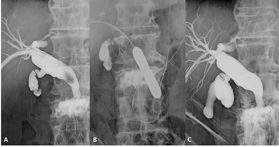
Percutaneous transhepatic biliary drainage was performed with a 21G Chiba needle (Cook Medical, Bloomington, IN, USA). The right hepatic duct B5 or B6 branch was punctured under ultrasonographic guidance and then an 8.5 Fr pigtail catheter (Cook Medical) was located in the common bile duct. A guidewire was inserted percutaneously into the duodenum and one week later the tract was dilated with a Thal-Quick drainage catheter (Cook Medical). The final diameter of PTBD tract after dilation was about 6 mm. PTCS-L was performed under 5 mg midazolam and 25 mg meperidine hydrochloride (intravenous) sedation using a choledochoscope (CHF-P20Q; Olympus Optical Co Ltd, Tokyo, Japan) 1 week after tract dilation. A 0.035-inch of guidewire (Boston Scientific Corp., Natick, MA, USA) was advanced through the papilla and into the duodenum under the cholangioscopic guidance and a dilation balloon catheter (Boston Scientific Corp.) was inserted over the guidewire passed into the duodenal papilla. The papilla was dilated by inflating the balloon gradually up to 12 mm or more using an inflation device (Indeflator; Abbott, Santa Clara, CA, USA) for 60 sec (Fig. 1A, B); balloons ranged in diameter from 12 to 20 mm. A cholangiogram was obtained after dilation of papilla in order to determine whether or not the bile duct perforation had occurred (Fig. 1C).
The scope-pushing and/or saline-flushing methods were used to facilitate stone removal through the dilated papilla. When the stone could not be retrieved using these methods after dilation of papilla, electrohydraulic lithotripsy (EHL), a stone basket, or some other options were used.
Measurement of outcomes
The primary endpoint for the study was overall adverse event rate which included pancreatitis, perforation, bleeding, and cholangitis. Secondary endpoints included total procedure time, initial procedural success rate, number of sessions required to clear the CBD completely, use of EHL, and use of a basket. Total procedure time was defined as the time from scope insertion to complete stone removal after PPLBD. Procedure-related adverse events were evaluated by cholangioscopic examination or cholangiography to rule out the possibility of distal CBD or papilla perforation. Patients were evaluated by physical examination, plain abdominal X-ray, and blood chemistry 12 hr after the procedure.
RESULTS
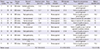
Among patients with large CBD stones in whom access to major duodenal papilla or bile duct had failed, poor surgical candidates or patients who refused to undergo surgical operation were enrolled prospectively. The clinical characteristics of study subjects and stones are shown in Table 1. Median maximum stone diameter was 13.7 mm (10.6-24.5), median maximum CBD diameter was 21.8 mm (13.2-26.5), and median balloon diameter was 13.5 mm (12-20). No adverse event occurred after PPLBD in any of the patients.
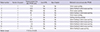
Median total procedure time after PPLBD was 17.8 min (4-42). Complete stone clearance in a single session of PTCS-L was achieved in all patients (100%). In no patient was a basket needed to remove a stone after PPLBD. Two patients (18.2%) required electrohydraulic lithotripsy (EHL). Direct scope-pushing and saline-flushing methods were used for the stone removal after PPLBD (Table 2).
DISCUSSION
PTCS-L is a useful method for treating CBD stones in patients with anatomical alterations or in whom selective bile duct cannulation fails, particularly in high-risk elderly patients. Since Ersoz et al. (6) first used a large diameter balloon catheter to dilate a papilla to remove difficult bile duct stones, several studies have demonstrated the efficacy and safety of EST plus EPLBD (7, 8, 9, 10, 11, 12). However, little data is available on EPLBD without preceding sphincterotomy for the removal of large CBD stones (13, 14). In these previous series, endoscopic retrograde dilation of the major duodenal papilla using a large diameter of balloon catheter was suggested to be a safe and effective treatment for large CBD stones in terms of immediate and long-term clinical outcomes.
When performing conventional PTCS-L for large CBD stones, stone-fragmentation procedures such as EHL or laser lithotripsy should be performed, and thus a basket may be required to retrieve fragmented stones. However, these procedures are laborious, take a long procedure time, and require accessory equipment. Recently, a report was issued on the use of a small balloon (maximum inflated diameter, 8 mm) for dilation of duodenal papillae through the percutaneous route to remove remnant stone fragments after mechanical lithotripsy (5), but, this still needs lithotripsy procedure. Accordingly, if PPLBD is found to be safe, its use could avoid the need for endoscopic accessories, shorten the procedure time, and be more convenient to endoscopists and patients.
Since non-surgical antegrade percutaneous removal of bile duct stones was first demonstrated (15), several studies have addressed antegrade papillary balloon dilation for the CBD stones (16, 17). However, in these studies, stones were relatively small and extraction balloon catheters were used to push out the stones into the duodenum. In the current study, only a cholangioscope was used to retrieve stones after PPLBD in most patients even though all stones were larger than 1 cm.
The mechanism or reason why the incidence of post-ERCP pancreatitis is low in PPLBD has not definitely been identified yet. However, anterograde approach could lower the post-ERCP pancreatitis compared to retrograde approach because there is no risk of pancreatic duct insult caused by selective bile duct cannulation. Moreover, papillary balloon dilatation itself has known to be not associated with pancreatitis (18, 19).
Of the eleven patients enrolled in this study, two patients (18.2%) needed EHL after PPLBD because the balloon diameter selected for papillary dilation was smaller than the maximum stone diameter. The precise measurement of stone diameter is often difficult on the cholangiograms during PTCS-L. In order to overcome this limitation, endoscopists should select the largest possible balloon catheter (13).
Despite the single-arm nature and small sample size of the present study, it was conducted on a prospective basis. No major adverse event occurred even after percutaneous antegrade large-balloon dilation of the papilla. In addition, the rate of complete stone removal in the index PTCS-L was 100% in the present study.
The present study suggests that PPLBD may be a safe and effective treatment for large CBD stones more than 1 cm in patients with anatomical alterations or in whom selective bile duct cannulation fails, particularly in high-risk elderly patients who is a high operation risk candidates or who refused surgery. We suggest that a large scale of prospective, randomized, and multi-center study be taken to validate this technique as an option for removing large CBD stones.

 XML Download