

 PDF
PDF ePub
ePub Citation
 Print
Print


INTRODUCTION
Microsporidia are eukaryotic organisms comprising over 150 genera and more than 1300 species (1). Eight genera have been described in human hosts: Enterocytozoon, Encephalitozoon, Pleistophora, Trachipleistophora, Vittaforma, Brachiola, Nosema, and Microsporidium (1). Among these, Enterocytozoon (Ent.) bieneusi and the three species of the genus Encephalitozoon, E. hellem, E. intestinalis, and E. cuniculi, are the most prevalent types primarily found in HIV-infected patients (2).
Since 1990, the prevalence of microsporidia infections in AIDS patients has been estimated to vary from 1.5% to 50%, depending on the geographic region and the diagnostic methods applied (1, 3). In HIV-positive patients, the most common clinical manifestation is chronic diarrhea and wasting due to enteric infection; however, the spectrum of disease caused by these pathogens is broad (4). There is increasing appreciation that these organisms can also cause gastrointestinal and ocular infections in immunocompetent individuals (5). Microsporidia species that infect humans have also been identified in animals and water sources, raising public health concerns regarding zoonotic and waterborne transmission (6, 7). To date, there have been several reports on microsporidia infections in animals such as cows, pigs, and birds in the Republic of Korea (8, 9, 10). However, there have been no reports on human infection or environmental contamination. In the present study, we detected E. intestinalis infections in human diarrheal stool specimens of unidentified etiology. We also report that we have identified E. hellem in farm soil samples.
MATERIALS AND METHODS
Stool and soil sample preparation
A total of 139 diarrheal stools were collected from May to June 2011 from seven different localities by the Korean Center for Disease Control (KCDC). All samples were of unknown etiology and derived from the routine monitoring program for waterborne protozoa (e.g., Cryptosporidium parvum, Giardia lamblia, and Entameba histolytica) infections performed by the KCDC. No clinical data besides the diarrhea diagnosis, e.g., HIV infection status of the patients, were available. To investigate environmental contamination as a mediator of human infection, 34 soil samples from seven different localities along the western side of the Korean Peninsula were collected in January 2012. The diarrheal stool samples were washed with distilled water twice by centrifugation at 1,000×g using a Union 32R Plus centrifuge (Hanil Science Industrial, Incheon, Korea). Before DNA extraction, pellets were freeze-thawed three times using liquid nitrogen and a 50℃ water bath.
The procedure of soil sample preparation was the same as described by Hong et al. (11). Briefly, 20 g of soil taken from each locality was sieved with gauze, mixed with 500 mL filtered (0.22 µm) distilled water and centrifuged at 2,000×g for 20 min using a Sorvall®RC6 Plus centrifuge (Thermo Scientific, Waltham, MA, USA). The pellet was transferred to a 50-mL tube, washed in distilled water, and centrifuged again at 1,000×g for 20 min. The supernatant was discarded, 40 mL of distilled water was added to the pellet, and the sample was mixed well. The mixed solution (1 mL) was transferred to a microcentrifuge tube and centrifuged using a microcentrifuge (5415R, Eppendorf, Hamburg, Germany). The pellet was collected and stored at 4℃ until DNA purification. For DNA extraction from the fecal materials and soil samples, the QIAquick stool mini kit (QIAGEN Inc., Valencia, CA, USA) was used according to the manufacturer's instructions.
Real-time polymerase chain reaction
For real-time quantitative polymerase chain reaction (qPCR), we used primers and probes designed for the small subunit rRNA gene of E. intestinalis or E. hellem (GenBank nos. L19567 and L19070, respectively) (Table 1). Absolute qPCR reactions were performed as described previously (12). Briefly, reaction mixtures included 0.1×LightCycler® FastStart HybProbe master mix (Roche, Mannheim, Germany); each primer set was used at a concentration of 0.5 µM (Bioneer, Daegeon, Korea), and the probe at a concentration of 0.1 µM (TIB MOLBIO, Berlin, Germany). qPCR was performed with a LightCycler®2.0 (Roche), and the qPCR for each mixture was performed as follows: initial denaturation at 95℃ for 10 min, followed by 55 cycles of denaturation at 95℃ for 5 sec, annealing at 55℃ for 15 sec, and extension at 72℃ for 8 sec, and a final cooling step at 40℃ for 30 sec. The results were analyzed using the LightCycler® software (version 4.05, Roche). DNase/RNase-free water was included as a negative control. The plasmid DNA standard for qPCR was prepared as previously described, using the 18S rRNA gene of E. intestinalis or E. hellem as the target (12). For genomic DNA preparations, both E. intestinalis and E. hellem were purchased from ATCC (http://www.atcc.org).
The nucleotide sequences of the qPCR products were confirmed using an ABI 7700 Sequence Detector and the SDS v.1.6.3 software (Applied Biosystems, Foster City, CA, USA) at the Cosmo Sequence Facility Service (Seoul, South Korea). The agarose gel extraction product using the QIAquick gel extraction kit (Qiagen) after electrophoresis on a 2.5% agarose gel (w/v) was used for this purpose. Gene sequences of E. intestinalis and E. hellem were aligned using Clone Manager Suite 7 (Sci-Ed Software, NC, USA).
Genotype analysis of E. hellem
The genotypes of E. hellem isolated from soil samples were analyzed by DNA sequencing after PCR against two target genes (internal transcribed spacer 1 [ITS1, GenBank no. AF272836] and polar tube protein [PTP, GenBank no. AF044915]), as previously reported (13, 14, 15). PCR reactions were performed in a C1000™ Thermal Cycler (BIO-RAD, Hercules, CA, USA) using previously described conditions and primers (14, 15). PCR products were purified using a gel extraction kit (QIAGEN) after 2% agarose gel electrophoresis. The DNA sequences of the PCR products were confirmed as described above.
RESULTS
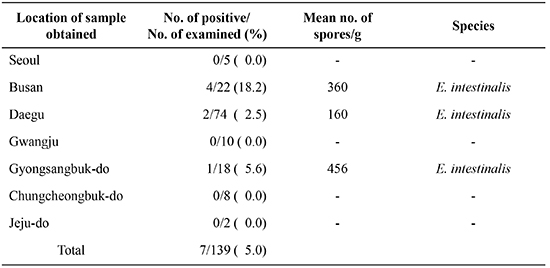
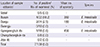
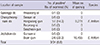
Among the 139 diarrheal stool samples, seven (5%) were positive for E. intestinalis, as determined by qPCR (Table 2). Among the seven localities, positive samples were found from Busan, Daegu, and Gyongsangbuk-do (Table 2). The mean number of spores per g of feces was 160-456. Sequence analysis showed that the seven positive qPCR products were 98% homologous to E. intestinalis (data not shown). With the exception of one case, most positive cases involved patients under 20 yr of age (Table 3).
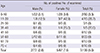
Of the 34 farm soil samples, E. hellem was detected in three soil samples (8.8%m Table 4). The number of spores detected from the soil samples ranged from 9,275 to 16,455 per g of soil. E. hellem was detected in samples from two out of seven locations (Hongseong-gun and Buan-gun, Table 4). Sequence analysis showed that the positive real-time PCR products were 99% identical to E. hellem (data not shown).
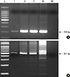
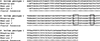
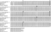
To evaluate the genotypes of E. hellem isolates from soil samples, PCR amplifications of the ITS1 and PTP genes were performed, and 208-bp and 521-bp PCR products were produced, respectively (Fig. 1). Alignment data for the ITS1 gene PCR products of three isolates of E. hellem from soil samples indicated that the isolates were genotype 1 and contained ATTT tetranucleotides followed by TTT sequences (Fig. 2). Further genotype analysis using PTP gene PCR products indicated that all three isolates were genotype 1B and contained seven copies of the 60-bp tandem repeat (Fig. 3).
DISCUSSION
Microsporidia have been identified as a cause of opportunistic infections associated with persistent diarrhea and weight loss in people with AIDS (16, 17). With heightened awareness and improved diagnostic methods, microsporidia infections have been detected in a wide range of human populations, including organ transplant recipients, travelers, children, contact lens wearers, and the elderly (18, 19).
People with AIDS who are infected with the Encephalitozoon species, particularly E. intestinalis, initially develop persistent diarrhea (1). In addition, Encephalitozoon species can disseminate and then cause clinical symptoms including sinusitis, keratoconjunctivitis, encephalitis, tracheobronchitis, interstitial nephritis, hepatitis, or myositis (1).
In the present study, we found that 5% of diarrheal stools were positive for E. intestinalis, which is the same percentage reported in a previous study on non-HIV infected patients with chronic diarrhea (20). To our knowledge, ours is the first report of human E. intestinalis infections in Korea. The results showed Busan is the highest endemic area among the seven places examined. And the most positive cases were under twenty in their ages. However, it is hard to explain these findings, as there was no available data for the infection sources or other clinical information of these positive cases.
Ground, surface, sewage, and swimming pool water have all been reported as an environmental sources of infection for E. intestinalis (1). However, it has remained unclear if soil is also a source of infection. In the present study, we therefore attempted to detect E. intestinalis in farm soil samples. We also tried to identify other Encephalitozoon species such as E. hellem in soil, as the standard genomic DNA of E. hellem was available from ATCC (http://www.atcc.org). As described in the results, we detected E. hellem, but not E. intestinalis in three of 34 soil samples. E. hellem infections were recently confirmed in pet parrots in Korea; however, human cases of infection have not been reported yet (8). E. hellem is a well-known cause of human keratoconjunctivitis in HIV patients, and to date, its environmental sources have not been reported (1, 21). Thus, our study is the first to show that farm soil can be a source of E. hellem infection. Among the seven localites examined, E. hellem was detected from two locations, such as Hongseong-gun, Chungcheognam-do and Buan-gun, Jeollabuk-do. The soil sample from Hongseong-gun was collected from near cattle farms, whereas those from Buan-gun was not close to cattle farms. This finding suggested that E. hellem infection could be established irrelevantly with livestock farms. We quantitated the spore number from human diarrhea and soil samples using qPCR. There have been no reports on the infectious dose of microsporidia yet. In addition, it is unclear that all the spores detected in this study have infectivity to the host or not. Therefore it is necessary to investigate more detail on the biological aspect of microsporidia spores such as viability and infectivity if we could interpret the meaning of the spore numbers detected here.
Molecular genotyping tools have been developed and employed to delineate the transmission of human microsporidiosis (14). For E. intestinalis, no intraspecific variation of the ITS gene has been reported (2, 22), E. hellem is known for its strong intraspecies genetic variability (2, 14, 15). On the basis of the ITS gene sequence of E. hellem ribosomal DNA, Mathis et al. (2) described three genotypes. Based on their classification, the three E. hellem isolates identified in our study are all genotype 1, because they all contained ATTT tetranucleotides followed by TTT in the ITS region. Xiao et al. (14) suggested using the PTP gene locus as an alternative genotyping tool and identified four PTP genotypes. According to PTP genotyping, the three E. hellem isolates found in our study are all genotype 1B, which has seven copies of the 60-bp tandem repeat. Based on ITS genotyping, Lee et al. (8) reported genotype 1A and 2B E. hellem in fecal materials of pet parrots in Korea. Hence, the genotypes of E. hellem detected in the farm soil samples in this study are different from those previously reported. The new finding of genotype 1B indicates that various genotypes of E. hellem exist in Korea. This finding will be useful for future epidemiological studies on microsporidia.
Ent. bieneusi was considered a major species as much as E. intestinalis as it is known to cause intestinal microsporidiosis. Although Ent. bieneusi infections were identified in fecal materials of animal hosts such as cattle and piglets in Korea (9, 10), human infection has not been reported yet. However, since Ent. bieneusi is a zoonotic organism, it is highly likely that human cases of infection exist. Therefore, human Ent. bieneusi infections should be investigated in Korea.
Our study was limited by the small sample sizes for both human and environmental samples as well as the locations of the soil sample collection, which comprised just a few areas located on the western side of the Korean Peninsula. Therefore, it is necessary to study these organisms on a larger scale to evaluate the epidemiological characteristics of microsporidia infection in Korea in more detail.
In conclusion, we detected E. intestinalis infections in human diarrheal stools using qPCR and nucleotide sequencing. We also identified E. hellem in environmental farm soil samples. To our knowledge, this is the first report on human microsporidia infections in Korea.

 XML Download