Journal List > J Korean Med Sci > v.30(12) > 1022808

Cho, Park, Park, Kim, Kang, Hong, and Kyung: Comparison of HE4, CA125, and Risk of Ovarian Malignancy Algorithm in the Prediction of Ovarian Cancer in Korean Women

Abstract

This study is a multi-center clinical study, which aimed to compare CA125, HE4, and risk of ovarian malignancy algorithm (ROMA) in predicting epithelial ovarian cancer of Korean women with a pelvic mass. Prospectively, serum from 90 Korean women with ovarian mass was obtained prior to surgery. For control group, serum from 79 normal populations without ovarian mass was also obtained. The HE4 and CA125 data were registered and evaluated separately and ROMA was calculated for each sample. Total 67 benign tumors and 23 ovarian cancers were evaluated. Median serum levels of HE4 and CA125, and ROMA score were significantly higher in patients with ovarian cancer than those with benign ovarian tumor and normal population (P < 0.001). In ROC curve analysis for women with a pelvic mass, area under the curve (AUC) for HE4 and ROMA was higher than CA125. Statistical differences in each study compared to CA125 were marginal (P compared to CA125; 0.082 for HE4 and 0.069 for ROMA). Sub-analysis revealed that AUC for HE4 and ROMA was higher than AUC for CA125 in post-menopausal women with a pelvic mass, but there were no statistically significant differences (P compared to CA125; 0.160 for HE4 and 0.127 for ROMA). Our data suggested that both HE4 and ROMA score showed better performance than CA125 for the detection of ovarian cancer in women with a pelvic mass. HE4 and ROMA can be a useful independent diagnostic marker for epithelial ovarian cancer in Korean women.

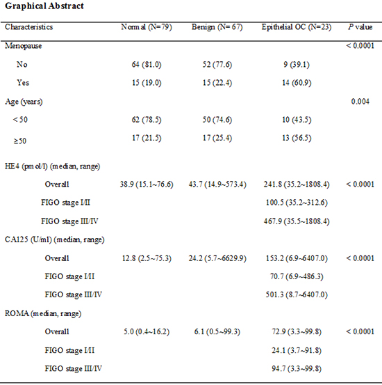
Graphical Abstract

jkms-30-1777-ab001

INTRODUCTION

Ovarian cancer is the leading cause of death from gynecologic cancer worldwide (1). More than 70% of ovarian cancers are detected at advanced stage, because most patients at earlier, more curable stage have no specific symptoms. The 5-yr survival rate is about 80% to 90% for patients with stage I disease, whereas only 30% for patients with stage III or IV (2). Despite several trials to improve survival rates in patients with advanced ovarian cancer, the results were not satisfied (34). Therefore, early detection is the key to the successful treatment.
Unfortunately, effective screening tools for early detection of ovarian cancer have not been established yet (56). Although most gynecologic oncologists are using multimodality screening with transvaginal ultrasonography and CA125 in early detection of ovarian cancer, it is expensive and not as sensitive or specific as necessary (5,7,8). Risk of malignancy index (RMI), where M is menopausal status, U is ultrasound findings and C is serum CA125 level, is the most widely used tool for the detection of ovarian cancer. Patients with pelvic mass can be stratified into high and low risk groups according to RMI. In two prospective multicenter studies, a sensitivity and specificity of RMI was approximately 81 to 92% and 82 to 85% respectively (910). CA125, which is a key factor in the RMI system, has been so far the best-performing single tumor marker in early detection and prediction of prognosis of ovarian cancer. However, CA125 can be elevated not even in ovarian cancer but also in benign gynecologic and non-gynecologic diseases. Furthermore, 20% of ovarian cancer patients presented normal or only slightly elevated serum CA125, especially in early stage disease (1112). Recently, several new markers have been proposed and investigated for early detection of ovarian cancer. Among them, special attention has been focused on Human Epididymis-specific protein 4 (HE4), a precursor of human epididymis protein. A number of studies reported that HE4 protein is frequently over-expressed in ovarian cancers (1314). Specifically in a study for premenopausal women, HE4 was reported to offer superior specificity in discrimination of benign from malignant ovarian masses compared to CA125 (15). Based on a meta-analysis approach, recent guidelines have suggested that HE4 could be used as an aid in ovarian cancer diagnosis (16). Similarly, some authors have proposed the use of HE4 in combination with a symptom index and CA125 as an annual first-line screen for ovarian cancer (17).
In addition, other studies have proposed the use of a risk of ovarian malignancy algorithm (ROMA) in predicting ovarian cancer. ROMA calculates the probability of risk for ovarian cancer, by considering CA125, HE4, and the menopausal status of patients. By the combined use of both tumor markers, ROMA was expected to improve the sensitivity and specificity in patients with pelvic masses (1819). Some authors reported that ROMA had better diagnostic performance than the widely used RMI (20). In contrast, other authors suggested that there is no benefit from combining HE4 and CA125 in ovarian cancer screening (21,22,23). A prospective cohort study including 160 women consisted of healthy controls, benign diseases, and borderline tumors/adenocarcinomas of ovarian, tubal, peritoneal and endometrial origin, showed that there was no other detection benefit from RMI compared to HE4 alone or included in ROMA (23). Other prospective study, which includes 389 women with pelvic mass showed that HE4 and ROMA did not perform significantly better than CA125 (AUC for HE4 0.857, AUC for ROMA 0.898, AUC for CA125 0.877) (22).
There have been few data of HE4 and ROMA based on Asian population (2425). Recently, a prospective trial including 319 Japanese women (131 benign, 19 borderline, 75 malignant, and 94 healthy controls) reported that ROMA showed better sensitivity than CA125 and HE4 in the diagnosis of type I and II epithelial ovarian cancer (25). However, there have been no data based on Korean women.
Therefore, in this study, we aimed to compare CA125, HE4, and ROMA and evaluate the efficacy of each diagnostic tool in predicting epithelial ovarian cancer of Korean women.

MATERIALS AND METHODS

Patients

This is a prospective, multicenter clinical study conducted at Hallym University Medical Center in Korea (Dongtan Sacred Heart Hospital, Kangnam Sacred Heart Hospital, Chuncheon Sacred Heart Hospital, and Pyeongchon Sacred Heart Hospital). From December 2014 to April 2015, 90 Korean women with pelvic mass and 79 Korean women without pelvic mass (for control group) were consecutively enrolled. Exclusion criteria were as follows: 1) Women with known relapse of a previous cancer,2) Women with co-existence of cancer in other sites. Prior to surgery, all patients underwent pelvic ultrasonography (transvaginal and/or transabdominal). Then, the ovarian masses were removed surgically and examined by a pathologist specialized in gynecologic pathology. In cases with ovarian cancer, full surgical staging and optimal debulking (residual tumor less than 1 cm in diameter) was performed. If optimal debulking surgery was not possible, biopsy for diagnosis and staging followed by neoadjuvant cheomotherapy and interval debulking were suggested. The International Federation of Gynecology and Obstetrics (FIGO) criteria for epithelial ovarian cancer staging were used to classify the patients (26).
All patients were registered online data sheet. Clinical data (age, menopausal status, serum CA125 and HE4 levels, and ROMA), treatment information, and survival status are updated continuously.

Methods

Blood samples were collected within 2 weeks prior to surgery (in women with a pelvic mass)/or at visit of enrollment (in women without a pelvic mass). Serum CA125 and HE4 levels were determined using the fully automated chemiluminescent microparticle immunoassays (CMIA) on the Architect i2000 system (Abbott Diagnostics Division, Illinois, U.S.A), according to manufacturer's instructions, and appropriate controls were included in each run. ROMA was calculated using the following algorithms proposed by Moore et al. (18):
Premenopausal: PI (predictive index) = -12+2:38 * LN(HE4)+0.0626 * LN(CA125)
Postmenopausal: PI (predictive index) = -8.09+1:04 * LN(HE4)+0.732 x * LN(CA125)
Then ROMA-value (predictive value) was calculated using the following equation:
ROMA (%) = ePI/(1+ePI) * 100
Menopausal status was defined as absence of periods from more than 1 yr.

Statistical analysis

Clinical data registered in online datasheet were used for statistical analyses. Categorical variables were compared by Fisher's exact test or chi-square test as appropriate. Continuous variables were compared by t-test. The accuracy of HE4, CA125, and ROMA in discrimination of cancer from benign disease was evaluated by using the receiver operating characteristics (ROC) curve analysis. Overall and menopausal status subgroup ROC curves and areas (accuracy) for CA125 vs. HE4 vs. ROMA were plotted and compared using the chi-square test. For all statistical tests, P value less than 0.05 was considered significant. Statistical analyses were performed using SPSS for Windows (version 21.0, SPSS Inc., NY, USA) and Medcalc software (version 15.2.2, Portland, USA).

Ethics statement

The study was approved by the institutional review board of Hallym University Hospital (IRB No. 2010-36). All patients were informed verbally and in writing with document, and invited to participate in the study after written consent.

RESULTS

Among the 90 women with a pelvic mass, 23 were malignant and 67 were benign. There were no cases of borderline ovarian cancer during the study period. Women with menopause and older age (≥50 yr) were frequently observed in ovarian cancer group than others (Table 1). Also, serum HE4 and CA125 levels, and ROMA score were all significantly higher in ovarian cancer group than others (P<0.001) (Table 1). Among 23 cancer patients, 9 were stage I-II and 14 were stage III-IV. Subgroup analysis revealed that HE4 level and ROMA score in advanced stage (FIGO stage III-IV) ovarian cancer group were about 4 times higher that early stage (FIGO stage I-II) group (Table 1). Most frequent histologic type was serous adenocarcinoma in ovarian cancer group and mature cystic teratoma in benign ovarian tumor group (Table 2).
In ROC curve analysis, area under the curve (AUC) in discriminating ovarian cancer from benign ovarian disease was 0.755 for CA125, 0.859 for HE4, and 0.860 for ROMA, and there were marginally statistical differences among three markers (Fig. 1A; P compared to CA125; 0.0818 for HE4 and 0.0690 for ROMA). In sub-group analysis, AUC was 0.766 for CA125, 0.746 for HE4, and 0.749 for ROMA in premenopausal women (Fig. 1B). And no statistical differences were found. Similarly, in postmenopausal women, AUC was 0.729 for CA125, 0.838 for HE4, and 0.795 for ROMA and there were no statistical differences among three markers (Fig. 1C).
In the detection of benign ovarian tumor from normal population, AUC was 0.711 for CA125, 0.590 for HE4, and 0.587 for ROMA and there were marginally statistical differences among three markers (Fig. 2A; P compared to CA125; 0.0826 for HE4 and 0.0656 for ROMA). In sub-group analysis, AUC was 0.787 for CA125, 0.733 for HE4, and 0.818 for ROMA in premenopausal women, which showed no significant statistical differences among three markers (Fig. 2B; P compared to CA125; 0.6401 for HE4 and 0.6097 for ROMA). However, in postmenopausal women, AUC was 0.720 for CA125, 0.618 for HE4, and 0.620 for ROMA and there were marginal statistical differences among three markers (Fig. 2C; P compared to CA125; 0.0908 for HE4 and 0.0873 for ROMA).
Suggested cut-off values for HE4 and ROMA determined by ROC curve analysis was 72.3 and 14.6, respectively. HE4 and ROMA provided better sensitivity, specificity, negative and positive predictive values comparing to CA125 (Table 3).

DISCUSSION

In this study, we aimed to prospectively assess whether preoperatively measured serum concentration of HE4 and ROMA score is superior to CA125 in the detection of ovarian cancer in Korean women with a pelvic mass.
Our data showed that both HE4 and ROMA score was not inferior to CA125 as an independent diagnostic tool in discriminating epithelial ovarian cancer from benign disease. Moreover, HE4 and ROMA offered higher sensitivity, specificity, positive and negative predictive values than CA125 in women with a pelvic mass.
HE4 and ROMA score has been in the limelight for early detection of ovarian cancer, which is generally detected in advanced stage disease. A number of data supports that HE4 seems to be more specific than CA125, which has low sensitivity in early stage ovarian cancer (1418,27,28,29,30,31,32). ROMA, which incorporates serum CA125 and HE4, and the menopausal status, has been also currently proposed as an effective diagnostic tool in ovarian cancer (1933). Moreover, HE4 and ROMA showed better accuracy than CA125 alone, in postmenopausal women (34).
In contrast, other studies showed no benefit of HE4 and ROMA (2324). A recent prospective study suggested that ROMA, and also HE4 alone, showed similar performance to CA125 alone in pre-menopausal women, but worse performance in postmenopausal women (22). In another retrospective study, HE4 was not superior to CA125 in predicting borderline ovarian tumors or presence of invasive implants (35).
These differences could be stem from the racial differences and related histologic types of ovarian cancer in study populations. Although data from Asian women is relatively rare, some studies suggested that the diagnostic role and appropriate cut-off values of HE4 and ROMA should be re-evaluated in Asian women with a pelvic mass. For an instance, Asian women showed a relatively higher incidence of mucinous epithelial ovarian cancer compared to Caucasian women and lower serum HE4 level (2436). A prospective, multi-centered study with patients from six Asian countries showed that median HE4 level in women with ovarian cancer was lower than the recommended cut-off (140 pM/L), which is based on studies in Caucasian populations (24). They used HE4 cut-off 70 pM/L for all women, which is similar to our cut-offs (72.3 pM/L for all women) (24). ROC curve analysis showed that ROMA had a better performance than CA125 in the detection of ovarian cancer (P value compared to CA125; 0.0102 for ROMA), which is similar to our data (24). In addition, they reported that ROMA showed a clear benefit in premenopausal women (24). ROMA offered improved specificity (87.4% vs. 63.2%) and positive predictive value compared to CA125 (34.69% vs. 16.8%), in premenopausal women (24). On the contrary, our data showed that menopausal status did not affect the performance of HE4 and ROMA in the detection of ovarian cancer. In addition, suggested cut-off values in pre- and post-menopausal women for HE4 (69.2 pM/L vs. 72.3 pM/L) and ROMA (15.1 vs. 19.3) in our data were almost same. It may be meaningless to use 2 cut-offs for pre- and postmenopausal women, since Moore et al. recently showed that HE4 increases with age, not with menopausal status (37). According to a cut-off suggested by ROC curve analysis, HE4 and ROMA score offered better sensitivity (78.3% and 78.3% vs. 69.6%), specificity (94.0% and 85.1% vs. 65.7%), negative (92.6% and 91.9% vs. 86.3%) and positive predictive values (81.8% and 64.3% vs. 59.0%) compared to CA125 in discriminating ovarian cancer from benign ovarian tumor.
There was limited literature about HE4 and ROMA algorithm in single-race Asian women. Specifically, there was only one prospective study for 159 Korean women, including 78 women with ovarian cancer (38). The authors suggested that ROMA score may provide higher accuracy for detecting ovarian cancer (87.5% sensitivity at a specificity of 93.8%) (38). But they compared serum CA125 only and ROMA score, not HE4 only, in predicting ovarian cancer.
In spite of small subjects including only 23 cancer patients, our data is worth focusing. This is the only prospective data comparing serum HE4, CA125 and ROMA scoring in the detection of ovarian cancer in single-race Asian women. In addition, we included normal population without pelvic mass for control group in our study. In our data, both HE4 and ROMA score showed better performances than CA125 for the detection of ovarian cancer in women with a pelvic mass, which suggested that HE4 and ROMA can be a useful independent diagnostic marker for epithelial ovarian cancer in Korean women.
Also, our data supported that those optimal cut-off values for serum HE4 and ROMA score should be fixed appropriately for Asian population. In addition, it may be re-considered to use 2 separate cut-offs for pre- and postmenopausal women. To clarify the diagnostic role of serum HE4 and ROMA in Asian women, more large-scaled prospective study will be required.

Figures and Tables

Fig. 1

ROC curves for ROMA algorithm, CA125, and HE4 in patients with pelvic masses. (A) ROC curves for overall patients. By comparing to AUC for CA125, AUC for HE4 (P = 0.082) and for ROMA (P = 0.069) were not inferior. (B) ROC curves for premenopausal patients. In premenopausal women, AUC for HE4 (P = 0.828) and for ROMA (P = 0.843) was not different from that for CA125. (C) ROC curves for postmenopausal patients. In postmenopausal women, AUC for HE4 (P = 0.160) and for ROMA (P = 0.127) was not different from that for CA125.

jkms-30-1777-g001
Fig. 2

ROC curves for ROMA algorithm, CA125, and HE4 in patients with normal population and ovarian cancer. (A) ROC curves for overall patients. AUC for CA125 showed marginal superiority to AUC for HE4 (P = 0.083) and for ROMA (P = 0.066). (B) ROC curves for premenopausal patients. In premenopausal women, AUC for HE4 (P = 0.640) and for ROMA (P = 0.610) was not different from that for CA125. (C) ROC curves for postmenopausal patients. In postmenopausal women, AUC for CA125 showed marginal superiority to AUC for HE4 (P = 0.091) and for ROMA (P = 0.087).

jkms-30-1777-g002
Table 1

Baseline data of the subjected patients

jkms-30-1777-i001
Parameters Normal (n = 79) Benign (n = 67) Epithelial OC (n = 23) P value
Menopause <0.001
 No 64 (81.0) 52 (77.6) 9 (39.1)
 Yes 15 (19.0) 15 (22.4) 14 (60.9)
Age (yr) 0.004
 <50 62 (78.5) 50 (74.6) 10 (43.5)
 ≥ 50 17 (21.5) 17 (25.4) 13 (56.5)
HE4 (pM/L) (median, range)
 Overall 38.9 (15.1-76.6) 43.7 (14.9-573.4) 241.8 (35.2-1,808.4) <0.001
  FIGO stage I/II 100.5 (35.2-312.6)
  FIGO stage III/IV 467.9 (35.5-1,808.4)
CA125 (U/mL) (median, range)
 Overall 12.8 (2.5-75.3) 24.2 (5.7-6,629.9) 153.2 (6.9-6,407.0) <0.001
  FIGO stage I/II 70.7 (6.9-486.3)
  FIGO stage III/IV 501.3 (8.7-6,407.0)
ROMA (median, range)
 Overall 5.0 (0.4-16.2) 6.1 (0.5-99.3) 72.9 (3.3-99.8) <0.001
  FIGO stage I/II 24.1 (3.7-91.8)
  FIGO stage III/IV 94.7 (3.3-99.8)
Table 2

Histologic types of women with pelvic masses

jkms-30-1777-i002
Histologic type No. (%)
Malignant ovarian tumor (N=23)
 Serous 12 (52.2)
 Endometrioid 4 (17.4)
 Mucinous 3 (13.1)
 Poorly differentiated 1 (4.3)
 Clear cell 2 (8.7)
 Hepatoid 1 (4.3)
Benign ovarian tumor (N=67)
 Serous cystadenoma 9 (13.4)
 Mucinous cystadenoma 10 (14.9)
 Endometriosis 16 (23.9)
 Mature cystic teratoma 17 (25.4)
 Hemorrhagic cyst 15 (22.4)
Table 3

Sensitivities, specificities, negative predictive values, and positive predictive values of CA125, HE4, and ROMA for ovarian malignancy in women with pelvic mass

jkms-30-1777-i003
Test Sensitivity (%) Specificity (%) Negative predictive value (%) Positive predictive value (%)
CA125 69.6 65.7 86.3 59.0
HE4 78.3 94.0 92.6 81.8
ROMA 78.3 85.1 91.9 64.3

Notes

Funding This research was supported by Hallym University Research Fund 2014 (HURF-2014-49).

DISCLOSURE The authors have no potential conflicts of interest to disclose.

AUTHOR CONTRIBUTION Study conception and design: Kyung MS. Data collection: Hong SH, Park SH, Park YH, Kim HB. Data analysis and interpretation: Cho HY, Kyung MS. Drafting and revision: Kyung MS, Cho HY, Kang JB. Final approval: all authors.

References

1. Siegel R, Ma J, Zou Z, Jemal A. Cancer statistics, 2014. CA Cancer J Clin. 2014; 64:9–29.
2. Barakat RR, Markman M, Randall ME. Principles and practice of gynecologic oncology. 6th ed. Philadelphia: Lippincott Williams & Wilkins;2009. p. 757–847.
3. Mehta DA, Hay JW. Cost-effectiveness of adding bevacizumab to first line therapy for patients with advanced ovarian cancer. Gynecol Oncol. 2014; 132:677–683.
4. Havrilesky LJ, Pokrzywinski R, Revicki D, Higgins RV, Nycum LR, Kohler MF, Berchuck A, Myers ER, Secord AA. Cost-effectiveness of combination versus sequential docetaxel and carboplatin for the treatment of platinum-sensitive, recurrent ovarian cancer. Cancer. 2012; 118:386–391.
5. Clarke-Pearson DL. Clinical practice. Screening for ovarian cancer. N Engl J Med. 2009; 361:170–177.
6. Buys SS, Partridge E, Black A, Johnson CC, Lamerato L, Isaacs C, Reding DJ, Greenlee RT, Yokochi LA, Kessel B, et al. Effect of screening on ovarian cancer mortality: the Prostate, Lung, Colorectal and Ovarian (PLCO) Cancer Screening Randomized Controlled Trial. JAMA. 2011; 305:2295–2303.
7. Gilbert L, Basso O, Sampalis J, Karp I, Martins C, Feng J, Piedimonte S, Quintal L, Ramanakumar AV, Takefman J, et al. Assessment of symptomatic women for early diagnosis of ovarian cancer: results from the prospective DOvE pilot project. Lancet Oncol. 2012; 13:285–291.
8. Valentin L, Jurkovic D, Van Calster B, Testa A, Van Holsbeke C, Bourne T, Vergote I, Van Huffel S, Timmerman D. Adding a single CA 125 measurement to ultrasound imaging performed by an experienced examiner does not improve preoperative discrimination between benign and malignant adnexal masses. Ultrasound Obstet Gynecol. 2009; 34:345–354.
9. Håkansson F, Høgdall EV, Nedergaard L, Lundvall L, Engelholm SA, Pedersen AT, Hartwell D, Høgdall C. Danish 'pelvic mass' ovarian cancer study. Risk of malignancy index used as a diagnostic tool in a tertiary centre for patients with a pelvic mass. Acta Obstet Gynecol Scand. 2012; 91:496–502.
10. van den Akker PA, Aalders AL, Snijders MP, Kluivers KB, Samlal RA, Vollebergh JH, Massuger LF. Evaluation of the Risk of Malignancy Index in daily clinical management of adnexal masses. Gynecol Oncol. 2010; 116:384–388.
11. Duffy MJ, Bonfrer JM, Kulpa J, Rustin GJ, Soletormos G, Torre GC, Tuxen MK, Zwirner M. CA125 in ovarian cancer: European Group on Tumor Markers guidelines for clinical use. Int J Gynecol Cancer. 2005; 15:679–691.
12. Rosen DG, Wang L, Atkinson JN, Yu Y, Lu KH, Diamandis EP, Hellstrom I, Mok SC, Liu J, Bast RC Jr. Potential markers that complement expression of CA125 in epithelial ovarian cancer. Gynecol Oncol. 2005; 99:267–277.
13. Drapkin R, von Horsten HH, Lin Y, Mok SC, Crum CP, Welch WR, Hecht JL. Human epididymis protein 4 (HE4) is a secreted glycoprotein that is overexpressed by serous and endometrioid ovarian carcinomas. Cancer Res. 2005; 65:2162–2169.
14. Hellström I, Raycraft J, Hayden-Ledbetter M, Ledbetter JA, Schummer M, McIntosh M, Drescher C, Urban N, Hellström KE. The HE4 (WFDC2) protein is a biomarker for ovarian carcinoma. Cancer Res. 2003; 63:3695–3700.
15. Holcomb K, Vucetic Z, Miller MC, Knapp RC. Human epididymis protein 4 offers superior specificity in the differentiation of benign and malignant adnexal masses in premenopausal women. Am J Obstet Gynecol. 2011; 205:358.e1–358.e6.
16. Redman C, Duffy S, Bromham N, Francis K. Guideline Development Group. Recognition and initial management of ovarian cancer: summary of NICE guidance. BMJ. 2011; 342:d2073.
17. Andersen MR, Goff BA, Lowe KA, Scholler N, Bergan L, Drescher CW, Paley P, Urban N. Use of a Symptom Index, CA125, and HE4 to predict ovarian cancer. Gynecol Oncol. 2010; 116:378–383.
18. Moore RG, McMeekin DS, Brown AK, DiSilvestro P, Miller MC, Allard WJ, Gajewski W, Kurman R, Bast RC Jr, Skates SJ. A novel multiple marker bioassay utilizing HE4 and CA125 for the prediction of ovarian cancer in patients with a pelvic mass. Gynecol Oncol. 2009; 112:40–46.
19. Lenhard M, Stieber P, Hertlein L, Kirschenhofer A, Fürst S, Mayr D, Nagel D, Hofmann K, Krocker K, Burges A. The diagnostic accuracy of two human epididymis protein 4 (HE4) testing systems in combination with CA125 in the differential diagnosis of ovarian masses. Clin Chem Lab Med. 2011; 49:2081–2088.
20. Moore RG, Jabre-Raughley M, Brown AK, Robison KM, Miller MC, Allard WJ, Kurman RJ, Bast RC, Skates SJ. Comparison of a novel multiple marker assay vs the Risk of Malignancy Index for the prediction of epithelial ovarian cancer in patients with a pelvic mass. Am J Obstet Gynecol. 2010; 203:228.e1–228.e6.
21. Molina R, Escudero JM, Augé JM, Filella X, Foj L, Torné A, Lejarcegui J, Pahisa J. HE4 a novel tumour marker for ovarian cancer: comparison with CA 125 and ROMA algorithm in patients with gynaecological diseases. Tumour Biol. 2011; 32:1087–1095.
22. Van Gorp T, Cadron I, Despierre E, Daemen A, Leunen K, Amant F, Timmerman D, De Moor B, Vergote I. HE4 and CA125 as a diagnostic test in ovarian cancer: prospective validation of the Risk of Ovarian Malignancy Algorithm. Br J Cancer. 2011; 104:863–870.
23. Jacob F, Meier M, Caduff R, Goldstein D, Pochechueva T, Hacker N, Fink D, Heinzelmann-Schwarz V. No benefit from combining HE4 and CA125 as ovarian tumor markers in a clinical setting. Gynecol Oncol. 2011; 121:487–491.
24. Chan KK, Chen CA, Nam JH, Ochiai K, Wilailak S, Choon AT, Sabaratnam S, Hebbar S, Sickan J, Schodin BA, et al. The use of HE4 in the prediction of ovarian cancer in Asian women with a pelvic mass. Gynecol Oncol. 2013; 128:239–244.
25. Fujiwara H, Suzuki M, Takeshima N, Takizawa K, Kimura E, Nakanishi T, Yamada K, Takano H, Sasaki H, Koyama K, et al. Evaluation of human epididymis protein 4 (HE4) and Risk of Ovarian Malignancy Algorithm (ROMA) as diagnostic tools of type I and type II epithelial ovarian cancer in Japanese women. Tumour Biol. 2015; 36:1045–1053.
26. Shepherd JH. Revised FIGO staging for gynaecological cancer. Br J Obstet Gynaecol. 1989; 96:889–892.
27. Moore RG, Brown AK, Miller MC, Skates S, Allard WJ, Verch T, Steinhoff M, Messerlian G, DiSilvestro P, Granai CO, et al. The use of multiple novel tumor biomarkers for the detection of ovarian carcinoma in patients with a pelvic mass. Gynecol Oncol. 2008; 108:402–408.
28. Wu L, Dai ZY, Qian YH, Shi Y, Liu FJ, Yang C. Diagnostic value of serum human epididymis protein 4 (HE4) in ovarian carcinoma: a systematic review and meta-analysis. Int J Gynecol Cancer. 2012; 22:1106–1112.
29. Shah CA, Lowe KA, Paley P, Wallace E, Anderson GL, McIntosh MW, Andersen MR, Scholler N, Bergan LA, Thorpe JD, et al. Influence of ovarian cancer risk status on the diagnostic performance of the serum biomarkers mesothelin, HE4, and CA125. Cancer Epidemiol Biomarkers Prev. 2009; 18:1365–1372.
30. Montagnana M, Lippi G, Ruzzenente O, Bresciani V, Danese E, Scevarolli S, Salvagno GL, Giudici S, Franchi M, Guidi GC. The utility of serum human epididymis protein 4 (HE4) in patients with a pelvic mass. J Clin Lab Anal. 2009; 23:331–335.
31. Wilailak S, Chan KK, Chen CA, Nam JH, Ochiai K, Aw TC, Sabaratnam S, Hebbar S, Sickan J, Schodin BA, et al. Distinguishing benign from malignant pelvic mass utilizing an algorithm with HE4, menopausal status, and ultrasound findings. J Gynecol Oncol. 2015; 26:46–53.
32. Chung SH, Lee SY, Ju W, Kim SC. Clinical efficacy of serum human epididymis protein 4 as a diagnostic biomarker of ovarian cancer: A pilot study. Obstet Gynecol Sci. 2013; 56:234–241.
33. Ruggeri G, Bandiera E, Zanotti L, Belloli S, Ravaggi A, Romani C, Bignotti E, Tassi RA, Tognon G, Galli C, et al. HE4 and epithelial ovarian cancer: comparison and clinical evaluation of two immunoassays and a combination algorithm. Clin Chim Acta. 2011; 412:1447–1453.
34. Sandri MT, Bottari F, Franchi D, Boveri S, Candiani M, Ronzoni S, Peiretti M, Radice D, Passerini R, Sideri M. Comparison of HE4, CA125 and ROMA algorithm in women with a pelvic mass: correlation with pathological outcome. Gynecol Oncol. 2013; 128:233–238.
35. Braicu EI, Van Gorp T, Nassir M, Richter R, Chekerov R, Gasimli K, Timmerman D, Vergote I, Sehouli J. Preoperative HE4 and ROMA values do not improve the CA125 diagnostic value for borderline tumors of the ovary (BOT) - a study of the TOC Consortium. J Ovarian Res. 2014; 7:49.
36. Tung KH, Goodman MT, Wu AH, McDuffie K, Wilkens LR, Kolonel LN, Nomura AM, Terada KY, Carney ME, Sobin LH. Reproductive factors and epithelial ovarian cancer risk by histologic type: a multiethnic case-control study. Am J Epidemiol. 2003; 158:629–638.
37. Moore RG, Miller MC, Eklund EE, Lu KH, Bast RC Jr, Lambert-Messerlian G. Serum levels of the ovarian cancer biomarker HE4 are decreased in pregnancy and increase with age. Am J Obstet Gynecol. 2012; 206:349.e1–349.e7.
38. Kim YM, Whang DH, Park J, Kim SH, Lee SW, Park HA, Ha M, Choi KH. Evaluation of the accuracy of serum human epididymis protein 4 in combination with CA125 for detecting ovarian cancer: a prospective case-control study in a Korean population. Clin Chem Lab Med. 2011; 49:527–534.
TOOLS
ORCID iDs

Hye Yon Cho
https://orcid.org/http://orcid.org/0000-0002-5697-8118

Sung Ho Park
https://orcid.org/http://orcid.org/0000-0003-2171-235X

Young Han Park
https://orcid.org/http://orcid.org/0000-0002-5114-4763

Hong Bae Kim
https://orcid.org/http://orcid.org/0000-0002-3720-1127

Jung Bae Kang
https://orcid.org/http://orcid.org/0000-0002-6989-6138

Seung Hwa Hong
https://orcid.org/http://orcid.org/0000-0001-8118-745X

Min Sun Kyung
https://orcid.org/http://orcid.org/0000-0001-5279-4584

Similar articles