

 PDF
PDF ePub
ePub Citation
 Print
Print


INTRODUCTION
Lung transplantation (LT) prolongs survival and improves pulmonary function of patients who have end-stage lung disease, thereby being a life-saving treatment for these patients. Approximately, 3,000 LTs per year are performed worldwide (1). Compared with western countries, Korea was not an early adopter for LT; the first LT was performed in July, 1996, and nine institutions have performed LT so far (23). Recently, the number of LT has been increased in Korea. However, the number of lung donors is not enough for patients who need LT, indicating that extensive effort for optimization of LT should be made to achieve successful clinical outcome. Moreover, post-transplantation complications and clinical outcome may be closely related with the primary disease of the lung.
Therefore, comprehensive understanding of primary pathology of explanted lungs from LT patients could be helpful for improving success rates of LT. To address this issue, we reviewed and analyzed pathology of the lungs from LT recipients at our center over the subsequent decade (2006-2014).
MATERIALS AND METHODS
Twenty-nine consecutive patients who underwent single or double-lung, or heart-lung transplantation at the Seoul National University Hospital (SNUH) between January, 2006 and December, 2014 were included in this study. Clinical information was collected from the medial records and included ages at time of transplantation, genders, underlying pulmonary diseases, referral diagnoses, types of transplantation, and post-transplantation clinical course.
The factors evaluated during perioperative period included intensive care unit (ICU) care, preoperative infection, dependence on mechanical ventilator (MV) or extracorporeal membrane oxygenation (ECMO) support, use of intraoperative support such as cardiopulmonary bypass (CPB) and/or ECMO, operation time, total ischemic time, and lengths of ECMO, MV support and hospital stay. The surgical pathology reports were also reviewed. At least three representative sections were taken from each lobe of the explanted lungs along with hilar nodes and vessels of LT recipients. Routine hematoxylin and eosinstain was prepared from formalin-fixed paraffin-embedded sections and various special stains for microorganisms were performed when indicated. Three pathologists reviewed all microsectioned slides of the lungs from LT recipients and compared with clinical information.
RESULTS
Clinical characterization of patients with lung transplantation
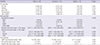
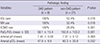
The clinico-pathological features of 29 patients who underwent deceased-donor LT are summarized in Table 1. Of 29 patients, one patient received single LT, 25 patients did bilateral sequential single LT, and three patients underwent heart-lung transplantation. These patients included eighteen males and eleven females with a median age of 49 yr (from 11 to 70 yr) at the time of transplantation. Table 2 showed perioperative clinical parameters of 29 patients. Nineteen patients had ICU care before operation. About one third of patients (11/29) had an episode of infection during pre-operative period and the most common pathogens were Vancomycin-resistant Enterococcus (n=5) and imipenem-resistant Acinetobacter baumannii (n=5). For respiratory support in preoperative management, 12 and 18 patients required tracheostomy and MV, respectively. During operation, majority of patients (27/29) got respiratory support using CPB (n=14), ECMO (n=3), and both CPB and ECMO (n= 10). Mean operation time was 647.7±136.8 min (range, 444-917 min), and total ischemic time was 627.2±174.8 min (range, 283-965 min) except one unavailable case. Mean ECMO and MV weaning times were 9.3 days (range, 0.5-80.1 days) and 14 days (range, 1-47 days) for 14 and 24 patients, respectively. Mean period of hospitalization was 88.3 days (range, 1-375 days). None of them had re-transplantation.
Clinico-pathological analysis of primary pulmonary diseases of patients with LT
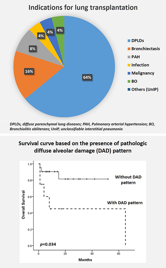
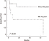
Diffuse parenchymal lung diseases (DPLDs) accounted for 55% (16/29) of lung pathology of recipients with LT (Table 1). Clinical or referral diagnosis of these patients included idiopathic pulmonary fibrosis (IPF, n=6), acute interstitial pneumonia (AIP, n=5), collagen vascular disease-related interstitial lung disease (CVD-ILD, n=4), and lymphangioleiomyomatosis (LAM, n=1). The final pathologic diagnosis of explanted lungs in all patients with IPF was usual interstitial pneumonia (UIP) (n=6), which was the most frequent underlying lung disease for LT in this cohort. Among five patients with clinical diagnosis of AIP, four cases showed diffuse alveolar damage (DAD) pattern, while one case demonstrated non-specific interstitial pneumonia (NSIP) with partial DAD pattern. Furthermore, patients with CVD-ILD exhibited either DAD (n=3) or UIP (n=1) pattern. There was minor discrepancy between referral and final diagnosis in one case; the patient was 63-yr old male with aggravated productive cough and sputum for 3 months was referred to our hospital under the clinical diagnosis of AIP. In contrast to clinical diagnosis, the explanted lungs showed diffuse interstitial thickening and inflammatory cell infiltration, which was consistent with NSIP, and partially combined with DAD pattern (Fig. 1).
Bronchiectasis was one of common indications for LT (n=4). Two patients (51-yr-old male and 48-yr-old female) underwent LT under the clinical diagnosis of bronchiectasis, which might be due to the sequelae of tuberculosis (Tb). However, there were no pathologic findings such as granulomas, suggesting lack of active tuberculosis in explanted lungs. A 23-yr-old male who had suffered from recurrent upper respiratory infection since 5-yr old age had been clinically suggested for Katagener's syndrome, but the genetic test was negative for Katagener's syndrome. The other patients (F/66 yr) had been clinically diagnosed with diffuse panbronchiolitis, which was confirmed by pathologic examination of explanted lungs.
Pulmonary arterial hypertension (PAH) (n=2) was rare primary indication for LT in our institute. An 11-yr-old female received bilateral sequential single LT due to primary pulmonary hypertension, while a 28-yr-old male underwent heart-lung transplantation for congenital heart disease and subsequent Eisenmenger's syndrome. In microscopic examination, the walls of pulmonary arteries were thickened with plexiform intimal proliferation, being consistent with PAH.
Infection and malignancy were also rare indication for LT. A 49-yr-old male had a medication for 6 months due to tuberculosis (Tb) pleurisy 15 yr ago, and one year later partial removal of pericardium was done due to constrictive pericarditis related with Tb complication. After surgery, generalized edema and recurrent pneumonia developed due to refractory constrictive pericarditis and dyspnea with pneumonic infiltration was aggravated a few months before heart-lung transplantation. However, examination of bacteria revealed no antibiotics-resistant strains in this patient. Microscopic examination revealed extensive granulomatous inflammation with caseation necrosis, indicating tuberculosis.
A 58-yr-old female with localized adenocarcinoma underwent left lower lobectomy. Eight months later, wedge resection of the lung was performed due to multiple nodules newly detected in both lungs. Microscopic examination showed invasive mucinous adenocarcinoma (i.e., mucinous bronchioloalveolar carcinoma [BAC]). However, there was no evidence of extrapulmonary tumor metastasis based on systemic evaluation, and thus she underwent bilateral sequential single LT to eliminate residual tumor in the lungs, which was mucinous type of adenocarcinoma involving both lungs in the absence of lymph node metastasis. Molecular studies revealed KRAS mutation in the tumor. The tumor recurred in the lungs 10 months after LT and the patient died of disease progression 16 months after LT.
Bronchiolitis obliterans (BO) (n=1) was rare indication for LT in our cohort. A 36-yr-old female who had undergone chemotherapy and allogenic peripheral blood stem cell transplantation (PBSCT) for T-cell prolymphocytic leukemia developed chronic graft-versus-host disease (GVHD) in the liver and lungs, which was attenuated by corticosteroids therapy. However, approximately 4 yr after PBSCT, the patient presented with dyspnea on exertion and further exacerbated due to influenza infection, thereby undergoing bilateral sequential single LT. The main histologic findings were luminal obstruction in a few small bronchioles with peribronchiolar lymphoplasmacytic infiltration, fibrosis, and foamy cell collection, being consistent with BO.
Four patients who received chemotherapy and autologous (n =1) or allogenic (n=3) PBSCT due to hematolymphoid malignancy underwent LT. Among them, two patients were referred under the clinical diagnosis of idiopathic pneumonia syndrome, whereas other two patients presented with rapidly progressive acute respiratory distress syndrome (ARDS). Histological examination of the lungs from four patients showed diffuse and marked pulmonary interstitial fibrosis with nearly complete loss or collapse of alveolar spaces and shrinkage of the lung tissue around a bronchus (Fig. 2).
Clinical outcome of patients with lung transplantation
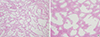
Clinical outcome of 29 recipients is briefly described in Table 1. The overall survival rate was 72.4% during a follow-up from 1 day to 65 months (mean 16.03 months). The underlying pulmonary diseases of eight patients who died after LT included AIP/DAD (n=2), CVD-ILD/DAD (n=3), Eisenmenger's syndrome (n=1), Tb (n=1), and lung cancer (n=1). Main causes of death for these patients were sepsis (n=4), disseminated intravascular coagulation (n=1), uncontrolled massive hemoptysis (n=1), disease recurrence (n=1), or chronic rejection (n =1). We compared the perioperative clinical factors of the patient according to clinical outcome (Table 2). However, there was no difference in these clinical parameters between two groups. The survival of patients who were pathologically diagnosed with DAD (8/29) was compared with the survival of those who were diagnosed with other disease (21/29), suggesting that DAD was one of factors to be associated with poor survival after LT (P=0.034) (Fig. 3). To characterize effect of DAD on patient's survival further, we compared preoperative clinical parameters between two groups (Table 3). All patients with DAD pattern took ICU care and used MV and ECMO during preoperative period. On the other hand, 11 out of 21 in patients without DAD pattern were treated in ICU. Among them, 11 and 6 patients used MV and ECMO, respectively. PaO2/FiO2 ratio and arterial pCO2 in patient with DAD pattern were significantly lower than in without the pattern (P=0.001 and P=0.032, respectively). These findings suggest that patients with DAD patterns showed worse clinical status in preoperative period than patient without DAD.
DISCUSSION
In this study, we performed clinic-pathological analysis of explanted lungs from patients with LT in our institute, although the number of LT is limited. In comparative analysis with clinical and pathology diagnosis in these patients with LT, there was a high concordance between the referral clinical diagnosis of patients before LT and the confirmed pathological diagnosis of explanted lung in our cohort. Three (10%) of 29 patients showed discrepancy between referral and final pathologic diagnosis, which was lower than those reported by two studies using large cohort (17% and 21%) (45). Among three patients, one had AIP vs. NSIP with DAD pattern and two presented with ARDS vs. diffuse interstitial fibrosing disease pattern at the end-stage. Furthermore, our data demonstrated that IPF/UIP, AIP/DAD, CVD-associated ILD, and bronchiectasis were the major indications for LT, accounting for 66% of LT cases in our institute. Pathologically, UIP (n=7) in IPF, CVD-ILD and DAD (n=8) in AIP, or CVD-ILD was frequently observed in explanted lungs from LT patients, indicating that IPF/UIP was the most common single disease of patients with LT in our cohort. In contrast to our results, The registry of the International Society for Heart and Lung Transplantation (ISHLT) reported that chronic obstructive pulmonary disease (COPD) (including emphysema) is the most common indications (38%) for adult LT, followed by ILD (mostly IPF) (28%) and bronchiectasis (19%) worldwide (1). Moreover, bronchiectasis was caused mostly by cystic fibrosis in ISHLT registry. Meanwhile, a Japanese cohort of LT recipients showed that primary pulmonary hypertension, ILD, LAM, and BO were common indications (6). These results suggest that the discrepancy between various registries might be due to difference in ethnics, socio-economic status, or regions of patients. Especially COPD is the rare indication in Asia compared with ISHLT data. There may be possible explanations for this discrepancy. First, incidence of α1-antitrypsin deficiency emphysema, one of the causes of COPD, is extremely rare in Asian population. Second, many COPD patients are old age thus, physical status of candidates deteriorated to receive LT. To achieve successful outcome in LT for old patients, preoperative respiratory rehabilitation programs is indispensable. However, this program is limited in Korea compared with Western countries. These factors might affect indication and/or frequency of patients with COPD for LT in various countries.
It has been reported that unstable clinical condition of patients (e.g., mechanical ventilation or extra-corporeal membrane oxygenation) is relative contraindication of LT (7), suggesting that pre-LT condition of patients including primary diseases of lungs might be critical factors to affect clinical outcomes of LT recipients. Consistent with this suggestion, patients with AIP has been rarely attempted for LT, especially during acute phase of disease because the outcome of these patients was poor (89). In this study, survival analysis revealed that presence of DAD pattern in explanted lung was a poor prognostic factor for LT (P=0.034) because patients with DAD frequently had poor preoperative clinical status. These findings suggest that careful consideration on LT indication should be needed for patients with refractory AIP/DAD because these patients might be expected for poor clinical outcome after LT.
One patient with BAC underwent LT died of recurrence of lung cancer 10 months after LT. The presence of malignancy in recipients might be considered as contraindication for LT (7). However, some clinic centers have performed LT for treatment of patients with pulmonary BAC. BAC, a pulmonary malignancy that does not widely invade stroma and spread outside of the lungs, has been thought as suitable disease for LT (10). Nevertheless, LT for BAC still remains controversial in terms of potential therapeutic modality for BAC. Garver et al. (11) reported that four of seven patients with BAC confined to lungs exhibited recurrent BAC after LT. An international survey reported that the recurrence-free survival at 5 yr was 35% and the median time of recurrence was 12 months in 26 patients who received LT for BAC (12). These findings suggest that indication of LT should be carefully considered for patients with BAC although LT is currently performed for patients with advanced BAC in a few centers.
Our study demonstrated that five patients who had been treated with chemotherapy and PBSCT due to hematolymphoid malignancy developed PBSCT-mediated complications such as BO, thereby undergoing LT in the course of treatment. However, microscopic examination revealed that destroyed lung parenchyma were found with marked interstitial fibrosis and thickening, and moderate interstitial inflammation in explanted lungs, which were unclassifiable into a specific histopathological category. We hypothesize that pathological processes in these patients may be partly attributable to previous chemotherapy because the pathologic findings in the lungs are similar to those observed in drug-induced pneumonitis (13). Furthermore, PBSCT has been known to be a condition to induce various pulmonary manifestations including chronic GVHD with BO, DPLD (e.g., NSIP), vasculopathies, veno-occlusive disease, and radiation fibrosis (1415). Thus, it is feasible that PBSCT may be one of factors to be related with complications mimicking BO, which led patients to undergo LT.
The strength of our study is that significant information of LT patients in single institute was provided by comprehensively analyzing clinicopathologic aspects in these patients. However, there is some limitation of our study for generalizing our results because the sample size of our study is too small.
In conclusion, current study demonstrated that pathology of the explanted lungs from patients with LT in our institute included idiopathic and CVD-associated IPF/UIP, AIP/DAD, and bronchiectasis, which were different from those of other countries. Thus, a nationwide registry on the lung pathology of patients with LT in Korea may provide important information further to achieve better outcome of LT in Korean patients.

 XML Download