Journal List > J Korean Med Sci > v.30(10) > 1022738

Kim, Shen, and Nam: Histone Deacetylases and Their Regulatory MicroRNAs in Hepatocarcinogenesis

Abstract

A growing body of evidence suggests that epigenetic modifications are promising potential mechanisms in cancer research. Among the molecules that mediate epigenetic mechanisms, histone deacetylases (HDACs) are critical regulators of gene expression that promote formation of heterochromatin by deacetylating histone and non-histone proteins. Aberrant regulation of HDACs contributes to malignant transformation and progression in a wide variety of human cancers, including hepatocellular carcinoma (HCC), gastric cancer, lung cancer, and other cancers. Thus, the roles of HDACs have been extensively studied because of their potential as therapeutic targets. However, the underlying mechanism leading to deregulation of individual HDACs remains largely unknown. Some reports have suggested that functional microRNAs (miRNAs) modulate epigenetic effector molecules including HDACs. Here, we describe the oncogenic or tumor suppressive functions of HDAC families and their regulatory miRNAs governing HDAC expression in hepatocarcinogenesis.

Graphical Abstract

jkms-30-1375-ab001

INTRODUCTION

Hepatocellular carcinoma (HCC) is the fifth frequently diagnosed malignancy and is the second leading cause of cancer death worldwide. Several environmental factors, such as hepatitis B and C viral infections, exposure to aflatoxin B1, and alcohol abuse, are major causes of HCC, but standardized therapeutics have not been established for HCC, except surgery and liver transplantation (1). A wide variety of genetic or epigenetic alterations and aberrant regulation of oncogenes or tumor suppressor genes are associated with the multistep progression of HCC, but the molecular pathogenesis of HCC remains poorly understood (23).
Histone modifications, such as acetylation, methylation, and phosphorylation of lysine residues, play critical roles regulating gene expression. Among these modifications, acetylation of histones is a major epigenetic alteration involved in transcriptional regulation. Acetylation of histones is balanced by the action of two enzyme families, such as histone acetyltransferases (HATs) and histone deacetylases (HDACs). HDACs, which remove acetyl groups from histone lysine residues, are implicated in transcriptional repression of a wide variety of genes (45). HDACs consist of four classes, such as class I (HDAC1, 2, 3, and 8), class II (HDAC4, 5, 6, 7, 9, and 10), class III (SIRT1-7 and sirtuins), and class IV (HDAC11), based on the homology of yeast histone deacetylases (4). HDACs induce the formation of heterochromatin without DNA binding by associating with other transcription factors. Interestingly, sirtuins deacetylate many non-histone proteins, such as p53, MyoD, FOXO3, NF-κB, and others (6). Because of their broad range of substrates, HDACs are implicated in the initiation and progression of human diseases including cancers. Aberrant regulation of oncogenic or tumor suppressive HDACs has been suggested in various human cancers, such as gastric cancer, lung cancer, and HCC (78910). Thus, understanding the regulatory mechanisms of HDACs in cancers is important for developing therapeutic agents to treat these cancers.
MicroRNAs (miRNAs) are small regulatory non-coding RNAs of 21-25 nucleotides. MiRNAs are generated from their precursor transcripts via a series of processing steps. Mature miRNAs mediate mRNA degradation or suppress mRNA translation by binding to the 3'-untranslated region (3'-UTR) of target mRNAs (11). Due to their wide variety of target genes, miRNAs affect many biological pathways, including cell proliferation, development, and differentiation. Deregulation of miRNAs facilitates cancer development by upregulating oncogenes or silencing tumor suppressor genes (12). Aberrant expression of miRNAs, such as miR-21, miR-29, and miR-221, regulates tumor cell growth, apoptosis, migration, and invasion in HCC by targeting proteins involved in those cellular pathways (131415). Furthermore, emerging evidence suggests that miRNAs inhibit expression of HDACs by directly targeting HDAC transcripts in human cancers.
In this review, we provide additional insights into the roles of HDACs, particularly in HCC. In addition, we describe the regulatory mechanisms of HDACs, focusing on post-transcriptional regulation by miRNAs and discuss the therapeutic potential of HDACs and HDAC-regulating miRNAs in liver malignancies.

HDACS IN HUMAN CANCERS

Aberrant HDAC expression in human cancers

Epigenetic regulators, such as HATs, HDACs and sirtuins, histone methyltransferases, histone demethylases, histone variants, and chromatin remodeling factors, have arisen as major regulators of gene expression in cancer research. The expression or activity of epigenetic regulators is disrupted in human cancers. In particular, three HDACs (HDAC1, HDAC2, and HDAC6) and four sirtuins (SIRT1, SIRT2, SIRT3, and SIRT7) are closely implicated in human tumors among HDACs (16).
Aberrant expression of HDACs generally disrupts cellular biosystems and occasionally initiates cancer and progression. That is, global rearrangements of DNA methylation and histone modifications are representative marker of cancer development. HATs and HDACs are the most frequent deregulated molecules among epigenetic modulators, as they are responsible for chromatin dynamics by adding or removing acetyl groups from lysine residues in the amino terminal tails. Hyperacetylated histones are generally located in active genes and hypoacetylated histones are found in silent regions of heterochromatin. Acetyl modification of histones reflects different cell status and, in some cases, the alterations may precede the cell transformation by changing the expression of tumor suppressors and oncogenes leading to initiation of cancer (16).
Aberrant expression of HDACs is correlated with cancer aggressiveness and poor prognosis (17). Many studies have suggested aberrant expression of HDACs in diverse cancers. Upregulation of HDACs is significantly correlated with key events during cancer onset and poor disease-free and overall survival. For example, functional loss of adenomatosis polyposis coli (APC), a tumor suppressor, increases HDAC2 expression and leads to tumor transformation, such as a lack of apoptosis in colorectal cancer (18). The HDAC2 protein level is overexpressed in lung cancer tissues and is associated with oncogenic properties, such as tumor cell growth and activation of cellular apoptosis (819). Similarly, HDAC2 is upregulated in gastric cancer tissues and targeted inactivation of HDAC2 reduces tumorigenesis by restoring p16INK4a activity (920). Moreover, our previous study reported that HDAC2 is negatively correlated with p21WAF1/Cip1 regulation and that aberrant expression of HDAC2 disturbs homeostasis by dysregulating gene expression of cell cycle components in liver cancer (10)
Some HDACs function as oncogenes or tumor suppressors, depending on the cellular context and tumor type. For example, SIRT3, a mitochondrial NAD-dependent deacetylase, is significantly overexpressed in oral squamous cell carcinoma cells and human tumor tissues, and inhibiting SIRT3 represses cell growth and proliferation in vitro and in vivo (21). In contrast to the oncogenic role of SIRT3 in oral cancer, SIRT3 transcript levels are decreased in breast cancer, and ectopic expression of SIRT3 suppresses cell growth by regulating hypoxia inducible factor target genes, supporting a tumor suppressive role for SIRT3 (22).
Although many studies have reported that abnormal expression and mutation of HDACs are closely related to tumorigenesis in many cancers, more effort is needed to delineate their biological roles and molecular mechanisms during cancer development.

Pivotal roles of individual HDACs in liver cancer

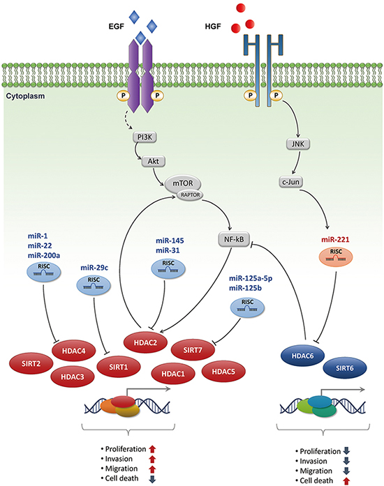
Emerging evidence indicates that expression of HDACs is remarkably disrupted in many human cancers. Lachenmayer et al. reported that expression of a subset of HDACs is increased in liver cancer compared to normal liver tissues with the presence of cirrhotic and dysplastic nodules (23). In addition, HDAC3 and HDAC5 DNA copy numbers are altered and their expression levels are significantly upregulated in HCC (24). Concomitantly, our group has described diverse roles of HDACs in HCC for several decades. In a comprehensive gene expression profile analysis of clinical samples of multi-step hepatocarcinogenesis, HDAC1, HDAC2, and SIRT7 were upregulated from pre-neoplastic lesions to high-grade HCCs, whereas HDAC6 was gradually downregulated (Fig. 1) (Table 1) (25).
In the previous study, HDAC8, a member of Class I HDACs, is overexpressed in liver cancer. HDAC8 knockdown repressed tumor cell growth and induced apoptosis through p53 expression and acetylation at Lys373 (26). Another study revealed that HDAC5 mRNA and protein levels are overexpressed in human HCC tissues and that inhibition of HDAC5 represses growth of HCC cell lines. Suppression of HDAC5 induces apoptotic cell death and G1/S cell cycle arrest by regulating apoptosis-associated molecules and cell cycle regulators (27). Similarly, depletion of HDAC2 selectively induces p16INK4a and p21WAF1/Cip1, leading to inhibited G1/S cell cycle transition. Furthermore, knockdown of HDAC2 causes hypophosphorylation of pRb and, consequently, E2F/DP1 target genes, such as CDC2, PCNA, and E2F1, are downregulated (10). mTROC1/NF-kBp50 signaling is related to growth factor-induced HDAC2 expression and is sustained in HCC. mTORC1 activity is maintained by HDAC2 stabilizing the mTOR/RAPTOR complex, and AKT (serine/threonine-specific protein kinase) is phosphorylated through a positive feedback loop triggered by HDAC2 (28). HDAC1 is overexpressed in a subset of human HCCs and liver cancer cell lines. Inactivating HDAC1 results in regressed tumor cell growth and activation of caspase-independent autophagic cell death, through the LC3B-II activation pathway in Hep3B cells (29). In this regard, overexpression of HDAC1 and HDAC2, which are class I HDACs, may play a pivotal role by regulating mitotic effectors during development of HCC.
SIRT7 is a class III HDAC, which requires the NAD+ cofactor for catalytic activity. SIRT7 was initially known as a nuclear protein associated with ribosomal gene transcription (30). SIRT7 expression was upregulated gradually in a large cohort of patients with HCC (25). Furthermore, depletion of SIRT7 inhibits in vitro and in vivo HCC tumorigenesis by selectively regulating the cell cycle and autophagic molecules in HCC cells. That is, SIRT7 may control tumor progression or development of cancer phenotype (31). SIRT2, another member of the sirtuin family, is also upregulated in HCC cell lines and in a subset of human HCCs. Overexpression of SIRT2 in HCC tissues is significantly associated with the presence of microscopic vascular invasion, an advanced tumor stage, and poor survival rates (32). Unlike the oncogenic functions of SIRT7 and SIRT2 in HCC, decreased SIRT6 expression is observed in primary human liver cancers and apoptosis-insensitive hepatoma cell lines. Ectopic expression of SIRT6 in the HepG2 liver cancer cell line increases apoptotic cell death. Moreover, loss of SIRT6 induces global hypomethylation and metabolic changes (2633).
Although anomalous expression of individual HDACs, such as HDAC1, HDAC2, and HDAC6, have been reported in many cancers, the roles of HDAC6 in cancer development are still hold both oncogene and tumor suppressor. In our recent study, HDAC6 expression was downregulated in patients with HCC, and was significantly related with poor prognosis for 5-yr overall, disease-free, and recurrence-free survival. Intriguingly, ectopic overexpression of HDAC6 significantly suppressed cell growth in liver cancer cell lines and induced LC3B-II conversion and caspase-independent autophagic cell death through the JNK/Beclin1 pathway. This finding suggests that HDAC6 is involved in tumor suppression by nonepigenetic regulation in hepatocarcinogenesis (7).

Regulation of HDAC expression by miRNAs and therapeutic approaches

MiRNAs are a class of small noncoding RNAs that negatively regulate gene expression. Many miRNAs have emerged as key regulators associated with liver tumorigenesis. For example, Pineau and his group profiled the expression of miRNAs in 104 HCC tissue samples and 35 HCC cell lines (15). Of the upregulated miRNAs, miR-221 was the most upregulated miRNA in tumor samples. MiR-221 targeted the CDK inhibitor p27Kip1 and DNA damage-inducible transcript 4 (DDIT4), inhibiting cell proliferation when an anti-miR specific for miR-221 was treated to liver cancer cell lines. Similarly, a variety of miRNAs were identified as oncogenes or tumor suppressors governing aberrant expression of key epigenetic regulators in hepatocarcinogenesis (Fig. 1) (Table 2). For example, HDAC4 has critical roles in cancer development and has been identified as a direct target of miR-1, miR-22 and miR-200a in HCC (223435). Additionally, miR-31 and miR-145 are significantly downregulated in a subset of primary HCCs, and ectopic expression of these miRNAs exerts an anti-tumor effect by directly targeting oncogenic HDAC2 in vivo and in vitro (3637). SIRT1 is aberrantly regulated in HCC and its overexpression stimulates HCC cell growth via inactivating transcription of p21Cip1, p27Kip1, p15INK4b and activating CDK2, CDK6, cyclin D3, and cyclin D1. Notably, although the SIRT1 protein is highly overexpressed in patients with HCC, SIRT1 mRNA expression is not significantly different between HCC and non-tumor groups. Additional research evidenced that miR-29c was an endogenous regulator of SIRT1 and its expression was significantly downregulated in a large cohort of HCC patients (38). Up-regulation of SIRT7 exerts the tumorigenic effect of HCC and its expression is negatively correlated with miR-125a-5p and miR-125b, which directly regulate translation of SIRT7 by binding the 3'-UTR of its transcript (31). In addition to these tumor suppressive miRNAs targeting oncogenic HDACs, a recent study identified that repression of the tumor suppressor HDAC6 was directly regulated by miR-221 via coordinated JNK/c-Jun- and NF-κB-signaling pathways in the progression of liver tumorigenesis (8).
Many groups have suggested possible clinical applications for miRNA-based cancer therapy. Although a plethora of inhibitors have been developed and utilized to treat HCC, suitable drugs for liver cancer therapy are needed because overall survival rates of patients with liver cancer remain low. For example, sorafenib is the standard treatment for advanced HCC, but there are many side effects to be solved, such as subcorneal pustular dermatosis (39). Furthermore, cancerous cells occasionally circumvent drug targeted signaling by adapting to diverse environments, which is linked to poor patient prognosis (40). Accumulating evidence indicates that strategies based on modulating miRNA are a possible approach for liver cancer therapy. Elmén et al. showed simple systemic delivery of anti-miR and effectively antagonized liver-expressed miR-122, a tumor suppressive microRNA, in non-human primates without toxicity (41). This treatment resulted in prolonged survival and reduced the size and the number of tumors. Furthermore, systemic delivery of miRNA using adeno-associated virus vehicle is another attractive method for liver cancer therapy. If miRNAs could be delivered in viral vectors and continuously transcribed, they may sustain high expression of miRNA mimics or anti-sense miRNAs in target tissues. One study reported that miR-26a, which is downregulated in HCC, was delivered successfully to the liver and reduced the tumor burden in a c-Myc-induced murine liver cancer model (42). However, many additional miRNAs that successfully inhibit liver tumorigenesis remain to be characterized. That is, a single miRNA or a cluster of miRNAs governing a subset of oncogenes initiating or developing cancer may be a promising candidate for cancer therapeutic delivery. For example, miR-188 efficiently blocks the G1/S cell cycle transition by directly targeting multiple cyclins, such as cyclin D, cyclin E, and cyclin A, in nasopharyngeal cancer cells (43). Similarly, miR-7 remarkably suppresses the downstream epidermal growth factor receptor pathway by regulating multiple molecules, such as PI3K, phosphorylated AKT, Raf-1, and phosphorylated MEK 1/2, in glioma cells (44). Similarly, because of the critical roles of HDACs in cancer development, if miRNAs that dominantly regulate these effectors could be found and specifically delivered to a target tissue, they may have novel cancer therapeutic applications.
In this review, we summarized the role of the HDAC families and HDACs targeting miRNAs during the development and progression of liver cancer. Accumulating evidence suggests that HDACs regulate the expression and activities of a variety of proteins involved in cancer development. Furthermore, certain HDACs are anomalously expressed in tumor tissues and have redundant functions in liver cancer (232425). Despite the many drugs that inhibit HDAC activities, overall survival rates and prognoses of patients with HCC remain poor. That is, liver cancer cells easily acquire drug resistance because they adjust to their environment through transformation (4546). Thus, miRNAs could be an efficient therapeutic approach for liver malignancies. Although technological progression asserts that using miRNAs or anti-miR as therapeutics is practical and safe, more studies are needed to move the field toward clinical applications.

Figures and Tables

Fig. 1

Schematic summary of the regulation of HDACs family and their regulatory miRNAs in liver cancer. HDACs are regulated by growth factors and that in turn induce initiation and progression of liver cancer. On the other hand, miRNAs also modulate HDACs family and closely associated with aberrant expression of HDACs. Therefore, these comprehensive HDACs and miRNAs network may contribute to liver tumorigenesis.

jkms-30-1375-g001
Table 1

Disrupted HDACs expression in hepatocellular carcinoma

jkms-30-1375-i001
Expression HDACs Functions Reference
Upregulated HDAC1 Autophagic cell death 29
HDAC2 G1/S cell cycle arrest 102528
HDAC3 Cell proliferation and migration 24
HDAC4 Cell proliferation and migration 223435
HDAC5 Apoptosis and G1/S cell cycle arrest 2427
HDAC8 Cell proliferation and inhibition of apoptosis 26
SIRT1 G1/S cell cycle arrest 38
SIRT2 Cell motility and invasiveness 32
SIRT7 G1/S cell cycle arrest 2531
Downregulated HDAC6 Autophagic cell death 7
SIRT6 Apoptotic cell death 33
Table 2

MiRNAs targeting HDACs in hepatocellular carcinoma

jkms-30-1375-i002
HDACs miRNAs Functions of miRNAs Reference
HDAC2 miR-145 G1/S cell cycle arrest 37
miR-31 Inhibition of cell proliferation and migration 36
HDAC4 miR-1 Inhibition of cell proliferation 22
miR-22 Inhibition of cell proliferation 34
miR-200a Inhibition of cell proliferation and migration 35
HDAC6 miR-221 Inhibition of autophagic cell death 8
SIRT1 miR-29c G1/S cell cycle arrest 38
SIRT7 miR-125a-5p G1/S cell cycle arrest 31
miR-125b

Notes

Funding This study was supported by grants from the National Research Foundation (NRF) of Korea (2012R1A5A2047939 and 2012M3A9D1054476), and by grants from the Korea Health Technology R&D Project through the Korea Health Industry Development Institute (KHIDI), funded by the Ministry of Health & Welfare, Republic of Korea (grant numbers: HI14C3298 and HI14C1920).

DISCLOSURE No potential conflicts of interest are declared.

AUTHOR CONTRIBUTION All authors participated in writing and revision and agreed to final manuscript.

References

1. Jemal A, Bray F, Center MM, Ferlay J, Ward E, Forman D. Global cancer statistics. CA Cancer J Clin. 2011; 61:69–90.
2. Whittaker S, Marais R, Zhu AX. The role of signaling pathways in the development and treatment of hepatocellular carcinoma. Oncogene. 2010; 29:4989–5005.
3. Liu M, Jiang L, Guan XY. The genetic and epigenetic alterations in human hepatocellular carcinoma: a recent update. Protein Cell. 2014; 5:673–691.
4. Peserico A, Simone C. Physical and functional HAT/HDAC interplay regulates protein acetylation balance. J Biomed Biotechnol. 2011; 2011:371832.
5. Yang XJ, Seto E. The Rpd3/Hda1 family of lysine deacetylases: from bacteria and yeast to mice and men. Nat Rev Mol Cell Biol. 2008; 9:206–218.
6. Blander G, Guarente L. The Sir2 family of protein deacetylases. Annu Rev Biochem. 2004; 73:417–435.
7. Jung KH, Noh JH, Kim JK, Eun JW, Bae HJ, Chang YG, Kim MG, Park WS, Lee JY, Lee SY, et al. Histone deacetylase 6 functions as a tumor suppressor by activating c-Jun NH2-terminal kinase-mediated beclin 1-dependent autophagic cell death in liver cancer. Hepatology. 2012; 56:644–657.
8. Bae HJ, Jung KH, Eun JW, Shen Q, Kim HS, Park SJ, Shin WC, Yang HD, Park WS, Lee JY, et al. MicroRNA-221 governs tumor suppressor HDAC6 to potentiate malignant progression of liver cancer. J Hepatol. 2015; 63:408–419.
9. Kim JK, Noh JH, Eun JW, Jung KH, Bae HJ, Shen Q, Kim MG, Chang YG, Kim SJ, Park WS, et al. Targeted inactivation of HDAC2 restores p16INK4a activity and exerts antitumor effects on human gastric cancer. Mol Cancer Res. 2013; 11:62–73.
10. Noh JH, Jung KH, Kim JK, Eun JW, Bae HJ, Xie HJ, Chang YG, Kim MG, Park WS, Lee JY, et al. Aberrant regulation of HDAC2 mediates proliferation of hepatocellular carcinoma cells by deregulating expression of G1/S cell cycle proteins. PLoS One. 2011; 6:e28103.
11. Bartel DP. MicroRNAs: genomics, biogenesis, mechanism, and function. Cell. 2004; 116:281–297.
12. Esquela-Kerscher A, Slack FJ. Oncomirs - microRNAs with a role in cancer. Nat Rev Cancer. 2006; 6:259–269.
13. Meng F, Henson R, Wehbe-Janek H, Ghoshal K, Jacob ST, Patel T. MicroRNA-21 regulates expression of the PTEN tumor suppressor gene in human hepatocellular cancer. Gastroenterology. 2007; 133:647–658.
14. Xiong Y, Fang JH, Yun JP, Yang J, Zhang Y, Jia WH, Zhuang SM. Effects of microRNA-29 on apoptosis, tumorigenicity, and prognosis of hepatocellular carcinoma. Hepatology. 2010; 51:836–845.
15. Pineau P, Volinia S, McJunkin K, Marchio A, Battiston C, Terris B, Mazzaferro V, Lowe SW, Croce CM, Dejean A. miR-221 overexpression contributes to liver tumorigenesis. Proc Natl Acad Sci U S A. 2010; 107:264–269.
16. Rodriguez-Paredes M, Esteller M. Cancer epigenetics reaches mainstream oncology. Nat Med. 2011; 17:330–339.
17. Bolden JE, Peart MJ, Johnstone RW. Anticancer activities of histone deacetylase inhibitors. Nat Rev Drug Discov. 2006; 5:769–784.
18. Zhu P, Martin E, Mengwasser J, Schlag P, Janssen KP, Göttlicher M. Induction of HDAC2 expression upon loss of APC in colorectal tumorigenesis. Cancer Cell. 2004; 5:455–463.
19. Jung KH, Noh JH, Kim JK, Eun JW, Bae HJ, Xie HJ, Chang YG, Kim MG, Park H, Lee JY, et al. HDAC2 overexpression confers oncogenic potential to human lung cancer cells by deregulating expression of apoptosis and cell cycle proteins. J Cell Biochem. 2012; 113:2167–2177.
20. Fornari F, Gramantieri L, Ferracin M, Veronese A, Sabbioni S, Calin GA, Grazi GL, Giovannini C, Croce CM, Bolondi L, et al. MiR-221 controls CDKN1C/p57 and CDKN1B/p27 expression in human hepatocellular carcinoma. Oncogene. 2008; 27:5651–5661.
21. Gramantieri L, Fornari F, Ferracin M, Veronese A, Sabbioni S, Calin GA, Grazi GL, Croce CM, Bolondi L, Negrini M. MicroRNA-221 targets Bmf in hepatocellular carcinoma and correlates with tumor multifocality. Clin Cancer Res. 2009; 15:5073–5081.
22. Datta J, Kutay H, Nasser MW, Nuovo GJ, Wang B, Majumder S, Liu CG, Volinia S, Croce CM, Schmittgen TD, et al. Methylation mediated silencing of MicroRNA-1 gene and its role in hepatocellular carcinogenesis. Cancer Res. 2008; 68:5049–5058.
23. Lachenmayer A, Toffanin S, Cabellos L, Alsinet C, Hoshida Y, Villanueva A, Minguez B, Tsai HW, Ward SC, Thung S, et al. Combination therapy for hepatocellular carcinoma: additive preclinical efficacy of the HDAC inhibitor panobinostat with sorafenib. J Hepatol. 2012; 56:1343–1350.
24. Wu LM, Yang Z, Zhou L, Zhang F, Xie HY, Feng XW, Wu J, Zheng SS. Identification of histone deacetylase 3 as a biomarker for tumor recurrence following liver transplantation in HBV-associated hepatocellular carcinoma. PLoS One. 2010; 5:e14460.
25. Nam SW, Park JY, Ramasamy A, Shevade S, Islam A, Long PM, Park CK, Park SE, Kim SY, Lee SH, et al. Molecular changes from dysplastic nodule to hepatocellular carcinoma through gene expression profiling. Hepatology. 2005; 42:809–818.
26. Wu J, Du C, Lv Z, Ding C, Cheng J, Xie H, Zhou L, Zheng S. The up-regulation of histone deacetylase 8 promotes proliferation and inhibits apoptosis in hepatocellular carcinoma. Dig Dis Sci. 2013; 58:3545–3553.
27. Fan J, Lou B, Chen W, Zhang J, Lin S, Lv FF, Chen Y. Down-regulation of HDAC5 inhibits growth of human hepatocellular carcinoma by induction of apoptosis and cell cycle arrest. Tumour Biol. 2014; 35:11523–11532.
28. Noh JH, Bae HJ, Eun JW, Shen Q, Park SJ, Kim HS, Nam B, Shin WC, Lee EK, Lee K, et al. HDAC2 provides a critical support to malignant progression of hepatocellular carcinoma through feedback control of mTORC1 and AKT. Cancer Res. 2014; 74:1728–1738.
29. Xie HJ, Noh JH, Kim JK, Jung KH, Eun JW, Bae HJ, Kim MG, Chang YG, Lee JY, Park H, et al. HDAC1 inactivation induces mitotic defect and caspase-independent autophagic cell death in liver cancer. PLoS One. 2012; 7:e34265.
30. Ford E, Voit R, Liszt G, Magin C, Grummt I, Guarente L. Mammalian Sir2 homolog SIRT7 is an activator of RNA polymerase I transcription. Genes Dev. 2006; 20:1075–1080.
31. Kim JK, Noh JH, Jung KH, Eun JW, Bae HJ, Kim MG, Chang YG, Shen Q, Park WS, Lee JY, et al. Sirtuin7 oncogenic potential in human hepatocellular carcinoma and its regulation by the tumor suppressors MiR-125a-5p and MiR-125b. Hepatology. 2013; 57:1055–1067.
32. Chen J, Chan AW, To KF, Chen W, Zhang Z, Ren J, Song C, Cheung YS, Lai PB, Cheng SH, et al. SIRT2 overexpression in hepatocellular carcinoma mediates epithelial to mesenchymal transition by protein kinase B/glycogen synthase kinase-3beta/beta-catenin signaling. Hepatology. 2013; 57:2287–2298.
33. Marquardt JU, Fischer K, Baus K, Kashyap A, Ma S, Krupp M, Linke M, Teufel A, Zechner U, Strand D, et al. Sirtuin-6-dependent genetic and epigenetic alterations are associated with poor clinical outcome in hepatocellular carcinoma patients. Hepatology. 2013; 58:1054–1064.
34. Zhang J, Yang Y, Yang T, Liu Y, Li A, Fu S, Wu M, Pan Z, Zhou W. microRNA-22, downregulated in hepatocellular carcinoma and correlated with prognosis, suppresses cell proliferation and tumourigenicity. Br J Cancer. 2010; 103:1215–1220.
35. Yuan JH, Yang F, Chen BF, Lu Z, Huo XS, Zhou WP, Wang F, Sun SH. The histone deacetylase 4/SP1/microrna-200a regulatory network contributes to aberrant histone acetylation in hepatocellular carcinoma. Hepatology. 2011; 54:2025–2035.
36. Kim HS, Lee KS, Bae HJ, Eun JW, Shen Q, Park SJ, Shin WC, Yang HD, Park M, Park WS, et al. MicroRNA-31 functions as a tumor suppressor by regulating cell cycle and epithelial-mesenchymal transition regulatory proteins in liver cancer. Oncotarget. 2015; 6:8089–8102.
37. Noh JH, Chang YG, Kim MG, Jung KH, Kim JK, Bae HJ, Eun JW, Shen Q, Kim SJ, Kwon SH, et al. MiR-145 functions as a tumor suppressor by directly targeting histone deacetylase 2 in liver cancer. Cancer Lett. 2013; 335:455–462.
38. Bae HJ, Noh JH, Kim JK, Eun JW, Jung KH, Kim MG, Chang YG, Shen Q, Kim SJ, Park WS, et al. MicroRNA-29c functions as a tumor suppressor by direct targeting oncogenic SIRT1 in hepatocellular carcinoma. Oncogene. 2014; 33:2557–2567.
39. Tajiri K, Nakajima T, Kawai K, Minemura M, Sugiyama T. Sneddon-Wilkinson disease induced by sorafenib in a patient with advanced hepatocellular carcinoma. Intern Med. 2015; 54:597–600.
40. Merlo LM, Pepper JW, Reid BJ, Maley CC. Cancer as an evolutionary and ecological process. Nat Rev Cancer. 2006; 6:924–935.
41. Elmén J, Lindow M, Schütz S, Lawrence M, Petri A, Obad S, Lindholm M, Hedtjärn M, Hansen HF, Berger U, et al. LNA-mediated microRNA silencing in non-human primates. Nature. 2008; 452:896–899.
42. Kota J, Chivukula RR, O'Donnell KA, Wentzel EA, Montgomery CL, Hwang HW, Chang TC, Vivekanandan P, Torbenson M, Clark KR, et al. Therapeutic microRNA delivery suppresses tumorigenesis in a murine liver cancer model. Cell. 2009; 137:1005–1017.
43. Wu J, Lv Q, He J, Zhang H, Mei X, Cui K, Huang N, Xie W, Xu N, Zhang Y. MicroRNA-188 suppresses G1/S transition by targeting multiple cyclin/CDK complexes. Cell Commun Signal. 2014; 12:66.
44. Liu Z, Jiang Z, Huang J, Huang S, Li Y, Yu S, Yu S, Liu X. miR-7 inhibits glioblastoma growth by simultaneously interfering with the PI3K/ATK and Raf/MEK/ERK pathways. Int J Oncol. 2014; 44:1571–1580.
45. Maluccio M, Covey A. Recent progress in understanding, diagnosing, and treating hepatocellular carcinoma. CA Cancer J Clin. 2012; 62:394–399.
46. Tsochatzis EA, Meyer T, Burroughs AK. Hepatocellular carcinoma. N Engl J Med. 2012; 366:92.
TOOLS
ORCID iDs

Hyung Seok Kim
https://orcid.org/http://orcid.org/0000-0003-2784-0109

Qingyu Shen
https://orcid.org/http://orcid.org/0000-0001-7294-0251

Suk Woo Nam
https://orcid.org/http://orcid.org/0000-0001-5767-8291

Similar articles