

 PDF
PDF Citation
 Print
Print


INTRODUCTION
Rejuvenation of the skin is a frequently used treatment within the protocols of aesthetic medicine. Currently, filler injections are a very popular method for the outpatient treatment of facial wrinkles because of their convenience and, along with botulinum toxin, their effectiveness. After the concept of filler injections for facial soft tissue defects was introduced in the 19th century, numerous filler materials were introduced without sufficiently proven safety that have caused various complications. It has been only 30 yr since the introduction of the first Food and Drug Administration (FDA)-approved filler material, bovine collagen filler; subsequently, extensive research into effective and safe filler materials has led to many other options for treatment. There are now over 35 major filler product companies worldwide. However, the increased use of these fillers has resulted in many unexpected complications. For example, Sung et al. (1) reported nasal skin necrosis after hyaluronic acid (HA) filler injection, and Do et al. (2) reported long-term complications, such as lump formation and inflammation, after treatment with polyacrylamide hydrogel (PAAG) filler.
One of the main indications for the use of a filler injection is to reduce facial wrinkles, among which crow's feet are a common concern. Crow's feet are the rhytids spreading from the lateral canthus to the temple and may caused by aging, habitual squinting, or sunlight exposure. On histological examination, crow's feet show a configurational change in the skin that deteriorates the elastic tissue network (3). In addition, areas of crow's feet show epidermal thinning, a more compact stratum corneum, increased perifollicular fibrosis and granular layer thickness, and increased solar elastosis, compared with skin with less sun exposure (4). Human skin contains free radicals that can damage cellular deoxyribonucleic acid (DNA) and lead to aging. Exposure to sunlight causes the number of free radicals to increase, increasing the possibility of damage. Volumizing the area under the wrinkles and rejuvenating the damaged tissue can improve crow's feet. Among the many filler materials available, hyaluronic acid fillers are now the most widely used, since hyaluronic acid increases the skin's capacity for holding water and its viscoelasticity, improving its overall appearance.
In addition to these materials, a new formulation has been developed, based on more than 40 yr of research, which is innovative, original, specific for rejuvenation of skin, consisting of macromolecules, specifically polynucleotides, at high concentration. In recent years, these new filler products, made from purified polynucleotides derived from germ cells of salmon and other fishes, have been used in central Europe. While previously existing filler products simply fill a contracted or depressed space, the polynucleotide-containing products not only fill the space, but also improve the regeneration environment of damaged tissue, resulting in more natural tissue regeneration (5). It has been reported that polynucleotides promote the growth of human corneal fibroblasts and increase reparation on ultraviolet B (UVB)-damaged dermal fibroblasts (3). They also appear to promote proliferation of human pre-adipocytes (6). In vivo studies have demonstrated therapeutic effects of polynucleotides on patients undergoing skin explants (7), and polynucleotides also promote fast corneal epithelization after photorefractive keratotomy (3). Polynucleotides have also been associated with an increase in the healing process in bone repair (6, 8) and have been shown to stimulate angiogenesis and wound healing via increased vascular endothelial growth factor production during pathologic conditions of low tissue perfusion such as diabetes mellitus and thermal injury (9). However, there are few articles about the impact of polynucleotides on superficial skin layers. In this study, we aimed to demonstrate the effects of polynucleotides in both animal and clinical studies. We selected a high-concentration, non-cross-linked HA filler (Yvoire-Hydro®: LG Life Sciences, Korea) as a control device and carried out an animal study and a clinical trial to measure the durability, efficacy and safety of the PRM-001 polynucleotide product (Rejuran®: Pharmaresearch Products, Inc., Seoul, Korea) on crow's feet.
MATERIALS AND METHODS
This study was approved by the Institutional Review Board and Ethics Committee and the Institutional Animal Care and Use Committee of Seoul National University Bundang hospital, and followed the guidelines regarding study of humn subjects in clinical trials of the 1975 Declaration of Helsinki. We aimed to demonstrate the durability, efficacy and safety of PRM-001 in the correction of crow's-feet in an animal model and in a clinical trial. The numbers of animals and clinical trial subjects required for sufficient statistical power in the present study were determined based on the suggestion of the statistics committee of Seoul National University Bundang Hospital.
Study devices
Animal study
Animal preparation
A total of 6-week old hairless mice were used. All mice were housed in separate cages after the procedure, with free access to food and water. They were randomly divided into three groups: Group 1 was injected with phosphate buffered saline (PBS) as a negative control group; Group 2 was injected with HA filler as a positive control group; and Group 3 was injected with PRM-001 as an investigational group. All mice were exposed to UV light 3 times per week; the exposure amount was checked using a Waldmann UV meter. UV exposure was maintained for 10 weeks and the exposure amount was increased from 1 minimal erythema dose (MED) to 4 MED after the exposure check.
Durability
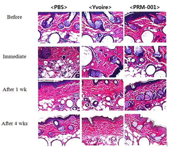
All mice were anesthetized using ketamine and Rompun. six hours before the procedure, and at one day, three days, one week, two weeks, and four weeks after the procedures, pictures with a stereoscopic microscope (SMZ1500, Nikon, Japan) were taken. Before and immediately after the procedures, as well as one week and four weeks after the injection of the fillers, mice were euthanized and each injection site was harvested and fixed with 10% neutral buffered formalin, and hematoxylin and eosin (H&E) staining was performed.
Efficacy
Tissue elasticity was measured using a Cutometer (MPA580, Courage Khazaka, Germany), six hours before injection and one day, three days, one week, two weeks, and four weeks after the injection. The skin was stretched with 50 mbar negative pressure for one second and released. This measurement was taken three times and analyzed.
Skin surface roughness was measured using Primos 3D stereoscopic images (Primos Pico, GFMesstechnik GmbH), while checking with the Cutometer.
Wrinkles were assessed after anesthesia using Silflo (Flexico, England), and mouse skin was molded before injection as well as one week and four weeks after injection. The skin moldings were analyzed with Skin Visioline VL650 (Courage Khazaka, Germany), measuring the surface and depth of wrinkle shadow followed by UV light exposure. Before the injection, as well as one week and four weeks after injection, mice were euthanized, the injection sites were harvested and fixed with 10% neutral buffered formalin, and Van Gieson staining was performed to check collagen synthesis. Statistical comparisons between the groups were performed using SPSS.
Clinical trial
Patient selection
Seventy-two adults, who were over 20 yr of age and presented for the correction of crow's feet, were enrolled. All subjects provided written informed consent for participation in this clinical trial. The exclusion criteria were a history of previous cosmetic procedures (including botulinum toxin and fillers), eyelid scars, bleeding tendency and suspected low compliance such as cases of cosmetic surgery addiction, alcoholism, and drug abuse (Table 1).
Study flowchart
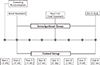
Screening and randomization of the subjects were performed, and the investigational and control fillers were injected immediately after randomization. At this time, to minimize the effects of treatment methods, two different investigators performed the procedures. All subjects received PRM-001 treatment on one side and HA filler on the contralateral side of the crow's feet in a ratio of 1:1. After observing for two minutes after a test injection whether an acute allergic reaction occurred, the main injections of the investigational and control fillers were given. All subjects were given a diary and then instructed to record the appearance and disappearance of adverse effects (AEs) for the following two weeks. The same investigator then performed repeated treatments every two weeks, for a total of three times. Repeated treatments were performed with the same device as for the initial treatment, and after the final treatment, a 12-week observation period was maintained for follow-up. A total of four injections were performed, every two weeks, by the same two investigators. All subjects visited the the study clinic at two, four, six, eight, ten and eighteen weeks after initial treatment (Fig. 1).
Treatment procedure
All treatments were performed using both linear threading and serial puncture techniques. Each device was injected up to a volume of 1.0 mL, and the dose was altered according to the judgment of the investigator.
Efficacy assessment
Primary objective
The non-inferiority of the efficacy of PRM-001 in improving wrinkles as compared with HA filler was based on the improvement ratio on clinical photography 12 weeks after the treatment of crow's feet compared screening.
Secondary objectives
Changes in improvement at two and four weeks after the final treatment compared with screening, according to the judgment of investigators.
Changes in wrinkle severity (lateral canthal line severity scale) scores at two and four weeks after the final treatment compared with screening, according to the judgment of investigators
Changes in resting state wrinkle severity (lateral canthal line severity scale) scores at two and four weeks after the final treatment compared with screening, according to the judgment of investigators
Changes in visual analogue scores at two, four, and twelve weeks after the final treatment compared with screening, according to the judgment of the study subjects.
Safety analysis
In the current study, based on adverse events, physical examination and clinical laboratory tests, and electrocardiogram (EKG), we compared the safety of PRM-001 and HA filler.
AEs
We evaluated all the AEs that occurred in the subjects who submitted a written informed consent, including those that did not occur after the subjects submitted a written informed consent and the AEs that were aggravated although they did not occur before the subjects submitted a written informed consent.
We reported the number of cases of these AEs and the proportion of the subjects who presented with AEs more than once. We also described their severity. In reporting the frequency of AEs, we analyzed the overall frequency of AEs, adverse device events (ADEs), serious adverse event (SAE). Then, we compared these values between the two groups.
Clinical laboratory parameters, vital signs and physical examination
We evaluated clinical parameters at 18 weeks and compared them with the same parameters at the time of screening. We provided descriptive statistics on continuous variables, using baseline and endpoint values, and then analyzed the difference between baseline and endpoint using the paired t-test or Wilcoxon's signed rank test. In addition, we also analyzed categorical variables using McNemar's test.
Data analysis
Statistical analysis
In the current study, unless otherwise noted, we performed two-sided tests with a statistical significance of 0.05. In addition, we reported the number of subjects, mean±standard deviation (SD), median, minimum and maximum values for the continuous variables. Moreover, we also reported the frequency and proportion of the subjects for the categorical variables. The worst observation carried forward (WOCF) method was applied to the primary objective only.
Efficacy set
In the current study, we performed the full analysis set (FAS) for the assessment of the efficacy of treatment modalities. Additionally, we also performed the per-protocol (PP) analysis.
Safety set
The safety analysis set comprises all the subjects who were enrolled in the current study and received a minimum of one treatment and the safety analysis after the treatment.
Full analysis set
The FAS comprises all subjects who were given a randomization number after being enrolled in the current study and who received more than one primary objective test.
Per-protocol set (PPS)
The PP set comprises all the FAS subjects who completed the current study without seriously violating the study protocol. However, we excluded subjects who were not included according to the study inclusion criteria and the subjects who underwent procedures or treatments that might affect the results of the efficacy analysis (including prohibited concomitant medications) during the study period.
RESULTS
Animal study
Durability
We examined 10 mice for the durability of the fillers using a stereoscopic microscope and for the shape of the injected filler and synthesis of inflammatory cells using H&E staining. Group 3 showed similar durability to Group 1 and Group 2 and also showed a similar inflammatory response compared to the control groups.
Efficacy
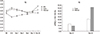
Tissue elasticity results are summarized in Fig. 2. Tissue elasticity was not different between groups before injection, but increased tissue elasticity was observed in Groups 1 and 2 six hours after injection. Four weeks after injection, Group 3 showed the greatest tissue elasticity compared to Group 1.
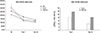
Skin surface roughness showed no statistical difference before injection between the groups, but four weeks after injection, Group 3 showed statistical differences compared to Group 1 (Fig. 3). One week after injection, Group 3 showed a statistical difference in wrinkle depth compared to Group 1, but otherwise the groups showed no difference for 4 weeks after injection (Fig. 4). After Van Gieson staining, elastic fibers, collagen fibers and fibroblasts were increased in Groups 2 and 3, and Group 3 showed the greatest increases compared to Group 1 (Fig. 5).
Clinical trial
Baseline characteristics
In the current study, we enrolled a total of 72 subjects; 68 were women and 4 were men. The safety set, which comprised all the subjects who were enrolled in the study and received a minimum of one treatment and the safety analysis after the treatment, included 72 subjects. However, two subjects dropped out or were excluded from the FAS, so the total FAS included 70 subjects. Within the FAS, 9 subjects dropped out from the study and 2 subjects were dropped due to taking concomitant medications, and a final number of 59 subjects were enrolled to the PPS. Within the FAS, 67 subjects were women and 3 were men. The age range of 40-50 yr included 31 subjects (44.3%), and the range of 50-60 yr included 30 subjects (42.9%).
Efficacy outcome
Primary objective outcome measure
In the FAS analysis, the improvement ratio on clinical photography at 12 weeks after the treatment of crow's feet as compared with screening, serving as the primary objective outcome measure, was 95.7% (67/70) in the PRM-001 group and 94.3% (66/70) in the HA filler group. Therefore, the difference in the rate of the improvement between the two groups was 1.5%, and the lower limit of the one-sided 97.5% confidence interval was -7.5%, which was less than the maximum allowance of -15% based on which the clinical non-inferiority could be determined. These results indicate that PRM-001 is non-inferior to HA filler. Also on the PP set analysis were 94.9% (56/59) in the PRM-001 group and 93.2% (55/59) in the HA filler group; the lower limit of the one-sided 97.5% confidence interval was -8.8%, which was less than the maximum allowance of -15% based on which the clinical non-inferiority could be determined (Table 2).
Secondary objective outcome measure
Changes in improvement at two and four weeks after the final treatment as compared with screening, according to the judgment of investigators
FAS: Two weeks after the final treatment, the investigational group showed 100% (62/62) improvement compared to screening, and the control group showed 98.4% (61/62) improvement. There was no statistically significant difference between the groups (P=0.315). Four weeks after the final treatment, there was 96.7% (59/61) improvement in both the investigational and control groups.
PPS: Two weeks after the final treatment, the investigational group showed 100% (58/58) improvement compared to screening, and the control group showed 98.3% (57/58) improvement. There was no statistically significant difference between the groups (P=0.315). Four week after the final treatment, there was 96.6% improvement in both the investigational and control groups.
Changes in wrinkle severity (lateral canthal line severity scale) scores at two and four weeks after the final treatment as compared with screening, according to the judgment of investigators
FAS: Two weeks after the final treatment, the changes in wrinkle severity score were 2.13±0.97 in the investigational group and 2.21±0.94 in the control group, and there was a statistically significant difference between the two groups.
Changes in resting state wrinkle severity (lateral canthal line severity scale) scores at two and four weeks after the final treatment as compared with screening, according to the judgment of investigators
Two weeks after the final treatment, resting state wrinkle severity scores were 1.13±0.95 in the investigational group and 1.05±0.91 in the control group (P=0.631), and four weeks after treatment, these scores were 1.05±0.94 in the investigational group and 1.05±0.88 in the control group (P=1.000).
Changes in visual analogue scores two, four and twelve weeks after the final treatment as compared with screening, according to the judgment of subjects
Two weeks after the final treatment, visual analogue scores were 52.23±24.11 in the investigational group, 52.13±28.02 in the control group (P=0.984); four weeks after treatment, these scores were 54.57±25.98 in the investigational group, 53.64±27.45 in the control group (P=0.847); and twelve weeks after treatment, these scores were 46.57±27.29 in the investigational group and 46.72±28.05 in the control group (P=0.975).
These results demonstrate that there was non-inferiority between the two groups (Table 3).
Safety outcomes
AEs
Treatment-emergent local AEs occurred at a frequency of 31.9% (23/72) in the PRM 001 group and 48.6% (35/72) in the Yvoire group. The difference between two groups was more than 10% and this was statistically significant (P=0.042).
In the current study, there were two case of SAE; pneumonia and ligament disorder occurred in two subjects. This corresponded to the SAE for which in-hospital treatment was needed or the length of hospital stay should be prolonged. However, there were no subjects who discontinued their participation in the current study because of the SAE. Moreover, there was no causal relationship between the SAE and study devices according to the judgment of the investigators. Finally, there were no unexpected ADEs or other notable AEs in the current study.
DISCUSSION
Polynucleotides are known to assist with wound healing and to produce a volumizing effect. However, there are few articles about the role of polynucleotides in treating skin wrinkles. In the present study, we investigated the effect of polynucleotides on skin wrinkles in an animal model and in a clinical trial. In the animal study, the polynucleotide group showed similar durability and inflammatory responses to a positive control, HA filler. The polynucleotide group also showed the greatest elasticity and collagen composition between test and control groups, and there was a significant decrease in the skin surface roughness and wrinkle depth after polynucleotide treatment. In the clinical trial, there was no significant difference in any outcome measure between the test and control groups, and there were no unexpected adverse effects.
There exist numerous challenges in overcoming the signs of aging in areas of exposed skin, such as the face, neck, décolletage and hands. The terms bio-stimulation and bio-revitalization have been used to describe the function of many aesthetic medical devices. Bio-stimulation refers to the anabolic function of dermal fibroblasts; the treatments aim to stimulate the synthesis of proteins and extracellular components. In contrast, bio-revitalization directly supplies synthetic materials to the skin, either alone or in conjunction with other added molecules (4). Regeneration refers to the process of renewal, restoration, and growth that makes genomes, cells, organisms, and ecosystems resilient to natural fluctuations or events that cause disturbance or damage. Several clinical reports exist that describe the therapeutic use of polynucleotides in revitalizing the skin (5), and improving wound healing (6). PRM-001 filler is a new formulation for the regeneration of skin, composed of macromolecules with a concentration of 20 mg/mL of highly purified polynucleotides of natural origin (Rejuran®, PharmaResearch Products, Inc., Seoul, Korea). Yvoire-Hydro® (LG Life Sciences, Seoul, Korea), which is filled with highly concentrated, non-cross linked hyaluronic acid filler, is known to exert a volumizing and hydrating effect on skin and was selected as a control device due to its similar mechanism of action to PRM-001. PRM-001 is targeted to inject into the superficial dermal layer. The polynucleotide is widely present in the human body and is physiologically present in the extracellular environment (7). High molecular weight polynucleotide chains have many effects on our skin. They can easily bind to water molecules and act as free radical scavengers. In addition, the gradual degradation of polynucleotide molecules by enzymes in the extracellular environment to free metabolites can accentuate the activities of protection against free radicals (3). A recent in vitro study of fibroblasts subjected to UVB radiation shows that polynucleotides are capable of exerting a protective effect against irradiated cells. Other main effect of high molecular weight polynucleotide chains is on the metabolic activity of fibroblasts, the main cells that control the renewal of various dermal components. Polynucleotides contribute to regenerate several autologous key skin components, such as glycosaminoglycan, proteins, glycoproteins, and fibrils, and help to maintain their physiological function. Prolonged iso-osmotic hydration and anti-free radical actions contribute to recreate the most favorable physiological conditions in the dermal matrix that stimulate fibroblast metabolic activity and regeneration. Rejuran® filler has strong hydrating properties which can facilitate more favorable physiological conditions for the growth of cells in skin, and assist to increase the production of amorphous extracellular matrix components and fibrillar substances, and finally can reduce the fine wrinkles and improve skin turgidity, elasticity, and tonicity.
In our animal study, PRM-001 showed greatest increase of skin elasticity and collagen synthesis followed by fibroblast stimulation compared to other groups with similar durability. In the past, numerous fillers have been developed and used to achieve a simple volumizing effect, but PRM-001 can also stimulate fibroblast growth for skin rejuvenation and is more suitable for the concept of skin regeneration. Repeated injection of PRM-001 can stimulate and optimize the vitality and secreting activity of fibroblasts, and maintain and balance the homeostasis of an individual's skin system. Moreover, these properties can provide the opportunity to combine therapy with other medical or surgical procedures by increased collagenic or non-collagenic components while minimizing the adverse effects. Polynucleotide fillers are more suitable for regeneration than bio-stimulation or bio-revitalization. In addition, modern trends in revitalization are changing from providing more synthetic components to the dermal layer (e.g. collagen, hyaluronic acid, glycoproteins, etc.) to stimulating cells such as fibroblasts and to supplying autologous components to the skin and its cells by duplicating and increasing their metabolic activity.
Our study is the first study to demonstrate the durability, efficacy and safety of a polynucleotide cosmetic filler in crow's-feet correction. In this study, there were no important adverse effects, and clinically significant phenomena of safety were evident after the application of experimental and control devices.


 XML Download