

 PDF
PDF Citation
 Print
Print


INTRODUCTION
Peripheral nerve injury is one of the most common comorbidities that occur in patients with trauma or systemic diseases (e.g., iatrogenic injury or diabetes mellitus) (1). Autologous nerve grafting has been considered the gold-standard in patients with traumatic peripheral nerve defects if they are not indicated in a direct end-to-end repair (2). But it shows complications such as donor site morbidity (3). Other alternative methods have therefore been attempted using the regeneration and replacement of peripheral nerves. The former is to restore the intrinsic regeneration capability using cells or growth factors and the latter is to achieve a prompt recovery of the nerve continuity for the purposes of preventing the adverse effects of delayed reinnervation, including the irreversible peripheral atrophy or nerve defects (4). To date, however, the former has been of greater interest than the latter. Venous conduits, acellularized muscle, acellular nerve matrix and xenografts have been attempted to overcome the limitations of nerve replacement (5, 6, 7). Nevertheless, these attempts have also caused such problems as donor site morbidity. This has led to the emergence of tissue engineering as a reasonable alternative approach, for which an ideal scaffold for nerve regeneration should be equipped with such biological properties as biodegradability, biocompatibility and porosity as well as mechanical ones that mimic the native extracellular matrix (8).
To date, considerable efforts have been made to develop a tissue-engineered peripheral nerve substitute involved in nerve regeneration. This has led to the development of many synthetic biomaterials such as silicone, poly-tetrafluorethylene (PTFE), poly-L-glycolic acid (PLGA), poly-caprolactone (PCL) and poly-L-lactic acid (PLLA) (9, 10). In addition, natural polymers, such as chitosan, collagen, chitin, gelatin and conducting polymers, such as polypyrrole, polyaniline or polythiophene derivatives, are also widely used for nerve regeneration (11, 12). Nevertheless, they have a lack of the effects in inducing the nerve regeneration. This has led to attempts to combine their use with nerve growth factors (13, 14).
Schwann cells (SCs) are peripheral glial ones that are mainly involved in the nerve generation. Nevertheless, their therapeutic applications are limited because there is the same concern for donor site morbidity as the nerve grafting. Recently, there is a worldwide growing interest in neurogenic differentiation potential of pluripotential stem cells. From this context, bone marrow-derived stem cells (BMSCs) have already shown promising results in the peripheral nerve regeneration (15). It is not easy, however, to apply them for clinical purposes because of the invasive harvesting technique and their limited availability. Adipose-derived stem cells (ADSCs) have been identified as having similar characteristics to BMSCs; they possess pluripotential differentiation capability (16, 17). Moreover, they can be easily harvested through a liposuction procedure because the fat tissue is abundantly present in human body. Furthermore, they are also advantageous in that they promptly proliferate in the culture environment and are successfully integrated into the host tissue with immunological tolerance. Their capability of inducing the neuronal differentiation has been demonstrated in vitro (4, 18).
Ongoing studies are currently conducted to examine the in vivo effects of ADSCs in inducing the nerve regeneration, for which various routes of delivery are attempted and these include direct microinjection or suspension within artificial tubes, the injection of whole adipose tissue preparation, the parenteral administration of ADSCs and seeding within devitalized muscle or nerve grafts. Controversial opinions also exist regarding the status of differentiation of ADSCs. That is, the undifferentiated ADSCs have a positive effect in inducing the nerve regeneration, but they rarely differentiate into SCs or other neural components (12). In addition, there is also a possibility that they might cause tumor formation (19). Presumably, their positive effects might be due to the immunomodulation and the autocrine factors released from the naïve precursor cells. On the other hand, the differentiated ADSCs increase the possibility of post-transplant cell death, possibly due to the expression of major histocompatibility complex (MHC) antigens or the decreased proliferation (20).
In this experimental study, we examined the in vivo effects of non-differentiated and neuronal differentiated ADSCs in inducing the neuronal regeneration in the Sprague-Dawley (SD) adult male rats undergoing nerve defect bridged with the PCL nanotubes. Then, we performed immunohistochemical and histopathologic examinations, as well as the electromyography for functional evaluation.
MATERIALS AND METHODS
Preparation of PCL nano-tubes
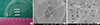
In situ polymerized multi-walled carbon nanotube/polycaprolactone nanocomposites (MWNT/PCL) were supplied by the Nanomaterial Application Division, Korea Institute of Ceramic Engineering and Technology (KICET). After the functionalization of MWNT by Friedel-Crafts acylation reaction, PCL was grafted with aromatic amine functionalized MWNT. Thus, as shown in Fig. 1, the MWNT/PCL composites were prepared with PCL solution in a 3:1 mixture of chloroform and methanol solution (21). This was followed by the application of electrospinning technique at a voltage of 15 kV to a 0.5-mm needle at room temperature. At a critical voltage, the jet of the polymer solution came out from the needle tip and then placed in a collector. The collector was 11 cm apart from the needle tip. To create tube-like implants, nanofibers were accumulated using a 16-G copper wire (diameter: 1.29 mm) held near the grounded target.
The culture and preparation of ADSCs
Harvesting of ADSCs
Human adipose tissue was obtained from patients undergoing unilateral breast reconstruction surgery using a transverse rectus abdominus myocutaneous (TRAM) flap. Thus, approximately 10 grams of normal fat tissue was harvested and then embedded in 1% antibiotic-antimycotic solution (Gibco BRL, Grand Island, NY) containing phosphate buffered saline (PBS) (Sigma Chemical Co., St. Louis, MO, USA). Then, the tissue sample was rinsed three times in a sterile environment. In the current experiment, we used discarded tissue and obtained a written informed consent from patients. The current experimental procedure was approved by the institutional review board (IRB) and Institutional Animal Care and Use Committee (IACUC) of Catholic University of Korea (IRB approval number: HC10EISI0047, IACUC approval number: HFH-006).
Isolation of ADSCs
The tissue sample was minced and then digested in an equal amount of PBS containing a 240 µg/mg of collagenase type I (Sigma Chemical Co., St. Louis, MO, USA). Then, the sample was incubated in a shaking incubator at a temperature of 37℃ for 10-15 min until it was homogenized. This was followed by the centrifugation at 1,500 rpm for five minutes, and the pellet was obtained. After the removal of the supernatant, the pellet was washed twice in low glucose Dulbecco's Modified Eagle Medium (DMEM) (Gibco Cat 11885) (Gibco BRL) and filtered through a 100-µm nylon mesh to eliminate the cell debris.
The ADSCs were transferred to plating medium (DMEM containing 10% fetal bovine serum [FBS] and 1% penicillin-streptomycin solution) at a density of 5×10⁴ cells/mL. They were adhered to tissue-treated T-flasks for six hours. Both non-adhered and floating ADSCs were removed by rinsing the cell culture plate with the PBS and covering it with the plating medium.
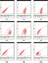
A flow cytometry was performed to confirm the characteristics of the cultured ADSCs. The cultured ADSCs were harvested by trypsin, washed twice with PBS, incubated with FITC-conjugated monoclonal antibodies, including CD13, CD29, CD90, CD105, CD49d, CD34, CD45, and HLA-DR and finally analyzed using the FACScan (all from BD Bioscience, MA) (Fig. 2).
Neuronal differentiation of ADSCs
To examine the effects of ADSCs in inducing the neuronal differentiation, the preconditioning was done. To do this, we treated them with 0.25% trypsin-EDTA and seeded into a 6-well plate containing a 20 mL of DMEM solution with a 1 mM of β-mercaptoethanol (BME) (Sigma Chemical Co., St. Louis, MO, USA) and 20% FBS for 24 hr and thereby harvested them from the culture flask. When they reached 80% confluence, the culture medium was replaced with neuronal induction medium containing a mixture of a 10 mM of BME and a 20 mL of DMEM for two hours. Then, the ADSCs were cultured in a mixture of 2% di-methyl sulfoxide (DMSA) (Sigma Chemical Co.) and a 20 µM of butylated hydroxyanisole (BHA) (Sigma Chemical Co.) containing a 20 mL of DMEM. The culture medium was changed at a 4-day interval. ADSCs for this experiment were chosen from the third subculture.
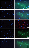
The morphology of ADSCs was examined using an inverted microscope. Thus, we examined whether they differentiated into the neuronal cells during the culture period (Fig. 3).
Twelve days later, we fixed the cultured ADSCs with 99% ethanol and treated them with 3% H2O2 for five minutes, thus attempting to remove the intracellular peroxidase activity. To examine the neuronal differentiation of ADSCs, we performed immunohistochemistry using neuronal cell-specific markers such as β-tubulin, neural cell adhesion molecule (NCAM) and s-100 (Dako, Glostrup, Denmark); neuronal nuclei (NeuN) (Chemicon Int., San Diego, CA, USA); and neuron-specific enolase (NSE), trkA and vimentin (Fig. 4). After confirmation of neuronal differentiation, ADSCs were kept 24 hr in culture medium before injection into the scaffold.
Animal experiments
We used 21 SD adult male rats (42 sciatic nerves), weighting 225-250 g (Orient, Korea). We randomly divided them into the three groups by using table of random numbers:
1) The control group (14 sciatic nerves): The SD rats were transplanted with the PCL nanotube scaffold.
2) The experimental group I (14 sciatic nerves): The SD rats were transplanted with the PCL nanotube scaffold with the non-differentiated ADSCs at a density of 7×10⁵ cells/0.1 mL.
3) The experimental group II (14 sciatic nerves): The SD rats were transplanted with the PCL nanotube scaffold with the neuronal differentiated ADSCs at a density of 7×10⁵ cells/0.1 mL.
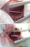
In the SD rats which were anesthetized an intra-peritoneal injection of ketamine (100 mg/kg) and xylazine (10 mg/kg), we dissected the segment of the sciatic nerve at a length of 15 mm and restored the gap using the PCL nanotube scaffold. The conduit was sutured using 3-4 stitches of 9-0 monofilament nylon for each nerve stump under an operating microscope (Zeiss OPMI 7) (Fig. 5).
Postoperatively, the SD rats were placed under a warm light, allowed to recover from anesthesia and housed separately with a free access to food and water in a cage at a constant temperature of 19-22℃) and a humidity of 40%-50% in a 12:12 light-dark cycle.
Six weeks later, we performed electromyography (EMG) and histological examinations for the SD rats.
Histologic and immunohistochemical examinations
Histologic examinations
Six weeks postoperatively, we carefully dissected the repaired nerve segments for histologic examinations. The sample was immediately fixed using 2.5% glutaraldehyde, rinsed with Sorenson's phosphate buffer (pH=7.4) and dehydrated using a series of ethanol. Then, the sample was rinsed with propylene oxide and then embedded in paraffin wax. This was followed by staining with toluidine blue and hematoxylin-eosin (H&E) dye. Perpendicularly to the main axis of the center of the sample, it was sectioned using an ultramicrotome at a thickness of 2 µm. Following this, we evaluated the morphology of regenerated axons.
Immunohistochemistry
To examine the degree of the neuronal differentiation, we performed immunohistochemistry using nestin, microtubule-associated protein 2 (MAP-2) and glial fibrillary acidic protein (GFAP), which were specific markers for neural stem cells, mature neurons and mature astrocytes, respectively.
Electromyography (EMG)
To evaluate the degree of the functional recovery, we performed EMG using the Medelec/TECA Synergy EMG System (Oxford Institute, UK). Bipolar hooked platinum recording electrode was inserted in the gastrocnemius muscle and stimulating electrode was applied to the sciatic nerve trunk at the proximal and distal ends of the graft. Reference electrode was inserted in the Achilles' tendon and ground electrode was applied to the ipsilateral thigh. After electrically stimulating the sciatic nerve at a voltage of 200 µV for ten minutes, we measured the evoked action potential (EAP) and nerve conduction velocity (NCV) using the MedLab V6.0 system (Meiyikeji, China).
Statistical analysis
Statistical analysis was done using the SPSS version 13.0 for windows (SPSS Inc., Chicago, IL, USA). All data was expressed as mean±SD (SD: standard deviation). We compared the differences between the three groups using the one-way analysis of variance (ANOVA). A P value of <0.05 was considered statistically significant.
RESULTS
Histologic and immunohistochemical examinations
The degree of the neuronal induction was significantly higher in the experimental group I and II as compared with the control group (H&E stain and toluidine blue-O stain). The degree of immunoreactivity to nestin, MAP-2 and GFAP was significantly higher in the experimental group I and II as compared with the control group (Fig. 6).
EMG findings
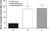
At six weeks, the NCV was 17.475±6.926 m/s in the experimental group I, 18.000±7.486 m/s in the experimental group II and 4.325±2.617 m/s in the control group (Fig. 7). These results indicate that it was significantly higher in the experimental group I and II as compared with the control group (P=0.021 and P=0.020, respectively). On the other hand, there was no significant difference in the NCV between the two experimental groups (P=0.473) (Fig. 8).
DISCUSSION
To prevent donor site morbidity after autologous nerve grafting for peripheral nerve injuries, the artificial nerve graft with cultured Schwann cells has been considered the second line of therapy. But its clinical application is also limited in that it is difficult to maintain a sufficient quantity of Schwann cells with an adequate condition and it is also necessary to obtain the donor site. Stem cells are a good source of Schwann cell-like cells. Earlier studies have therefore examined embryonic stem cells and BMSCs. Due to many ethical and practical issues, however, ADSCs have become an effective, minimally invasive source of stem cells. In vitro studies have demonstrated that the ADSCs are capable of differentiating into Schwann cell-like cells (4). This has provided diverse perspectives regarding the applicability of ADSCs to the peripheral nerve regeneration. Thus, we chose ADSCs for a potential tool of treatment in peripheral nerve injuries (13).
In the regeneration of peripheral nerve injuries, the clinical applicability of ADSCs might be based on their transplantation into the nerve gap using a tissue-engineered nerve substitute with or without the process of in vitro differentiation after harvested from liposuction (22, 23).
Accumulated evidence has shown that a tissue-engineered nerve substitute should be equipped with not only a tubular scaffold which bridges the gap of sectioned nerve, protects the nerve from scar formation and guides the regenerating fibers into the distal nerve stump but also a filler that is mainly involved in the biological mechanisms associated with the axon regeneration and the migration of Schwann cells.
To date, there have been attempts to use such materials as fibrin, laminin, silicone tube, allogeneic and xenogeneic acelluar nerve graft, artery, vein, muscle, polyglycolic acid (PGA), glycolidetrimethylene carbonate (Maxon), poly e-caprolactone and collagen as nerve conduits (4, 9, 24). Ideal materials for nerve tubulization should be equipped with the biocompatibility and a lack of adverse effects. In addition, they should have a slender structure, flexibility, sterility and bioresorbability. Furthermore, they should be beneficial for nerve healing and regeneration. In a clinical setting, however, other factors are also needed. They should be stiff enough not to collapse in vitro, pliable enough to manipulate and bend in vivo and easy to fabricate to the desired topography and dimensions.
The use of non-absorbable synthetic materials has been decreased due to the local fibrosis and nerve compression (25). By contrast, many experimental studies have demonstrated the bioabsorbable tubes are more effective than autografts or allografts (26). In particular, resorbable guides made of collagen or polylactate caprolactone (PLC) are more effective than non-resorbable guides such as silicone, Teflon, or polysylfone (27). From this context, the PCL has nearly all the ideal properties that have been mentioned above.
From mechanical perspective, the PCL tube has higher tensile strength as compared with fresh rat sciatic nerve; it has a wall thickness of 120 µm and retains its flexibility and integrity. It is therefore useful for suturing the nerve gaps (28). In addition, its surface characteristics may also be important for the adhesion and permeability of the cells. In other words, the PCL tube provides a porous hydrophilic surface that spreads the cells and it has a semi-permeable property due to the sparse distribution of penetrating holes. Thus, the semi-permeable property of the PCL tube allows the influx of nutrients and growth factors to the lumen of the conduit, thus acting as not only a barrier against the ectopic tissue infiltration but also a modulator of changes in the intraluminal pressure due to fluid collection. Recently, the electrical conduction property of the PCL and polypyrrole (PPy) substrates has been attempted to enhance the neural cell growth in the electrical field (29). This leads to the speculation that the conducting polymers such as PCL might be beneficial for enhancing the potential and guidance of regenerating nerve from intrinsic or extrinsic electric stimulation.
To provide an adequate microenvironment for the regeneration of the native nerve tissue, the bands of Büngner should be formed. Longitudinally aligned PCL nanotubes could provide adhesion sites for Schwann cells or neurites of regenerating neurons and they could also construct aligned basal lamina tubes (23). The PCL tubes can be synthesized with electrospinning technique without heat or chemical reactions that cause a loss of its desirable mechanical and chemical properties. As a whole, the PCL nanotube ideally meets the chemical and structural needs for the peripheral nerve regeneration.
Filling materials can be classified into cellular, structural and neurotrophic components (30). Schwann cell is one of the most promising cellular components; most of the studies about neuronal differentiation of stem cells have been conducted to examine the peripheral nerve regeneration of Schwann cells. There is a great controversy as to the status of the differentiation of transplanted ADSCs and methods for prolonging their survival. Undifferentiated ADSC is readily available but it carries a risk of teratoma formation or differentiation into unwanted cell types. Previous observations have shown that undifferentiated ADSCs have positive effects on the nerve regeneration; this originates from the trophic support rather than the differentiation into Schwann cells or myelin formation. On the other hand, differentiated ADSCs are more involved in the nerve regeneration of endogenous Schwann cells. Still, however, there is a lack of more profound information about the actual mechanisms by which Schwann cells are derived from differentiated ADSCs as well as their best final phenotype that is the most effective in promoting the nerve regeneration with less immunogenicity.
In the current study, we succeeded in repairing 15-mm nerve defects using both undifferentiated and differentiated neural stem cells, thus showing that they were differentiated towards a Schwann cell-like phenotype, expressing markers like S-100 and neurite outgrowth in vitro. Histological examinations shows more myelinated axons and better electrophysiological results in the experimental groups as compared with the control group. But there was no significant difference in the NCV between two experimental groups. This might be due to following two reasons: 1) More precise neuronal induction of stem cells would be essential for Schwann cells, which should be accompanied by the combination of various types of native nerve tissue; 2) As compared with the modification of the cellular component, the fabrication of the structural component using both conduit and filling material may be more likely to be effective. Filling the tube with nanofibers or microbeads and aligning by electrical field stimulation in vitro or in vivo could have mimicked microenvironment more perfectly.
Furthermore, neurotrophic additives such as fibroblast growth factor (FGF), nerve growth factor (NGF), glial growth factor (GGF), ciliary neurotrophic factor (CNTF), vascular endothelial growth factor (VEGF), glial-derived neurotrophic factor (GDNF), neurotropin-3 (NT-3) and brain-derived neurotrophic factor (BDNF) could be used with ADSCs for the modulation of microenvironment and pharmacological modulation of the precursor cells to improve the survival and neurotropic potential of the cells (13).
To summarize, our results indicate that ADSCs have effects on inducing the differentiation of grafted cells into Schwann like-supportive cells in an animal experimental model of nerve defects, thus promoting the peripheral nerve regeneration. Still, however, little is known about the underlying mechanisms by which trophic factors and cytokines might have an impact on the survival of host cells in vivo. In addition, the differentiated ADSCs show a higher degree of the recovery despite a lack of statistical significance. Thus, our results suggest not only that it would be mandatory to develop more delicate methods for inducing neuronal differentiation of differentiated ADSCs but also that the differentiation period is somewhat time-consuming. Further studies are therefore warranted to analyze the fate of grafted cells and to clarify the mechanisms by which the nerve regeneration of Schwann cells.


 XML Download