Journal List > J Korean Med Sci > v.29(7) > 1022590

Han and Mook-Jung: Diverse Molecular Targets for Therapeutic Strategies in Alzheimer's Disease

Abstract

Alzheimer's disease (AD) is the most common form of dementia caused by neurodegenerative process and is tightly related to amyloid β (Aβ) and neurofibrillary tangles. The lack of early diagnostic biomarker and therapeutic remedy hinders the prevention of increasing population of AD patients every year. In spite of accumulated scientific information, numerous clinical trials for candidate drug targets have failed to be preceded into therapeutic development, therefore, AD-related sufferers including patients and caregivers, are desperate to seek the solution. Also, effective AD intervention is desperately needed to reduce AD-related societal threats to public health. In this review, we summarize various drug targets and strategies in recent preclinical studies and clinical trials for AD therapy: Allopathic treatment, immunotherapy, Aβ production/aggregation modulator, tau-targeting therapy and metabolic targeting. Some has already failed in their clinical trials and the others are still in various stages of investigations, both of which give us valuable information for future research in AD therapeutic development.

INTRODUCTION

Alzheimer's disease (AD) is a pernicious neurodegenerative disease which is incurable with remedies developed up to date. The number of patients increases every year worldwide and 5.2 million people in the United States are suffering from AD. This number is expected to expand to 36 million to 115 million in worldwide around the year of 2050 and the estimated economic cost and suffering is increasing greatly (1, 2). Therefore, AD is seemingly insurmountable disease and the increasing numbers of patients produce diverse societal concerns in different aspects.
Senile plaques of amyloid β (Aβ) in the brain parenchyma have been regarded as not only the main pathological phenomena (3, 4) but also the culprit of this disease according to amyloid cascade hypothesis based on molecular information found in AD study (5). Abnormal production and accumulation of Aβ in brain parenchyma result in AD pathologies through sequential events by aggregated forms of this protein and the amyloid plaque. Aβ is generated as a consequence of sequential cleavages of amyloid precursor protein (APP) by β- and γ-secretases (6, 7, 8). APP is first cleaved by either α- or β-secretase, and then, the remaining remnants of C83 or C99, respectively, are vulnerable to intramembrane proteolysis by γ-secretase. Amyloidogenic process of γ-secretase cleavage followed by β-secretase produces aggregation-prone Aβ which are in the center of AD etiology (4, 9). Disrupted synaptic plasticity, reduced dendritic spine density and memory impairment were proven in rodent model by extraction of Aβ oligomers from human patients (10).
Along with Aβ, microtubule-associated protein tau is another major factor of AD pathogenesis as a component of neurofibrillary tangles (NFTs) (11). Tau stabilizes microtubule protein and microtubule-associated processes in normal condition. During AD pathogenesis, tau becomes hyperphosphorylated, aggregated and finally accumulated as neurofibrillary tangles (12). Tau hyperphosphorylation and NFT formation is tightly related to the existence of excessive Aβ and plaques, proving the tau pathology in AD (13, 14). Not only as axonal protein but also as regulator of dendritic function, tau plays a pivotal role, especially mediating early Aβ toxicity during AD progress (15). Therefore, inevitably, Aβ and tau became the main targets in drug development. Many clinical trials aiming these two proteins have been performed whereas several lines of targets are still under investigations.
Up to date, only a few AD medications have been proved as improving AD symptoms, but none of them modify disease progress or pathological cascades (16). Researchers and clinicians suspect that the reason for many drug target candidates to fail in their clinical trials reside in the improper time of drug treatment, in fairly late stage of AD progression where irreversible damages have already occurred, including excessive Aβ deposits, neuronal impairment, death and blood brain barrier (BBB) disruption (17). Therefore, finding a diagnostic biomarker, especially for the early stages of AD pathology, is desperately needed for developing a valuable therapeutic target at the early stage and preventing progression of the disease.
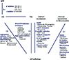
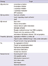
Diverse approaches for AD therapeutic strategy have arisen along with better understanding of cellular and molecular mechanism of AD pathogenesis. In Fig. 1 and Table 1, we listed possible strategies of drug development target based on accumulated scientific findings. In this review, we summarize the recent status of some AD drug targets using different strategies among them from published reports and ongoing clinical studies. In each category, we stated the representative drug targets, preclinical and clinical trials.

ALLOPATHIC TREATMENT FOR AD

In spite of better understanding for molecular mechanism during AD pathogenesis, the available medications for AD up to date provide only symptomatic benefit, not regulate or delay the progression of disease pathology. The US Food and Drug Administration (FDA) has approved only five medications for AD, including acetylcholinesterase inhibitors (AChEIs: donepezil, rivastigmine, galantamine and tacrine) and N-methyl-D-aspartate (NMDA) receptor antagonist, such as memantine (18, 19, 20, 21). These medications enhance cognitive function via increased acetylcholine level or glutamatergic receptor blocking, respectively. Combined administration of these two medications accelerates symptomatic improvement, representing slow progress of cognitive and functional impairment and delayed time for nursing home admission (22, 23). Besides, several lines of regulators for neurotransmission were suggested for symptomatic therapies in AD, including neuronal nicotinic acetylcholine receptor activation (ABT-418), GABAB receptor antagonism (SGS-742) and serotonergic modulation (Lu AE58054). However, they were insufficient to show significant efficacies of symptomatic improvement in clinical trials (24, 25, 26, 27). Recently, high affinity 5-hydroxytryptamine (HT) 6 receptor antagonist Lu AE58054 ([2-(6-fluoro-1H-indol-3-yl)-ethyl]-[3-(2,2,3,3,-tetrafluoropropoxy)-benzyl]-amine) was reported to improve novel object recognition task in a rat model with cognitive impairment induced by phencyclidine (28). The combined treatment of Lu AE58054 and donepezil is under phase III clinical trial (ClinicalTrials.gov identifier: NCT01955161). Additionally, even drugs with uncertain mechanism were reported to be effective in symptomatic improvement and protection against neurotoxicity by Aβ, including ethanolic extract of Angelica gigas (INB-176) and Ginkgo biloba (EGb761) respectively, however, none of which showed successful effectiveness in their preclinical and clinical trials (29, 30, 31).

Aβ PRODUCTION/AGGREGATION MODULATOR

Abnormal Aβ production and accumulation in brain parenchyma have been regarded as the central etiological hypothesis in AD pathogenesis (5, 10, 32). Therefore, the first line of strategy was inhibition of Aβ generation processes to prevent or cure the disease. The tight relevance of α-, β- and γ-secretases to Aβ production made researchers to discover modulating drugs for these enzyme activities in order to reduce intracellular and extracellular Aβ level. Whereas effective α-secretase activator was rarely identified, several types of β-secretase inhibitors were discovered and tested, starting with first-generation potent inhibitor OM99-2, OM00-3 (33, 34). Since then, numerous reports and patents of β-secretase inhibition were published, however, finding drug candidate with desirable potencies and efficacy has been fairly challenging (35). Recently discovered MK-8931 (Merck) is a promising β-secretase inhibitor whose result of phase I clinical trial was released in April, 2012. MK-8931 is now under phase II/III trial which was initiated in 2012 (ClinicalTrials. gov identifier: NCT01739348).
Gamma-secretase plays the critical role in Aβ generation, in charge of the rate-limiting cleavage of APP into Aβ. However, modulating this enzyme activity may cause diverse side effects because of its multiple cleavage actions on diverse substrates which are physiologically important, including notch receptor signaling. For this reason, modulating γ-secretase activity seems to be greatly complicated, requiring restricted substrate specificity for APP to reduce Aβ only, not affecting other substrate processing such as notch signaling (36, 37). Consequently, substrate specificity is the critical issue in the development of AD therapy using γ-secretase inhibition. Semagacestat (LY450139, Eli Lilly) was a promising drug candidate targeting γ-secretase inhibition (38), tested in two Phase III clinical trials. Even though both trials finished with a disappointing result of insufficient efficacy it showed a breakthrough for possible utilization of γ-secretase modulation in AD therapeutic development.
Mostly, Aβ elicits its toxicity by aggregated forms (10, 39, 40). Therefore, the inhibition of Aβ aggregation is one of the most effective strategies in order to inhibit Aβ toxicity. Therefore, diverse candidates for inhibition of Aβ aggregation have attracted attention. Curcumin and β-sheet breaker such as RS-0406 were discovered to inhibit polymerization of Aβ into oliogmer and fibril forms (41, 42). Compound D737 showed the most effective inhibition of Aβ aggregation among a collection of 65,000 small molecule candidates and elicited increased lifespan in a Drosophila melanogaster model of AD as well as reduction of Aβ toxicity in cell culture system (43). Indirect inhibition of Aβ aggregation was suggested by metal hypothesis of AD (44). Cupper/zinc ionophore, PBT2, which target the copper and zinc ions that mediate Aβ aggregation was proven to facilitate the aggregated Aβ clearance in the cortex, to lower Aβ level of cerebrospinal fluid (CSF) and to restore the cognitive impairment in AD patients (44, 45, 46). PBT2 completed phase II clinical trial (ClinicalTrials.gov identifier: NCT00471211) and are now under phase II clinical trial for Huntington disease as well. Additional large-scale clinical tests and high throughput screening for candidates of Aβ aggregation inhibitor are strongly encouraged in further investigation.
Various modifications of Aβ peptide have influence on its aggregation and toxicity. Especially, pyroglutamyl modification in N-terminus of Aβ is critical alteration because pyroglutamated Aβ (pGlu-Aβ) species readily accumulated into senile plaque and vasculature deposit due to increased stability and aggregation velocity (47, 48, 49). Glutaminyl cyclase (QC) was demonstrated as the main catalytic enzyme responsible for this pyroglutamyl modification of Aβ and intracortical microinjection of QC inhibitor, PBD150, significantly decreased pGlu-Aβ formation (50, 51).

IMMUNOTHERAPY

Since inflammation response and activation of phagocytic cells such as microglia and astrocytes had been appreciated as a pivotal contributor to AD pathogenesis, immune system became one of the most prominent targets in the aspect of AD therapeutic invention (52). Cytokines and other neurotoxic adducts secreted by immune-related cells were suspected as possible mediators of neuronal degeneration and cell death (53, 54). Furthermore, data analysis using genome wide association study (GWAS) supported this idea by proving that specific over-representation of genes related to immune pathway linked to AD risk (55). The protection effect of non-steroidal anti-inflammatory drugs (NSAIDs), especially ibuprofen, against AD proved that the suppression of immune response should be beneficial in AD (56). Many factors seemed to be tightly related to the protective effect of NSAIDs against AD, including age of cohort, apolipoprotein E (APOE) genotype, the duration of NSAIDs usage and NASAIDs types, showing significant effect in APOE ε4 allele carrier (56, 57, 58, 59).
Unfortunately, diverse clinical trials with different types of NSAIDs concluded not only beneficial effects but also insufficient efficacies and negative results (57, 60, 61, 62, 63, 64, 65). Narrowing down the target along with amyloid hypothesis, immunotherapy against Aβ peptide attracted a great deal of attention because it is direct resolution of the seemingly main cause of pathogenesis and progression of disease in AD. Several strategies for Aβ peptide immunotherapy has been tested, including passive immunization with monoclonal antibody against different regions of Aβ42 as well as active immunization using synthetic Aβ42 (66). Because Aβ peptide, the major component of senile plaques in AD brain, is regarded as the critical contributor in AD pathogenesis, enhanced clearance of Aβ via the administration of anti-Aβ monoclonal antibody, including bapineuzumab and solanezumab (passive vaccination), or Aβ antigen with adjuvant such as AN1792 (active vaccination) seemed fairly promising (67, 68). Preclinical trials for both active and passive immunotherapies against Aβ represented diverse beneficial effects of ameliorated brain Aβ burden, prevention of memory loss and improved cognitive function in different animal models of AD (67, 69). Preliminary test on human patients exerted promising outcomes of reduced plaque burden and cognitive benefit (70, 71), suggesting multiple mechanisms of actions including modulation of Aβ equilibrium balance between the central nervous system and plasma (72) or improved peripheral clearance and sequestration of brain Aβ (73). However, unavoidable side effects found in clinical trials hindered the further clinical development into AD therapeutic treatment on human, including meningoencephalitis, microhemorrhages and vasogenic edema (68, 74). Also, clinical evaluation in human AD patients failed to replicate the identical results as in the animal AD model, showing unsynchronized phenomena between reduced Aβ plaque and rescue of neurodegeneration during AD progression (75). Immunotherapy is regarded as one of the most promising therapeutic strategies in AD and some immunotherapeutic drug candidates are still under clinical trials, including the first monoclonal antibody for Aβ protofibril (BAN2401, ClinicalTrials.gov identifier: NCT01767311) and immunoglobulins combined with albumin by means of diverse application methods (ClinicalTrials. gov identifier: NCT01561053) (68).

TAU-TARGETING THERAPY

Intracellular neurofibrillary tangle (NFT) is another hallmark in AD pathogenesis, cytoskeletal inclusions consisted of hyperphosphorylated microtubule associated protein tau with paired helical filament structure (14). Especially, modulating endogenous tau level in APP-overexpressing mice halted Aβ-induced behavioral deficit in spite of maintaining high Aβ level, suggesting the relevance of tau during AD pathogenesis and implying the possibility for tau-targeting immunotherapy in AD (76, 77). Moreover, tight relationship between Aβ and tau pathologies during AD strengthened the rationale for tau-aiming therapeutic strategy in AD. It was proven by that Aβ immunotherapy reduced not only the extracellular Aβ plaques but also intracellular Aβ accumulation which resulted in the absence of early tau pathology (78). Different mechanistic approaches of tau-targeting therapies were tried, including reducing tau level itself, preventing tau hyperphosphorylation and inhibiting the aggregation (26). Many researches have focused on preventing hyperphosphorylation of tau, earlier event that cause detachment of tau protein from microtubule. Kinases responsible for tau phosphorylation (glycogen synthase kinase, GSK-3β, cyclin-dependent kinase-5, cdk5 and microtubule affinity-regulating kinase) and phosphatase (protein phosphatase 2A, PP2A) are the possible targets to achieve tau-aiming therapeutics, altering tau phosphorylation by modulating activity of the enzymes (79, 80, 81). Especially, GSK-3β inhibition is implicated in both Aβ and tau pathway concomitantly and is greatly appreciated in AD therapeutic development. GSK-3β inhibition by lithium, valproate, caffeine were also tested in preclinical and clinical studies and their efficacies were needed to be confirmed in further study because of inconsistent outcomes in different studies (82, 83, 84, 85). AZD 1080 (AstraZeneca) and NP-12/Tideglusib (Noscria) were the most promising GSK-3β inhibitor, however, AZD 1080 was withdrawn from AD therapeutic development due to the nephrotoxic side effect in phase I clinical trials (68, 86, 87). Since then, NP-12/Tideglusib has been recognized as an effective GSK-3β inhibitor and completed not only pilot clinical study using small sample with the result of positive trends in mini-mental state examination (MMSE), Alzheimer's disease assessment scale-cognitive subscale (ADAS-cog), Global deterioration scale (GDS) and Global cortical atrophy (GCA) (88) but also phase II clinical trial (ClinicalTrials.gov identifier: NCT01350362).
In addition to tau phosphorylation, several agents were also suggested to prevent tau aggregation. Methylthioninium chloride (methylene blue, MTC) was the first tau aggregation inhibitor discovered and reduced version of MTC, TRx0237, is now in the process of phase III clinical trial (89) (ClinicalTrials.gov identifier: NCT01689246). Also, diverse possible candidates were suggested as tau aggregation inhibitor, including anthraquinones, aminothienopyridazines, polyphenols and phenothiazines (68, 90, 91, 92). These compounds, however, need more verification because they failed to show consistent efficacies in in vivo studies.

METABOLIC TARGETING

Since type 2 diabetes mellitus (DM2) was found to be related to AD, glucose metabolism has emerged as a new interest in AD research. It was widely known that glucose metabolism and insulin signaling are impaired in AD brain (93, 94, 95). Insulin-degrading enzyme (IDE) was revealed to be responsible for Aβ degradation as well, more efficiently intracellular Aβ than extracellular form (96, 97, 98). Even though AD and DM2 share IDE as the key metabolic enzyme for their main etiological proteins, Aβ for AD and insulin for DM2, it is not enough to explain all the AD-mimic pathological phenomena found in diverse mechanism-driven diabetes mouse model with insulin resistance (98, 99, 100). Insulin has effect on cerebral function per se and is also tightly involved in inflammation and oxidative stress, representing enhanced inflammatory response and markers of oxidative stress by hyperinsulinemia (101). In other studies, increased autophagosome was suggested to accelerate the amyloidogenic APP processing in insulin-resistant condition (102). Thereby, insulin itself represented tight relevance to AD and became a new therapeutic target in AD. Metformin, a peripheral insulin sensitizer drug approved by the FDA, was reported to sensitize brain insulin action and prevent AD-associated pathological alteration in in vitro AD model (103). Also, other diverse insulin sensitizers are needed to be tested because they may have possibility to show valuable efficacy in AD as well. MSDC-0160 (mTOT modulator, Metabolic Solution Development Company, MSDC) was recently performed successful phase IIa clinical trial for type 2 diabetes (104). MSDC-0160 was demonstrated to elicit its insulin-sensitizing effect through newly discovered mitochondrial target of thiazolinedione (mTOT) located in the mitochondrial inner membrane (104, 105).
Mitochondria are one of the most devastated organelle in the process of AD development. During AD pathogenesis, mitochondrial impairment occurs in various brain areas, representing not only morphological alteration but also physiological dysfunction (106). Aβ was detected inside mitochondria compartment of AD mouse model expressing human mutant amyloid precursor proteins (APP), mostly within membrane and membrane-associated region (107, 108). Aβ seems to accumulate within mitochondria precedent to extracellular Aβ deposition. Study using mitochondria-specific targeting Aβ proved that mitochondrial accumulation of Aβ induced not only morphological alteration but also physiological dysfunction which was fatal enough to induce neuronal apoptosis (109, 110). Other findings also showed morphological alteration of mitochondria and physiological dysfunction, especially electron transport chain through cytochrome C oxidase and increased oxidative stress in in vitro and in vivo AD-mimic system and patients (110, 111, 112). Therefore, mitochondrial dysfunction by Aβ is a critical contributor during AD pathogenesis, and in the same line of thought, mitochondrial protection becomes a new strategy in AD treatment. Also, mitochondrial dysfunction and oxidative stress is more evidently the common linker between AD and abnormal glucose metabolism (113, 114). Therefore, mitochondrial recovery drug was proposed as a new concept for AD therapy and treatment of insulin-resistance. Dimebon (Pfizer), originally allergy-treating drug used in Russia, was known to improve ATP generation and energy metabolism in mitochondria and was tested its effectiveness in clinical trial for AD therapeutics (115). Unexpectedly, phase III trial performed with 598 patients ended up as failure with the lack of improvement. This failure leads researcher to suspect the novel mitochondrial mechanism of action of Dimebon, rather than promiscuous clinical effects, including inhibition of histamine H1 and serotonin receptors. In extended thoughts, other medicines with the effect of mitochondrial rejuvenation have been investigated as probable candidates for AD therapeutics. Piracetam is a nootropic drug and has effect on cognitive impairment during aging and dementia (116). The mechanism of action for piracetam was controversial, including effects on glutamate receptors, GABA-mimetic action and activation of calcium influx into neuronal cells (116). Recently, mitochondrial relevance of this drug has been found to enhance the membrane fluidity in brain mitochondria and consequently improve membrane potential, ATP generation and decrease apoptotic vulnerability in aging and AD model (117). These findings suggested the possibility that this drug exerted therapeutic effect through mitochondrial recovery in AD. In addition to mitochondrial function itself, axonal transport of mitochondria through microtubule protein was observed to be impaired in AD (118). This could be another strategic target in AD therapeutic development. The acetylation status of α-tubulin by histone deacetylation (HDAC6) is highly related to cargo transport along microtubules (119). Restored α-tubulin acetylation by HDAC6 inhibitor improves both anterograde and retrograde motility of mitochondria and, furthermore, rescued mitochondrial morphology in hippocampal neurons under AD-mimic condition of Aβ-induced impairment of mitochondria function (120). These diverse mitochondria-targeting mechanisms of action are all likely to be probable therapeutic mechanism for AD treatment and deserve to be evaluated.
Besides the relevance to mitochondrial dysfunction, disturbed glucose metabolism could be directly connected to Aβ generation and accumulation by modulating the enzyme activity of Aβ-generating enzyme through post-translational modification. Addition of O-linked β-N-acetylglucosamine (O-GlcNAc) to protein is a glucose level-dependent post-translational modification and the specific inhibition of O-GlcNAcase, 1,2-dideoxy-2γ-propyl-α-D-glucopyranoso-[2,1-d]-Δ2γ-thiazoline (NButGT), ameliorated Aβ generation by modulating nicastrin activity, a component of γ-secretase, through S708 site O-GlcNAcylation (121). Different from other O-GlcNAcylation modulating drugs, such as O-(2-Acetamido-2-deoxy-D-glucopyranosylidene) amino N-phenyl carbamate (PUBNAc) and Streptozotocin (STZ), NButGT was specific to O-GlcNAcase and showed the lack of cellular toxicity nor insulin resistance (122). Additionally, O-GlcNAcase inhibitor was reported to reduce the tau phosphorylation and improve long-term potentiation (LTP) as well, which are possibly beneficial in AD (123, 124). O-GlcNAcase inhibitor seems to be effective not only in Aβ generation but also in memory impairment and taupathies, which is needed to be further verified in future clinical studies. In addition to O-GlcNAcylation, diverse post-translational modification could be another valuable target for AD therapeutics, especially specific modification for AD-related proteins, including γ-secretase and β-secretase.

CONCLUSION

Recent findings suggest that Aβ accumulation is fairly slow and time-consuming process, likely to require more than two decades (125). During this long process, more than one physiological system seems to be linked each other to harmonize in order to induce pernicious AD pathology. Taken information together, it is unlikely that a single remedy could cure AD because of its complexity and intricate relationship among the multitude of pathological components during pathogenesis. For this reason, therapeutic targets for AD should include multiple strategies and combinational remedy, not single, for the maximum effectiveness and better consequences. Also, not only direct therapeutic treatment for pathological intervention but also delaying this long pathogenic process would contribute to reduce the number of AD patients and increase prognostic benefit (26).
Up to date, researchers are desperate to find new ways for AD treatment and tune the drug candidates for the maximum efficacies. For instance, in developing Aβ synthesis modulator as AD therapeutic target, researchers has to consider diverse questions and concerns, including the right margins of decreased Aβ production level, maintaining proper physiological level and off-target effects by influencing other substrates besides Aβ. Because it is hard to tune the enzyme activity within the right physiological catalytic range, the successful development of AD therapeutics with enzyme modulator is dependent upon its efficacy on aiming action, specificity and selectivity to target substrate. Also, as everyone agrees that the right timing of drug treatment is essential for the evaluation of efficacies for therapeutic targets, improvement in early diagnostic tool for AD have to be pursued along with AD therapeutic development.

Figures and Tables

Fig. 1
Diverse AD therapeutic strategies and their example chemicals tested in pre-clinical or clinical studies. APP, Amyloid precursor protein; Aβ, Amyloid β; QC, Glutaminyl cyclase; AchEI, Acetylcholinesterase inhibitors; nAchRc, Nicotinic acetylcholine receptor; 5-HT6 Rc, 5-hydroxytryptamine 6 receptor; NSAIDS, Nonsteroidal anti-inflammatory drugs; O-GlcNAc, O-linked β-N-acetylglucosamine; NButGT, 1, 2-dideoxy-2'-propyl-α-D-glucopyranoso-[2,1-d]-Δ2'-thiazoline; HDAC, Histone deacetylation.
jkms-29-893-g001
Table 1
Diverse strategies for Alzheimer's disease therapeutic target. This table was modified from the box table by Grill and Cummings (26)
jkms-29-893-i001

Aβ, Amyloid β; APP, Amyloid precursor protein; ApoE, Apolipoprotein E; CNS, Central Nervous System; BBB, Blood Brain Barrier.

ACKNOWLEDGEMENTS

This is an invited review a part of which has been presented at the Fourth Academic Forum of the National Academy of Medicine of Korea, held on September 26, 2013 in Seoul, Korea.

Notes

Funding: This work was supported by grants from the NRF (2012R1A2A1A01002881), the KNIH ROAD R&D Program Project (A092058 to I. M-J) and KIST ORP program.

References

1. Thies W, Bleiler L. Alzheimer's Association. 2013 Alzheimer's disease facts and figures. Alzheimers Dement. 2013; 9:208–245.
2. Wimo A, Winblad B, Jönsson L. The worldwide societal costs of dementia: estimates for 2009. Alzheimers Dement. 2010; 6:98–103.
3. Hardy JA, Higgins GA. Alzheimer's disease: the amyloid cascade hypothesis. Science. 1992; 256:184–185.
4. Hardy J, Selkoe DJ. The amyloid hypothesis of Alzheimer's disease: progress and problems on the road to therapeutics. Science. 2002; 297:353–356.
5. Karran E, Mercken M, De Strooper B. The amyloid cascade hypothesis for Alzheimer's disease: an appraisal for the development of therapeutics. Nat Rev Drug Discov. 2011; 10:698–712.
6. Vassar R, Bennett BD, Babu-Khan S, Kahn S, Mendiaz EA, Denis P, Teplow DB, Ross S, Amarante P, Loeloff R, et al. Beta-secretase cleavage of Alzheimer's amyloid precursor protein by the transmembrane aspartic protease BACE. Science. 1999; 286:735–741.
7. Wolfe MS, Xia W, Ostaszewski BL, Diehl TS, Kimberly WT, Selkoe DJ. Two transmembrane aspartates in presenilin-1 required for presenilin endoproteolysis and gamma-secretase activity. Nature. 1999; 398:513–517.
8. De Strooper B, Saftig P, Craessaerts K, Vanderstichele H, Guhde G, Annaert W, Von Figura K, Van Leuven F. Deficiency of presenilin-1 inhibits the normal cleavage of amyloid precursor protein. Nature. 1998; 391:387–390.
9. Mucke L. Neuroscience: Alzheimer's disease. Nature. 2009; 461:895–897.
10. Shankar GM, Li S, Mehta TH, Garcia-Munoz A, Shepardson NE, Smith I, Brett FM, Farrell MA, Rowan MJ, Lemere CA, et al. Amyloid-beta protein dimers isolated directly from Alzheimer's brains impair synaptic plasticity and memory. Nat Med. 2008; 14:837–842.
11. Mandelkow EM, Mandelkow E. Tau in Alzheimer's disease. Trends Cell Biol. 1998; 8:425–427.
12. Geschwind DH. Tau phosphorylation, tangles, and neurodegeneration: the chicken or the egg? Neuron. 2003; 40:457–460.
13. Götz J, Chen F, van Dorpe J, Nitsch RM. Formation of neurofibrillary tangles in P301l tau transgenic mice induced by Abeta 42 fibrils. Science. 2001; 293:1491–1495.
14. Lewis J, Dickson DW, Lin WL, Chisholm L, Corral A, Jones G, Yen SH, Sahara N, Skipper L, Yager D, et al. Enhanced neurofibrillary degeneration in transgenic mice expressing mutant tau and APP. Science. 2001; 293:1487–1491.
15. Ittner LM, Ke YD, Delerue F, Bi M, Gladbach A, van Eersel J, Wölfing H, Chieng BC, Christie MJ, Napier IA, et al. Dendritic function of tau mediates amyloid-beta toxicity in Alzheimer's disease mouse models. Cell. 2010; 142:387–397.
16. Buckholtz NS. Perspective: in search of biomarkers. Nature. 2011; 475:S8.
17. Callaway E. Alzheimer's drugs take a new tack. Nature. 2012; 489:13–14.
18. Raskind MA, Peskind ER, Wessel T, Yuan W. Galantamine in AD: a 6-month randomized, placebo-controlled trial with a 6-month extension: the Galantamine USA-1 Study Group. Neurology. 2000; 54:2261–2268.
19. Van Dyck CH, Tariot PN, Meyers B, Malca Resnick E. Memantine MEM-MD-01 Study Group. A 24-week randomized, controlled trial of memantine in patients with moderate-to-severe Alzheimer disease. Alzheimer Dis Assoc Disord. 2007; 21:136–143.
20. Tariot PN, Farlow MR, Grossberg GT, Graham SM, McDonald S, Gergel I. Memantine Study Group. Memantine treatment in patients with moderate to severe Alzheimer disease already receiving donepezil: a randomized controlled trial. JAMA. 2004; 291:317–324.
21. Winblad B, Kilander L, Eriksson S, Minthon L, Båtsman S, Wetterholm AL, Jansson-Blixt C, Haglund A. Severe Alzheimer's Disease Study Group. Donepezil in patients with severe Alzheimer's disease: double-blind, parallel-group, placebo-controlled study. Lancet. 2006; 367:1057–1065.
22. Atri A, Shaughnessy LW, Locascio JJ, Growdon JH. Long-term course and effectiveness of combination therapy in Alzheimer disease. Alzheimer Dis Assoc Disord. 2008; 22:209–221.
23. Lopez OL, Becker JT, Wahed AS, Saxton J, Sweet RA, Wolk DA, Klunk W, Dekosky ST. Long-term effects of the concomitant use of memantine with cholinesterase inhibition in Alzheimer disease. J Neurol Neurosurg Psychiatry. 2009; 80:600–607.
24. Sabbagh MN. Drug development for Alzheimer's disease: where are we now and where are we headed. Am J Geriatr Pharmacother. 2009; 7:167–185.
25. Froestl W, Gallagher M, Jenkins H, Madrid A, Melcher T, Teichman S, Mondadori CG, Pearlman R. SGS742: the first GABA(B) receptor antagonist in clinical trials. Biochem Pharmacol. 2004; 68:1479–1487.
26. Grill JD, Cummings JL. Current therapeutic targets for the treatment of Alzheimer's disease. Expert Rev Neurother. 2010; 10:711–728.
27. Potter A, Corwin J, Lang J, Piasecki M, Lenox R, Newhouse PA. Acute effects of the selective cholinergic channel activator (nicotinic agonist) ABT-418 in Alzheimer's disease. Psychopharmacology (Berl). 1999; 142:334–342.
28. Arnt J, Bang-Andersen B, Grayson B, Bymaster FP, Cohen MP, DeLapp NW, Giethlen B, Kreilgaard M, McKinzie DL, Neill JC, et al. Lu AE58054, a 5-HT6 antagonist, reverses cognitive impairment induced by subchronic phencyclidine in a novel object recognition test in rats. Int J Neuropsychopharmacol. 2010; 13:1021–1033.
29. DeKosky ST, Williamson JD, Fitzpatrick AL, Kronmal RA, Ives DG, Saxton JA, Lopez OL, Burke G, Carlson MC, Fried LP, et al. Ginkgo biloba for prevention of dementia: a randomized controlled trial. JAMA. 2008; 300:2253–2262.
30. Park SJ, Jung HJ, Son MS, Jung JM, Kim DH, Jung IH, Cho YB, Lee EH, Ryu JH. Neuroprotective effects of INM-176 against lipopolysaccharide-induced neuronal injury. Pharmacol Biochem Behav. 2012; 101:427–433.
31. Shi C, Zhao L, Zhu B, Li Q, Yew DT, Yao Z, Xu J. Protective effects of Ginkgo biloba extract (EGb761) and its constituents quercetin and ginkgolide B against beta-amyloid peptide-induced toxicity in SH-SY5Y cells. Chem Biol Interact. 2009; 181:115–123.
32. LaFerla FM, Green KN, Oddo S. Intracellular amyloid-beta in Alzheimer's disease. Nat Rev Neurosci. 2007; 8:499–509.
33. Ghosh AK, Gemma S, Tang J. Beta-Secretase as a therapeutic target for Alzheimer's disease. Neurotherapeutics. 2008; 5:399–408.
34. Ghosh AK, Bilcer G, Harwood C, Kawahama R, Shin D, Hussain KA, Hong L, Loy JA, Nguyen C, Koelsch G, et al. Structure-based design: potent inhibitors of human brain memapsin 2 (beta-secretase). J Med Chem. 2001; 44:2865–2868.
35. Ghosh AK, Brindisi M, Tang J. Developing β-secretase inhibitors for treatment of Alzheimer's disease. J Neurochem. 2012; 120:71–83.
36. Sisodia SS, St George-Hyslop PH. Gamma-Secretase, Notch, Abeta and Alzheimer's disease: where do the presenilins fit in? Nat Rev Neurosci. 2002; 3:281–290.
37. Louvi A, Artavanis-Tsakonas S. Notch signalling in vertebrate neural development. Nat Rev Neurosci. 2006; 7:93–102.
38. Henley DB, May PC, Dean RA, Siemers ER. Development of semagacestat (LY450139), a functional gamma-secretase inhibitor, for the treatment of Alzheimer's disease. Expert Opin Pharmacother. 2009; 10:1657–1664.
39. Walsh DM, Selkoe DJ. A beta oligomers - a decade of discovery. J Neurochem. 2007; 101:1172–1184.
40. Glabe CG. Structural classification of toxic amyloid oligomers. J Biol Chem. 2008; 283:29639–29643.
41. Nakagami Y, Nishimura S, Murasugi T, Kaneko I, Meguro M, Marumoto S, Kogen H, Koyama K, Oda T. A novel beta-sheet breaker, RS-0406, reverses amyloid beta-induced cytotoxicity and impairment of long-term potentiation in vitro. Br J Pharmacol. 2002; 137:676–682.
42. Yang F, Lim GP, Begum AN, Ubeda OJ, Simmons MR, Ambegaokar SS, Chen PP, Kayed R, Glabe CG, Frautschy SA, et al. Curcumin inhibits formation of amyloid beta oligomers and fibrils, binds plaques, and reduces amyloid in vivo. J Biol Chem. 2005; 280:5892–5901.
43. McKoy AF, Chen J, Schupbach T, Hecht MH. A novel inhibitor of amyloid β (Aβ) peptide aggregation: from high throughput screening to efficacy in an animal model of Alzheimer disease. J Biol Chem. 2012; 287:38992–39000.
44. Bush AI. Drug development based on the metals hypothesis of Alzheimer's disease. J Alzheimers Dis. 2008; 15:223–240.
45. Faux NG, Ritchie CW, Gunn A, Rembach A, Tsatsanis A, Bedo J, Harrison J, Lannfelt L, Blennow K, Zetterberg H, et al. PBT2 rapidly improves cognition in Alzheimer's disease: additional phase II analyses. J Alzheimers Dis. 2010; 20:509–516.
46. Adlard PA, Cherny RA, Finkelstein DI, Gautier E, Robb E, Cortes M, Volitakis I, Liu X, Smith JP, Perez K, et al. Rapid restoration of cognition in Alzheimer's transgenic mice with 8-hydroxy quinoline analogs is associated with decreased interstitial Abeta. Neuron. 2008; 59:43–55.
47. Lemere CA, Blusztajn JK, Yamaguchi H, Wisniewski T, Saido TC, Selkoe DJ. Sequence of deposition of heterogeneous amyloid beta-peptides and APO E in Down syndrome: implications for initial events in amyloid plaque formation. Neurobiol Dis. 1996; 3:16–32.
48. Saido TC, Yamao-Harigaya W, Iwatsubo T, Kawashima S. Amino- and carboxyl-terminal heterogeneity of beta-amyloid peptides deposited in human brain. Neurosci Lett. 1996; 215:173–176.
49. Kuo YM, Emmerling MR, Woods AS, Cotter RJ, Roher AE. Isolation, chemical characterization, and quantitation of A beta 3-pyroglutamyl peptide from neuritic plaques and vascular amyloid deposits. Biochem Biophys Res Commun. 1997; 237:188–191.
50. Schilling S, Appl T, Hoffmann T, Cynis H, Schulz K, Jagla W, Friedrich D, Wermann M, Buchholz M, Heiser U, et al. Inhibition of glutaminyl cyclase prevents pGlu-Abeta formation after intracortical/hippocampal microinjection in vivo/in situ. J Neurochem. 2008; 106:1225–1236.
51. Schilling S, Hoffmann T, Manhart S, Hoffmann M, Demuth HU. Glutaminyl cyclases unfold glutamyl cyclase activity under mild acid conditions. FEBS Lett. 2004; 563:191–196.
52. Sastre M, Klockgether T, Heneka MT. Contribution of inflammatory processes to Alzheimer's disease: molecular mechanisms. Int J Dev Neurosci. 2006; 24:167–176.
53. Rubio-Perez JM, Morillas-Ruiz JM. A review: inflammatory process in Alzheimer's disease, role of cytokines. ScientificWorldJournal. 2012; 2012:756357.
54. Weninger SC, Yankner BA. Inflammation and Alzheimer disease: the good, the bad, and the ugly. Nat Med. 2001; 7:527–528.
55. Lambert JC, Grenier-Boley B, Chouraki V, Heath S, Zelenika D, Fievet N, Hannequin D, Pasquier F, Hanon O, Brice A, et al. Implication of the immune system in Alzheimer's disease: evidence from genome-wide pathway analysis. J Alzheimers Dis. 2010; 20:1107–1118.
56. Vlad SC, Miller DR, Kowall NW, Felson DT. Protective effects of NSAIDs on the development of Alzheimer disease. Neurology. 2008; 70:1672–1677.
57. Imbimbo BP, Solfrizzi V, Panza F. Are NSAIDs useful to treat Alzheimer's disease or mild cognitive impairment? Front Aging Neurosci. 2010; 2:pii: 19.
58. Szekely CA, Breitner JC, Fitzpatrick AL, Rea TD, Psaty BM, Kuller LH, Zandi PP. NSAID use and dementia risk in the Cardiovascular Health Study: role of APOE and NSAID type. Neurology. 2008; 70:17–24.
59. Szekely CA, Thorne JE, Zandi PP, Ek M, Messias E, Breitner JC, Goodman SN. Nonsteroidal anti-inflammatory drugs for the prevention of Alzheimer's disease: a systematic review. Neuroepidemiology. 2004; 23:159–169.
60. Scharf S, Mander A, Ugoni A, Vajda F, Christophidis N. A double-blind, placebo-controlled trial of diclofenac/misoprostol in Alzheimer's disease. Neurology. 1999; 53:197–201.
61. Reines SA, Block GA, Morris JC, Liu G, Nessly ML, Lines CR, Norman BA, Baranak CC. Rofecoxib Protocol 091 Study Group. Rofecoxib: no effect on Alzheimer's disease in a 1-year, randomized, blinded, controlled study. Neurology. 2004; 62:66–71.
62. Soininen H, West C, Robbins J, Niculescu L. Long-term efficacy and safety of celecoxib in Alzheimer's disease. Dement Geriatr Cogn Disord. 2007; 23:8–21.
63. De Jong D, Jansen R, Hoefnagels W, Jellesma-Eggenkamp M, Verbeek M, Borm G, Kremer B. No effect of one-year treatment with indomethacin on Alzheimer's disease progression: a randomized controlled trial. PLoS One. 2008; 3:e1475.
64. Green RC, Schneider LS, Amato DA, Beelen AP, Wilcock G, Swabb EA, Zavitz KH. Tarenflurbil Phase 3 Study Group. Effect of tarenflurbil on cognitive decline and activities of daily living in patients with mild Alzheimer disease: a randomized controlled trial. JAMA. 2009; 302:2557–2564.
65. Pasqualetti P, Bonomini C, Dal Forno G, Paulon L, Sinforiani E, Marra C, Zanetti O, Rossini PM. A randomized controlled study on effects of ibuprofen on cognitive progression of Alzheimer's disease. Aging Clin Exp Res. 2009; 21:102–110.
66. Delrieu J, Ousset PJ, Caillaud C, Vellas B. 'Clinical trials in Alzheimer's disease': immunotherapy approaches. J Neurochem. 2012; 120:186–193.
67. Fu HJ, Liu B, Frost JL, Lemere CA. Amyloid-beta immunotherapy for Alzheimer's disease. CNS Neurol Disord Drug Targets. 2010; 9:197–206.
68. Panza F, Frisardi V, Solfrizzi V, Imbimbo BP, Logroscino G, Santamato A, Greco A, Seripa D, Pilotto A. Immunotherapy for Alzheimer's disease: from anti-β-amyloid to tau-based immunization strategies. Immunotherapy. 2012; 4:213–238.
69. Schenk D, Barbour R, Dunn W, Gordon G, Grajeda H, Guido T, Hu K, Huang J, Johnson-Wood K, Khan K, et al. Immunization with amyloid-beta attenuates Alzheimer-disease-like pathology in the PDAPP mouse. Nature. 1999; 400:173–177.
70. Das P, Howard V, Loosbrock N, Dickson D, Murphy MP, Golde TE. Amyloid-beta immunization effectively reduces amyloid deposition in FcRgamma-/- knock-out mice. J Neurosci. 2003; 23:8532–8538.
71. Bayer AJ, Bullock R, Jones RW, Wilkinson D, Paterson KR, Jenkins L, Millais SB, Donoghue S. Evaluation of the safety and immunogenicity of synthetic Abeta42 (AN1792) in patients with AD. Neurology. 2005; 64:94–101.
72. DeMattos RB, Bales KR, Cummins DJ, Dodart JC, Paul SM, Holtzman DM. Peripheral anti-A beta antibody alters CNS and plasma A beta clearance and decreases brain A beta burden in a mouse model of Alzheimer's disease. Proc Natl Acad Sci U S A. 2001; 98:8850–8855.
73. Dodart JC, Bales KR, Gannon KS, Greene SJ, DeMattos RB, Mathis C, DeLong CA, Wu S, Wu X, Holtzman DM, et al. Immunization reverses memory deficits without reducing brain Abeta burden in Alzheimer's disease model. Nat Neurosci. 2002; 5:452–457.
74. Orgogozo JM, Gilman S, Dartigues JF, Laurent B, Puel M, Kirby LC, Jouanny P, Dubois B, Eisner L, Flitman S, et al. Subacute meningoencephalitis in a subset of patients with AD after Abeta42 immunization. Neurology. 2003; 61:46–54.
75. Holmes C, Boche D, Wilkinson D, Yadegarfar G, Hopkins V, Bayer A, Jones RW, Bullock R, Love S, Neal JW, et al. Long-term effects of Abeta42 immunisation in Alzheimer's disease: follow-up of a randomised, placebo-controlled phase I trial. Lancet. 2008; 372:216–223.
76. Roberson ED, Scearce-Levie K, Palop JJ, Yan F, Cheng IH, Wu T, Gerstein H, Yu GQ, Mucke L. Reducing endogenous tau ameliorates amyloid beta-induced deficits in an Alzheimer's disease mouse model. Science. 2007; 316:750–754.
77. Santacruz K, Lewis J, Spires T, Paulson J, Kotilinek L, Ingelsson M, Guimaraes A, DeTure M, Ramsden M, McGowan E, et al. Tau suppression in a neurodegenerative mouse model improves memory function. Science. 2005; 309:476–481.
78. Oddo S, Billings L, Kesslak JP, Cribbs DH, LaFerla FM. Abeta immunotherapy leads to clearance of early, but not late, hyperphosphorylated tau aggregates via the proteasome. Neuron. 2004; 43:321–332.
79. Vogelsberg-Ragaglia V, Schuck T, Trojanowski JQ, Lee VM. PP2A mRNA expression is quantitatively decreased in Alzheimer's disease hippocampus. Exp Neurol. 2001; 168:402–412.
80. Sontag E, Luangpirom A, Hladik C, Mudrak I, Ogris E, Speciale S, White CL 3rd. Altered expression levels of the protein phosphatase 2A ABalphaC enzyme are associated with Alzheimer disease pathology. J Neuropathol Exp Neurol. 2004; 63:287–301.
81. Schneider A, Mandelkow E. Tau-based treatment strategies in neurodegenerative diseases. Neurotherapeutics. 2008; 5:443–457.
82. Hampel H, Ewers M, Bürger K, Annas P, Mörtberg A, Bogstedt A, Frölich L, Schröder J, Schönknecht P, Riepe MW, et al. Lithium trial in Alzheimer's disease: a randomized, single-blind, placebo-controlled, multicenter 10-week study. J Clin Psychiatry. 2009; 70:922–931.
83. Tariot PN, Aisen PS. Can lithium or valproate untie tangles in Alzheimer's disease? J Clin Psychiatry. 2009; 70:919–921.
84. Arendash GW, Mori T, Cao C, Mamcarz M, Runfeldt M, Dickson A, Rezai-Zadeh K, Tane J, Citron BA, Lin X, et al. Caffeine reverses cognitive impairment and decreases brain amyloid-beta levels in aged Alzheimer's disease mice. J Alzheimers Dis. 2009; 17:661–680.
85. Noble W, Planel E, Zehr C, Olm V, Meyerson J, Suleman F, Gaynor K, Wang L, LaFrancois J, Feinstein B, et al. Inhibition of glycogen synthase kinase-3 by lithium correlates with reduced tauopathy and degeneration in vivo. Proc Natl Acad Sci U S A. 2005; 102:6990–6995.
86. Domínguez JM, Fuertes A, Orozco L, del Monte-Millán M, Delgado E, Medina M. Evidence for irreversible inhibition of glycogen synthase kinase-3β by tideglusib. J Biol Chem. 2012; 287:893–904.
87. Eldar-Finkelman H, Martinez A. GSK-3 inhibitors: preclinical and clinical focus on CNS. Front Mol Neurosci. 2011; 4:32.
88. Del Ser T, Steinwachs KC, Gertz HJ, Andrés MV, Gómez-Carrillo B, Medina M, Vericat JA, Redondo P, Fleet D, León T. Treatment of Alzheimer's disease with the GSK-3 inhibitor tideglusib: a pilot study. J Alzheimers Dis. 2013; 33:205–215.
89. Wischik CM, Harrington CR, Storey JM. Tau-aggregation inhibitor therapy for Alzheimer's disease. Biochem Pharmacol. 2014; 88:529–539.
90. Pickhardt M, Gazova Z, von Bergen M, Khlistunova I, Wang Y, Hascher A, Mandelkow EM, Biernat J, Mandelkow E. Anthraquinones inhibit tau aggregation and dissolve Alzheimer's paired helical filaments in vitro and in cells. J Biol Chem. 2005; 280:3628–3635.
91. Taniguchi S, Suzuki N, Masuda M, Hisanaga S, Iwatsubo T, Goedert M, Hasegawa M. Inhibition of heparin-induced tau filament formation by phenothiazines, polyphenols, and porphyrins. J Biol Chem. 2005; 280:7614–7623.
92. Crowe A, Huang W, Ballatore C, Johnson RL, Hogan AM, Huang R, Wichterman J, McCoy J, Huryn D, Auld DS, et al. Identification of aminothienopyridazine inhibitors of tau assembly by quantitative high-throughput screening. Biochemistry. 2009; 48:7732–7745.
93. Blass JP. Brain metabolism and brain disease: is metabolic deficiency the proximate cause of Alzheimer dementia? J Neurosci Res. 2001; 66:851–856.
94. Blass JP, Gibson GE, Hoyer S. The role of the metabolic lesion in Alzheimer's disease. J Alzheimers Dis. 2002; 4:225–232.
95. Hoyer S. Brain glucose and energy metabolism abnormalities in sporadic Alzheimer disease: causes and consequences: an update. Exp Gerontol. 2000; 35:1363–1372.
96. Kurochkin IV, Goto S. Alzheimer's beta-amyloid peptide specifically interacts with and is degraded by insulin degrading enzyme. FEBS Lett. 1994; 345:33–37.
97. McDermott JR, Gibson AM. Degradation of Alzheimer's beta-amyloid protein by human and rat brain peptidases: involvement of insulin-degrading enzyme. Neurochem Res. 1997; 22:49–56.
98. Sudoh S, Frosch MP, Wolf BA. Differential effects of proteases involved in intracellular degradation of amyloid beta-protein between detergent-soluble and -insoluble pools in CHO-695 cells. Biochemistry. 2002; 41:1091–1099.
99. Farris W, Mansourian S, Chang Y, Lindsley L, Eckman EA, Frosch MP, Eckman CB, Tanzi RE, Selkoe DJ, Guenette S. Insulin-degrading enzyme regulates the levels of insulin, amyloid beta-protein, and the beta-amyloid precursor protein intracellular domain in vivo. Proc Natl Acad Sci U S A. 2003; 100:4162–4167.
100. Vekrellis K, Ye Z, Qiu WQ, Walsh D, Hartley D, Chesneau V, Rosner MR, Selkoe DJ. Neurons regulate extracellular levels of amyloid beta-protein via proteolysis by insulin-degrading enzyme. J Neurosci. 2000; 20:1657–1665.
101. Bosco D, Fava A, Plastino M, Montalcini T, Pujia A. Possible implications of insulin resistance and glucose metabolism in Alzheimer's disease pathogenesis. J Cell Mol Med. 2011; 15:1807–1821.
102. Son SM, Song H, Byun J, Park KS, Jang HC, Park YJ, Mook-Jung I. Accumulation of autophagosomes contributes to enhanced amyloidogenic APP processing under insulin-resistant conditions. Autophagy. 2012; 8:1842–1844.
103. Gupta A, Bisht B, Dey CS. Peripheral insulin-sensitizer drug metformin ameliorates neuronal insulin resistance and Alzheimer's-like changes. Neuropharmacology. 2011; 60:910–920.
104. Colca JR, McDonald WG, Cavey GS, Cole SL, Holewa DD, Brightwell-Conrad AS, Wolfe CL, Wheeler JS, Coulter KR, Kilkuskie PM, et al. Identification of a mitochondrial target of thiazolidinedione insulin sensitizers (mTOT): relationship to newly identified mitochondrial pyruvate carrier proteins. PLoS One. 2013; 8:e61551.
105. Divakaruni AS, Wiley SE, Rogers GW, Andreyev AY, Petrosyan S, Loviscach M, Wall EA, Yadava N, Heuck AP, Ferrick DA, et al. Thiazolidinediones are acute, specific inhibitors of the mitochondrial pyruvate carrier. Proc Natl Acad Sci U S A. 2013; 110:5422–5427.
106. Hirai K, Aliev G, Nunomura A, Fujioka H, Russell RL, Atwood CS, Johnson AB, Kress Y, Vinters HV, Tabaton M, et al. Mitochondrial abnormalities in Alzheimer's disease. J Neurosci. 2001; 21:3017–3023.
107. Manczak M, Anekonda TS, Henson E, Park BS, Quinn J, Reddy PH. Mitochondria are a direct site of A beta accumulation in Alzheimer's disease neurons: implications for free radical generation and oxidative damage in disease progression. Hum Mol Genet. 2006; 15:1437–1449.
108. Caspersen C, Wang N, Yao J, Sosunov A, Chen X, Lustbader JW, Xu HW, Stern D, McKhann G, Yan SD. Mitochondrial Abeta: a potential focal point for neuronal metabolic dysfunction in Alzheimer's disease. FASEB J. 2005; 19:2040–2041.
109. Du H, Guo L, Yan S, Sosunov AA, McKhann GM, Yan SS. Early deficits in synaptic mitochondria in an Alzheimer's disease mouse model. Proc Natl Acad Sci U S A. 2010; 107:18670–18675.
110. Cha MY, Han SH, Son SM, Hong HS, Choi YJ, Byun J, Mook-Jung I. Mitochondria-specific accumulation of amyloid β induces mitochondrial dysfunction leading to apoptotic cell death. PLoS One. 2012; 7:e34929.
111. Correia SC, Santos RX, Carvalho C, Cardoso S, Candeias E, Santos MS, Oliveira CR, Moreira PI. Insulin signaling, glucose metabolism and mitochondria: major players in Alzheimer's disease and diabetes interrelation. Brain Res. 2012; 1441:64–78.
112. Valla J, Schneider L, Niedzielko T, Coon KD, Caselli R, Sabbagh MN, Ahern GL, Baxter L, Alexander G, Walker DG, et al. Impaired platelet mitochondrial activity in Alzheimer's disease and mild cognitive impairment. Mitochondrion. 2006; 6:323–330.
113. Parihar MS, Brewer GJ. Mitoenergetic failure in Alzheimer disease. Am J Physiol Cell Physiol. 2007; 292:C8–C23.
114. Swerdlow RH. Brain aging, Alzheimer's disease, and mitochondria. Biochim Biophys Acta. 2011; 1812:1630–1639.
115. Bezprozvanny I. The rise and fall of Dimebon. Drug News Perspect. 2010; 23:518–523.
116. Malykh AG, Sadaie MR. Piracetam and piracetam-like drugs: from basic science to novel clinical applications to CNS disorders. Drugs. 2010; 70:287–312.
117. Leuner K, Kurz C, Guidetti G, Orgogozo JM, Müller WE. Improved mitochondrial function in brain aging and Alzheimer disease - the new mechanism of action of the old metabolic enhancer piracetam. Front Neurosci. 2010; 4:pii: 44.
118. Calkins MJ, Manczak M, Mao P, Shirendeb U, Reddy PH. Impaired mitochondrial biogenesis, defective axonal transport of mitochondria, abnormal mitochondrial dynamics and synaptic degeneration in a mouse model of Alzheimer's disease. Hum Mol Genet. 2011; 20:4515–4529.
119. Chen S, Owens GC, Makarenkova H, Edelman DB. HDAC6 regulates mitochondrial transport in hippocampal neurons. PLoS One. 2010; 5:e10848.
120. Kim C, Choi H, Jung ES, Lee W, Oh S, Jeon NL, Mook-Jung I. HDAC6 inhibitor blocks amyloid beta-induced impairment of mitochondrial transport in hippocampal neurons. PLoS One. 2012; 7:e42983.
121. Kim C, Nam DW, Park SY, Song H, Hong HS, Boo JH, Jung ES, Kim Y, Baek JY, Kim KS, et al. O-linked β-N-acetylglucosaminidase inhibitor attenuates β-amyloid plaque and rescues memory impairment. Neurobiol Aging. 2013; 34:275–285.
122. Lester-Coll N, Rivera EJ, Soscia SJ, Doiron K, Wands JR, de la Monte SM. Intracerebral streptozotocin model of type 3 diabetes: relevance to sporadic Alzheimer's disease. J Alzheimers Dis. 2006; 9:13–33.
123. Tallent MK, Varghis N, Skorobogatko Y, Hernandez-Cuebas L, Whelan K, Vocadlo DJ, Vosseller K. In vivo modulation of O-GlcNAc levels regulates hippocampal synaptic plasticity through interplay with phosphorylation. J Biol Chem. 2009; 284:174–181.
124. Yuzwa SA, Macauley MS, Heinonen JE, Shan X, Dennis RJ, He Y, Whitworth GE, Stubbs KA, McEachern EJ, Davies GJ. A potent mechanism-inspired O-GlcNAcase inhibitor that blocks phosphorylation of tau in vivo. Nat Chem Biol. 2008; 4:483–490.
125. Villemagne VL, Burnham S, Bourgeat P, Brown B, Ellis KA, Salvado O, Szoeke C, Macaulay SL, Martins R, Maruff P, et al. Amyloid β deposition, neurodegeneration, and cognitive decline in sporadic Alzheimer's disease: a prospective cohort study. Lancet Neurol. 2013; 12:357–367.
TOOLS
Similar articles