

 PDF
PDF ePub
ePub Citation
 Print
Print


INTRODUCTION
The fibular diaphysis can be excised along with the long peroneal artery and vein for use as a fibular free flap (1). The fibula has a bicortical structure that enables internal fixation and reconstruction with osteointegrated implants (2). Furthermore, the rich periosteal blood supply of the fibula enables multiple osteotomies for precise fabrication (3). Therefore, the fibular free flap is commonly used for reconstruction of extensive defects of the long bones and facial bones that cannot be reconstructed with nonvascularized bone (4).
Despite the common use of the fibular free flap, severe donor site morbidity is uncommon. Donor site compartment syndrome is especially uncommon after fibular free flap surgery, with only 10 previously reported cases (1-3, 5-11). Furthermore, to our best knowledge, there have been no reports of severe compartment syndrome of the donor leg that necessitated amputation (12).
Foot fillet flaps are a reconstructive option for below-knee amputation and may be used acutely as a free or island pedicled flap (13). However, in a chronically inflamed wound, dissection of the vascular pedicle is very difficult and the blood vessels are vulnerable to injury because of inflammation and adhesions to surrounding tissue. Literature review showed no reports of the use of the nonisland pedicled foot fillet flap for a below-knee amputation stump wound with typical bone length (14, 15).
We treated a patient who had an extensive chronic soft tissue defect of the leg that was caused by compartment syndrome after a fibular osseous free flap transfer. Successful coverage of the below-knee amputation stump was accomplished with a nonisland pedicled foot fillet flap.
CASE DESCRIPTION
A 66-yr-old man, who had developed compartment syndrome after a fibular osseous free flap transfer from the left leg to the mandible on April 24, 2013, was referred to our department on postoperative day 26. The donor site, which had been closed primarily, was opened on postoperative day 7. Evaluation showed a 20-cm fibular defect, a residual 8-cm proximal fibular segment, an intact tibia, and extensive necrosis in all compartments of the leg. The foot was well vascularized through the posterior tibial vessels but was insensate. Treatment included several debridements with irrigation in the operation room for 3 weeks. A suction pressure wound therapy system was applied on the wound and changed every 3 days.
After the wound improved, 3 treatment options were considered including above-knee amputation, below-knee amputation, or flap reconstruction without amputation. Considering the patient's age, his general condition, and the wound condition, we selected a below-knee amputation to preserve knee joint function. A foot fillet flap was considered to reduce donor site morbidity, but dissection of the posterior tibial vessels was not possible because of inflammation, scar formation, and adhesion to surrounding tissue. Therefore, a nonisland pedicled flap including skin and soft tissue in the pedicle was planned.
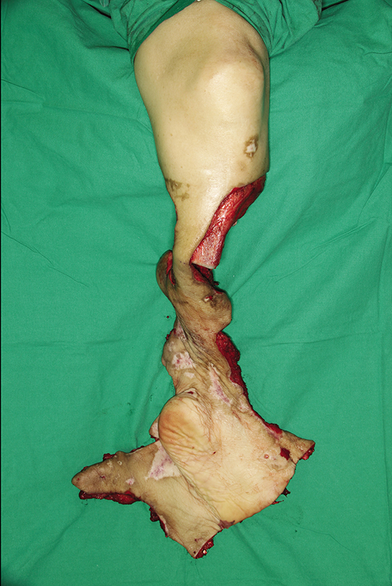
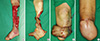
At 47 days after the initial fibular free flap transfer, below-knee amputation and coverage of the stump were performed under general anesthesia with loupe magnification, a pneumatic thigh tourniquet without limb exsanguinations. After devitalized tissue in the wound was debrided completely, the tibia was cut 13.5 cm distal to the tibial tuberosity. The dorsum of the foot was incised from the distal margin of the wound near the lateral ankle to the wound at the dorsum of the foot (Fig. 1A). The toes were amputated through the metatarsophalangeal joints. The remaining skin and soft tissue, including the vascular pedicle, were elevated at the subperiosteal level. The distal tibial and fibular segments and all tarsal and metatarsal bones were removed. Dense fibrous tissue at the plantar surface of the foot, large tendons, and volar plates of the metatarsophalangeal joints were excised. The intrinsic muscles of the foot were preserved, and the posterior tibial neurovascular bundle was not debulked (Fig. 1B). The flap was rotated 180° and folded, and the posterior plantar part of the flap was placed at the weight bearing aspect of the distal tibia. The anterior plantar part of the flap was placed in the soft tissue defect at the posterior calf, and the calf part of the flap was placed at the soft tissue defect of the anterior calf below the patella. After insertion of suction drainage tubes, the flap was sutured to the wound and remaining flap margins (Fig. 1C). Complete survival of the flap was achieved and the knee joint was mobilized early.
At 84 days after below-knee amputation and flap coverage, a debulking procedure was performed to reduce the excess volume of the healed flap. The posterior tibial neurovascular pedicle was inevitably transected and immediate neurorrhaphy of the posterior tibial nerve was performed. At 2 months after the debulking procedure, the patient noted tingling sensation at the flap. Follow-up at 7 months after below-knee amputation and nonisland pedicled foot fillet flap showed that the flap was well healed with improving sensation to the skin flap and the patient was satisfied with the result (Fig. 1D).
DISCUSSION
All of the previously reported cases of compartment syndrome after fibular free flap surgery were performed in an osteocutaneous fashion. They were not osseous free flaps, as was our case (8). The cause of the compartment syndrome in these previous cases may have been closure of the skin under excessive tension. Therefore, surgeons have been advised that primary closure of the donor site should be restricted to skin paddles of less than 4-6 cm in width. When the skin paddle width is more than 4-6 cm, primary closure should not be attempted, instead, the donor site should be covered with a split-thickness skin graft (1, 12). However, in the present patient, only the fibular bone was used as an osseous free flap without skin paddle. Compartment syndromes have never been described after harvesting of bone only, which does not result in any skin or fascia defect. The present case provides the lesson that primary closure of the donor leg without skin defect can also lead to excessive tension, resulting in compartment syndrome. Compartment syndrome can develop whenever the leg fascia is closed primarily. Potential causes for increased intracompartmental pressure even in cases where skin and fascia closures are performed without tension may be hematoma, inadequate drainage, severe edema, and tight dressing.
In all of the previous cases of compartment syndrome after fibular free flap surgery, the leg defects were treated with debridement, dressing changes, primary closure, skin graft, and/or flap surgery but not amputation (1, 3, 9). However, in the present case, severe compartment syndrome developed and the leg defect was treated with amputation even though the donor leg was closed without tension. Due to the rarity of the medical records about the microvascular fibular transfer, it was difficult to track the exact cause of this severe complication. However, because fasciotomy was performed at 7 days after the initial fibular free flap transfer in the present case, we presume that the cause was associated with delayed diagnosis and treatment.
For the treatment of the present case, various flaps are available as coverage options for the stump wound after below-knee amputation. Various free flaps may be used to cover the stump, including latissimus dorsi, rectus abdominis, scapular, and groin flaps (16, 17). However, they are time-consuming procedures and they always result in additional donor site morbidity. Therefore, they had not been appropriate treatment options for the present patient, who was exhausted because of the previous surgeries. To avoid additional donor site morbidity, various foot fillet flaps may be used as a free or island pedicled flap (13). The free foot fillet flap may have been an option as it requires only limited dissection of the vascular pedicle. The flap may be performed with 2 operative teams and have little blood loss during flap dissection, but microvascular anastomoses may be impossible because the chronically inflamed vessels are vulnerable to surgical injury. The island pedicled foot fillet flap may avoid the need for a microvascular anastomosis and may preserve continuity of the tibial nerve. However, the dissection of a lengthy vascular pedicle may be difficult in the chronically inflamed wound because of the fragility and adhesions to the surrounding tissue. If an island pedicled foot fillet flap would have been performed for the present patient, potential complications including venous insufficiency of the flap and problems associated with pedicle redundancy such as pedicle kinking during flap inset and compression of the coiled pedicle in the stump pocket may have comprised the final outcome. Therefore, both the free and the island pedicled foot fillet flaps were not considered appropriate treatment options for the present patient.
The nonisland pedicled foot fillet flap that was used in the present patient did not require skeletonization or debulking of the vascular pedicle. Therefore, this flap may be useful in patients who have chronic extensive soft tissue defects. In addition, this flap does not require microvascular anastomosis, has minimal venous congestion, and avoids problems associated with pedicle redundancy. However, a debulking procedure is frequently needed for reduction of flap volume as was performed in the present patient.
The nonisland pedicled foot fillet flap was used successfully to treat the below-knee amputation stump wound in the present patient. Although a debulking procedure was required, the safety is higher with the nonisland pedicled foot fillet flap than a free or islanded pedicled foot fillet flap. Therefore, the nonisland pedicled foot fillet flap may be a useful option for treatment of a chronically inflamed stump wound after below-knee amputation.
 XML Download