

 PDF
PDF ePub
ePub Citation
 Print
Print


INTRODUCTION
Trichophyton mentagrophytes is the second most common dermatophyte in Korea (1-3). Its subtypes include T. mentagrophytes var. interdigitale, T. mentagrophytes var. mentagrophytes, T. mentagrophytes var. nodulare, T. mentagrophytes var. goetzii, T. mentagrophytes var. granulosum and T. nentagrophytes var, erinacei (4, 5). T. mentagrophytes var. interdigitale, an anthropophilic isolate, is a frequent cause of chronic dermatophyte infection of feet, nails, and the groin (6). T. mentagrophytes var. mentagrophytes, a zoophilic isolate, is more often associated with inflammatory lesions of the scalp, the glabrous skin, the nails, and the beard region (6). Although T. mentagrophytes is a common dermatophyte in Korea, few reports have been issued on its epidemiological and mycological characteristics based on long-term large-scale study. Accordingly, this study describes the annual incidence and the distribution of subjects by age, sex, season, involved sites, and place of residence, and the fungal colony appearance of T. mentagrophytes in Korea.
MATERIALS AND METHODS
Patients
We retrospectively investigated the medical records and the epidemiological and mycological statuses of 6,250 patients with T. mentagrophytes infection diagnosed mycologically at the Catholic Skin Disease Clinic from 1992 to 2012.
Methods
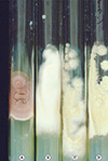
Based on medical records, 6,250 patients with T. mentagrophytes were retrospectively surveyed with respect to annual incidence and the distribution of the subjects by age, sex, season, involved sites, and place of residence. The presence of T. mentagrophytes infection was confirmed by fungal culture using potato dextrose agar corn meal Tween 80 media. In addition, microscopic examination was performed using lactophenol cotton blue stain, and colony appearance was classified as granular, persicolor, powdery, or downy (Fig. 1).
RESULTS
Annual incidence
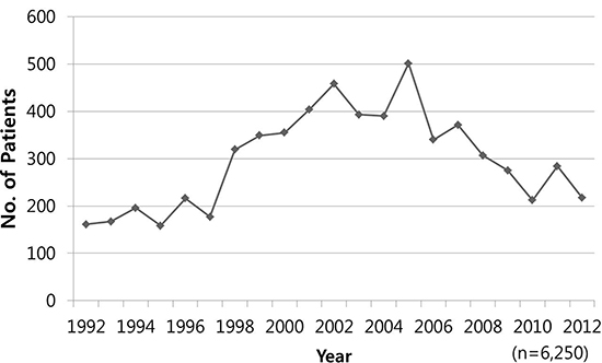
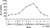
Annual numbers of patients presenting with a T. mentagrophytes infection fluctuated between 158 and 501 during the period 1992 to 2005. Number peaked in 2005 (n=501) and subsequently gradually decreased (Fig. 2).
Age and sex distribution
There was 1,610 patients (25.8%) in forties, which represented the peak in the age distribution, 1,424 (22.8%) were in their thirties, and 1,011 (16.2%) were in their fifties. The eldest patient was 98 yr old and the youngest 1 yr old. Of the 6,250 study subjects, 2,716 (43.5%) were <40 yr old (young), 2,621 (41.9%) were aged from 40 yr to <60 yr old (middle-aged), and 913 (14.6%) were ≥60 yr old (old). Out of the 6,250 study subjects, 3,527 (56.4%) were men and 2,723 (43.6%) were women (Fig. 3).
Monthly and seasonal distribution
Among 6,250 study population, 1,009 (16.1%) patients visited our hospital in July and 912 (14.6%) in June. Seasonally, 2,675 patients (42.8%) visited during the summer months, which was more than in any other season, and 871 (13.9%) in winter, showing the lowest seasonal number (Fig. 4).
Topographical distribution
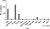
T. mentagrophytes infection developed in 2,415 patients (38.6%) in toewebs (tinea pedis, interdigital subtype), which was the most common infection site, followed by 1,739 (27.8%) in soles (tinea pedis, hyperkeratotic, and vesicular subtype), and in 897 (14.4%) in toenails (tinea unguium) (Fig. 5, 6).
Residency distribution
Of the 6,250 study subjects, 4,362 (69.8%) lived in Daegu, 1,534 (25.5%) in Gyeongbuk, 192 (3.1%) in Gyeongnam, 51 (0.8%) in Busan, and 111 (1.8%) in other areas.
Fungal culture
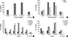
T. mentagrophytes exhibited variable overall colony appearance, which were classified into four variants: powdery, persicolor, downy, and granular. Identification of T. mentagrophytes was supported by gene analysis of the ribosomal internal transcribed spacer region. Of the 6,250 T. mentagrophytes samples, 2,403 (38.5%) exhibited a powdery, 2,215 (35.4%) a persicolor, 977 (15.6%) a granular, and 655 (10.5%) a downy colony appearance (Fig. 7A). The most common colony appearance before 2005 was powdery and the most common after 2005 was persicolor. Of the 3,527 male patients, 1,391 (39.4%) had a powdery colony appearance, and of the 2,723 female patients, 1,012 (37.2%) had a powdery colony appearance (Fig. 7B). According to age, the most common colony appearance was granular (n=320) in patients <20 yr old, persicolor (n=260) in patients from 20 yr old to <30 yr old, powdery (n=634) in patients from 30 yr old to <40 yr old, persicolor (n=667) in patients form 40 yr old to <50 yr old, powdery (n=404) in patients from 50 yr old to <60 yr old, powdery (n=225) in patients from 60 yr old to <70 yr old, and powdery (n=102) in patients aged ≥70 yr old. A powdery colony appearance accounted for; 20.8% of patients <20 yr old, 35.8% in patients from 20 yr old to <30 yr old, 44.5% in patients from 30 yr old to <40 yr old, 41.1% in patients from 40 yr old to <50 yr old, 40.0% in patients from 50 yr old to <60 yr old, 35.1% in patients from 60 yr old to <70 yr old, and for 37.5% in patients aged ≥70 yr old (Fig. 7C). In terms of seasonal distribution, the most common colony appearance was powdery (n=275) in winter, persicolor (n=676) in spring, and powdery in summer (n=1,099) and in fall (n=360) (Fig. 7D). In terms of topographical distribution, the most common colony appearances were powdery (n=1,040) for toewebs, granular (n=343) for toenails, and granular (n=54) for dorsal feet.
DISCUSSION
The fungal universe is composed of more than 1.5 million species (6). Dermatophytes are represented by approximately 40 species divided into three genera: Epidermopyton, Microsporum, and Trichophyton (6). In Korea, Trichophyton species, such as T. rubrum and T. mentagrophytes are mostly commonly isolated. However, although T. mentagrophytes is a common dermatophyte in Korea, few investigative reports have been issued on its epidemiological and mycological characteristics based on long-term large-scale analysis.
The prevalence of dermatophytosis in skin has changed over the last 50 yr. The incidence of dermatophytoses, with the exception T. rubrum infections, are decreasing, and the present study also shows that the incidence of T. mentagrophytes had decreased in Korea since 2005. Choe et al. (7) reported that the incidence of T. verrucosum infection decreased in the 2000s much more so than in the late 1900s. Lee et al. (8) also reported that the prevalence of Microsporum canis infection decreased markedly in the 2000s. In addition, the incidences of T. schoenleinii, T, violaceum, M. gypseum, M. ferrugineum, and Epidermophyton floccosum are also decreasing, and that of T. tonsurans infection has shown a recent decrease. The first report on T. tonsurans infection in Korea was issued by Suh et al. (9) in 1995. Since T. tonsurans was isolated from a middle school wrestler, it spread to amateur wrestlers and judoists as it was referred to as trichophytosis gladiatorum and to the general population (10). The observed decrease in T. mentagrophytes infection may have been caused by public health education, improved hospital accessibility, environmental improvements, and increased lifestyle diversity.
T. mentagrophytes is a causative fungus of tinea facieii, tinea capitis, tinea cruris, tinea pedis, tinea unguium, and tinea manus (11). In this study, tinea pedis, especially the interdigital type, was the most common T. mentagrophytes infection. The spreading of tinea pedis was accompanied by a parallel increase in the frequency of onychomycosis - a high proportion of onychomycosis was also revealed by the present study.
T. mentagrophytes is further classified as anthropophilic or zoophilic (6). Anthropophilic isolates, such as, T. mentagrophytes var. interdigitale, infect humans, and include the powdery, persicolor, and downy colony appearance types of T. mentagrophytes in fungal culture. Powdery colonies are composed of fine powder, whereas persicolor colonies are composed of thin, white, cottony strands, and the downy colonies exhibit white aerial mycelium. Zoophilic isolates of T. mentagrophytes, such as, T. mentagrophytes var. mentagrophytes also exhibit a granular colony appearance (12, 13), which is observed under the microscope as conspicuous sandy-brown clumps of conidiation, which give the colony surface a coarsely granular appearance.
In this study, powdery and persicolor colony appearances were much more common than the granular colony appearance of T. mentagrophytes, which supports the notion that occupational diversity has driven the observed marked decrease in the incidence of zoophilic T. mentagrophytes infections. However, in the present study, patients aged less than 20 yr old were most commonly infected with zoophilic T. mentagrophytes, indicating that pets should be considered as a major source of zoophilic T. mentagrophytes. Zoophilic T. mentagrophytes is associated with a wide range of rodents, such as lagomorphs (rabbits and relatives), hedgehogs, and other small mammals (14). Although these smaller mammals are probably the primary reservoir, zoophilic T. mentagrophytes may also cause infection in horses and other large mammals (14).
In the present study, the incidence of T. mentagrophytes was highest in the fourth decade till 2001 and highest in the fifth from 2001. Contrary to a previous report, which found that the prevalence of T. mentagrophytes increases with age (15), we found that T. mentagrophytes infection was greatest among the middle-aged, which suggests that social interactions may be an important factor of T. mentagrophytes infection in Korea. Furthermore, the incidence rate in men was much higher than that in women. In addition, as was expected based on the relation between fungal growth, humidity, and temperature, its incidence was highest in summer (16).
This study provides considerable information on T. mentagrophytes infection in Korea. In particular, it confirms a continuing reduction in its annual incidence and shows that a powdery colony phenotype is the most common.

 XML Download