

 PDF
PDF ePub
ePub Citation
 Print
Print


INTRODUCTION
Angiogenesis is essential for many physiological processes including wound healing, cell regeneration, tissue growth and development, and embryonic development, and plays a very important role in the development of proliferative diabetic retinopathy, a serious complication of diabetes (1-3). Angiogenesis or the formation of new blood vessels from existing vessels has recently been widely studied; it has been found that various types of cytokines and growth factors, particularly vascular endothelial growth factor (VEGF), are involved in angiogenesis (4, 5).
It has been observed that retinal capillary closure and retinal ischemia can result in spread of angiogenic factors such as VEGF into the ischemic retina, leading to the formation of abnormal blood vessels (6-8). The mechanism of angiogenesis is similar in both corneal and retinal tissues. It has been reported that the VEGF is also involved in the angiogenesis of the cornea (9, 10). Therefore, it is very important to identify the factors associated with the angiogenesis, and also studies of genes related to angiogenesis are expected to be very useful in the treatment of corneal neovascular diseases.
Accordingly, we investigated the differences in angiogenesis gene expression of keratocytes between the genetic model for OLETF in diabetic rats and the control group of normal rats. In addition, an experiment was performed to observe the angiogenesis-related genes in the inflamed cornea by treating with interleukin-1α (IL-1α) and tumor necrosis factor-α (TNF-α).
MATERIALS AND METHODS
Cell culture and cytokine treatment
Fifty-week-old, genetically affected type 2 diabetic (OLETF, Otsuka Long-Evans Tokushima Fatty) and normal rats were purchased from Otsuka Pharmaceutical Company (Tokushima, Japan). The corneal epithelium was removed; the corneal stroma was isolated, and washed several times with saline containing antibiotics. The corneal endothelial layer was removed, and then the corneal stromal tissue was cut into small pieces and cultured serially in Dulbecco's modified Eagle medium (DMEM, Gibco BRL, Grand Island, NY, USA) containing 10% fetal bovine serum (FBS, Gibco BRL), 100 units/mL penicillin (Gibco BRL), and 100 mg/mL streptomycin (Gibco BRL). The culture medium was changed at 2- to 3-day intervals. When the cells became confluent, the culture medium was removed, the cells were washed once with Dulbecco's phosphate-buffered saline (D-PBS, Gibco BRL) and dissociated with 0.25% trypsin-0.02% EDTA. For the microarray experiments, cells between the third and fourth generation of serial culture were collected and stored at -70℃. The corneal stromal cells of diabetic and normal rats were treated with the cytokines, IL-1α and TNF-α (R&D Systems, Minneapolis, MN, USA), at a concentration of 20 ng/mL for 6 hr.
RNA extraction
Total cellular RNAs were extracted from the corneal stromal cells of diabetic and normal rats in primary culture using the RNeasy mini kit (QIAGEN Inc, Valencia, CA, USA) for gene microarrays. First, denaturing solution was added to cell pellets on ice for 5 min, and then phenol and chloroform were added to all samples. The samples were centrifuged, and the supernatant containing RNA was transferred to a new tube. The process was repeated. Subsequently, isopropanol at 20℃ was added to precipitate the RNA, and then the mixture was centrifuged and the supernatant was discarded carefully. The pellet was washed with 80% ethanol at 20℃ and then air-dried. Extraction of RNA was quantitated and confirmed by electrophoresis.
DNA gene microarrays
Sixty-mer oligonucleotides corresponding to each gene were synthesized and subsequently placed on a slide using a robotic gene microarray. The robotic gene microarray places 0.25-1 nL DNA samples on a slide sample in spots averaging 100-150 nm. cDNA was synthesized from the isolated RNA samples using RT primers (Genisphere Inc, Hatfield, PA, USA) and SuperScript II reverse transcriptase (Invitrogen, Grand Island, NY, USA). The purified cDNA was hybridized to an Agilent Rat oligo 22K chip (Agilent tech, Santa Clara, CA, USA). For hybridization, slides were placed in a dark hybridization chamber at 62℃ for 16 hr. The slides were removed, washed three times, and dried using a centrifuge. A fluorescent dye was added to DNA capture reagents that bind RT primers. Thereafter, the second hybridization was performed for 4 hr. The slides were removed, washed, and dried using a centrifuge. Afterward, the fluorescence level was measured using an Axon Laser Fluorescence Scanner (Axon Instruments Inc, Foster City, CA, USA). The microarray analyzer we used in our study has 105 rows and 215 columns. A total of 22,575 genes pertinent to angiogenesis were included.
Analysis
The hybridized slides were scanned by an Axon GenePix Laser Fluorescence Scanner and analyzed by GenePix Pro 5.1 (Axon Instruments) and GeneSpring 7.0 (Silicongenetics, Redwood City, CA, USA). Gene expression was considered as significant for genes showing more than a twofold difference in expression level between normal and diabetic rats. Genes showing similar patterns were distinguished by a Pearson's correlation analysis. The images thus obtained show that genes that are up-regulated are shown in red, whereas genes that are down-regulated are shown in green. First of all, we investigated the differences in angiogenesis gene expression in keratocytes between diabetic and normal rats. In addition, the angiogenesis-related genes that show different expressions between normal and diabetic keratocytes after treatment with IL-1α and TNF-α were also studied.
Real-time polymerase chain reaction
Real-time polymerase chain reaction (RT-PCR) was performed with SYBR Green I immunofluorescence dye and the HotStarTaq DNA polymerase QuantiTect SYBR Green PCR Kit (QIAGEN GmbH, Hamburg, Germany). To quantitate the control group, a standard curve for the genes of interest and glyceraldehydes 3-phosphate dehydrogenase (GAPDH) was prepared. GAPDH was reacted to three genes: Il6, Itga1, and Agtr1. The reactions were carried out using the ABI PRISM 7900 Sequence Detection System version 1.6 software (Perkin-Elmer Biosystems, Foster City, CA, USA). Relative quantificational measurements were performed using the relative standard curve method according to the manufacturer's instructions.
RESULTS
The subjects used in our study were 50-week-old diabetic OLETF rats. Mean blood glucose was 115.0±10.8 mg/dL in the control group and 193.8±18.9 mg/dL in the OLETF group, which demonstrated statistically significant differences between the two groups. Mean body weight was significantly higher in the OLETF group (control group: 518.0±13.3 g; OLETF group: 628.5±63.2 g). These are the typical characteristics of diabetic rats.
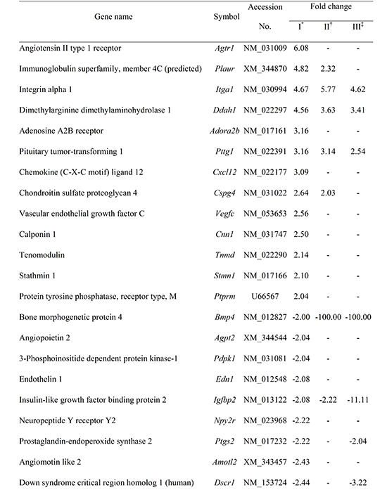
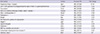
Diabetic keratocytes showed upward expression in 13 genes related to angiogenesis, compared to normal keratocytes, including the Vegfc, Agtr1, Itga1, Ddah1, Adora2b, and Cspg4. Twenty-two genes including the Mmp3, Dcn, Amotl2, and Pdgfa were down-regulated (Table 1).
Three genes in diabetic keratocytes, namely Itga1, Ddah1, and Pttg1, which promoted angiogenesis, showed an increased expression after both cytokine treatments, as well as compared to normal keratocytes. Genes that showed a decreased expression after both cytokine treatments were Mmp3 and Pdgfa.
Some genes showed different patterns of expression in response to two cytokines. Itga1 showed no reaction to TNF-α and responded to IL-1α only. Conversely, Esm1 did not show a reaction to IL-1α and indicated a decreased expression to TNF-α compared to the control group (Table 1).
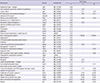
Newly expressed genes after IL-1α treatment are shown in Table 2. After IL-1α treatment, 15 genes showed different expression in diabetic keratocytes as compared to control group. Among them, eight genes showed an increased expression including Egr1, Cxcl12, and Tn. Seven genes showed a decreased expression including Il1b, Pdgfra, and Klf5.
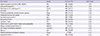
Newly expressed genes after TNF-α treatment are shown in Table 3. After TNF-α treatment, 14 genes showed different expression in diabetic keratocytes as compared to control group. TNF-α treatment increased gene expression in the Agtr1, Tnmd, and C1galt1. However, 11 genes including Edn1, Mmp9, and Prok1 showed a decreased expression compared to the control group.
Newly expressed genes after both IL-1α and TNF-α treatment are shown in Table 4. Five genes showed decreased gene expression, including tissue inhibitor of Timp3 and Mmp14. Conversely, for Ctss, Hmox1, and Il6, which control and induce angiogenesis, expression was increased by IL-1α stimulation and decreased by TNF-α treatment.
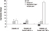
The RT-PCR was performed on three genes: Il6, Itga1, and Agtr1 that showed significantly different expression between normal and diabetic keratocytes, or were newly expressed after cytokine treatment. The primer preparation and annealing temperature for these genes are shown in Table 5.
After cytokine treatment, the Itga1 significantly showed an increased expression in diabetic rats. The Agtr1 showed an increased expression compared to normal rats, but showed no expression by IL-1α treatment and increased expression by TNF-α treatment. Il6 showed no expression in the keratocytes of diabetic rats, but increased expression by IL-1α treatment and decreased expression by TNF-α treatment (Fig. 1).
DISCUSSION
Thirty-five genes that showed different expressions in diabetic keratocytes compared to normal keratocytes, are related to VEGF. The VEFG is one of the most important regulators of angiogenesis, and is associated with endothelial cell proliferation of angiogenesis process (11). Activated angiogenic process from decreased blood supply to organs and tissue in diabetes, is related to the increased gene expression related to VEGF.
The angiotensin II type 1 receptor gene (Agtr1) is observed to have the highest increased expression in diabetic rats compared to normal rats. Angiotensin II up-regulates mRNA expression of VEGF and potentiates VEGF-mediated angiogenic activity through an up-regulation in kinase domain region in endothelial cells (12, 13). Therefore, up-regulated expression of Agtr1 in corneal stromal cells results in the activation of angiogenesis through the reinforcement of the VEGF action. Fernadez et al. (14) also reported that angiotensin II induces angiogenesis in the rabbit cornea, which coincides with the result of our study.
Twenty-two genes show decreased expression in diabetic keratocytes compared to normal keratocytes, and the representative gene is decroin (Dcn). This gene stabilizes the extracellular matrix assembly to provide a template for endothelial cells to form capillary tubes by interacting with specific other angiogenesis-associated extracellular matrix molecules such as type I collagen and fibronectin. In addition, Dcn is also known to prevent apoptosis of endothelial cells and to be involved in maturation of the blood vessels (15-18). Therefore, down-regulated expression of Dcn in diabetes suggests that normal blood vessel growth and maturation is impossible in diabetic tissues (incomplete angiogenesis). Furthermore, decorin is known to be involved in angiogenesis, particularly in condition in which the inflammatory component is dominant (19). This coincides with the result of our study, in which expression of Dcn increases when inflammation is induced by IL-1α and TNF-α treatment. In addition, angiopoietin 2 (Agpt2) and platelet-derived growth factor, alpha (PDGF-α, Pdgfa) show a decreased expression, which are related to stabilization and maturation of blood vessels through the recruitment of pericytes and vascular smooth muscle cells. The decreased expression of these genes is related to the fact that although neovascularization is stimulated in diabetes, the newly formed blood vessels are immature and are easily damaged by external stimulation.
Genes that show a significant increased expression after IL-1α and TNF-α treatment include Itga1, Ddah1, and Pttg1. Integrin alpha 1 (Itga1) is found to be directly involved in the process of angiogenesis by facilitating endothelial cell migration (20). In addition, dimethylarginine dimethylaminohydrolase 1 (Ddah1) is known to stimulate the process of angiogenesis (21) and that pituitary tumor-transforming 1 (Pttg1) induces basic fibroblast growth factor expression and thereby promoting angiogenesis (22).
The genes that show a decreased expression, Mmp3 and Pdgfa, exhibited greater action with cytokines. The decreased expression of these genes implies that the recovery of diabetic tissues and blood vessels may be delayed or cause damage due to inflammation. PDGF-α (Pdgfa) plays a critical role in vessel maturation by recruiting vascular pericytes which aid in basal lamina assembly. And it also prevents vascular endothelial cell apoptosis after VEGF withdrawal (23-25). An experiment on mice showed that 50% reduction of the pericyte density can cause retinopathy which is similar to diabetic retinopathy (26). Decreased expression of these genes in diabetic keratocyte might be related to the dysplasia of the vessel wall.
Among the eight angiogenesis-related genes that show an increased expression after IL-1α treatment, early growth response 1 (Egr1) has a direct reaction on angiogenesis through the formation, proliferation, and migration of endotheliocytes in microblood vessels (27). Chemokine (CXC motif) ligand 12 (Cxcl12) is a potent promoter of angiogenesis that promotes recruitment of endothelial progenitor cells from the bone marrow, and has an important role in the pathogenesis of chronic inflammatory disorders (28). Conversely, among the angiogenesis-related genes that are newly expressed after IL-1α treatment, platelet-derived growth factor receptor, alpha polypeptide (Pdgfra) and kruppel-like factor 5 (Klf5) exhibited a decreased expression. If Pdgfra and Klf5 are insufficient during embryogenesis, the incomplete formation of blood vessels or vascular malformation is induced by endothelial cell necrosis, significant reduction in the numbers of vessel wall pericytes and smooth muscle cells, and decreased deposition of extracellular matrix (29). This result shows that the retinal neovascularization is activated in proliferative diabetic retinopathy, but the formation of immature blood vessels promotes complications of diabetes such as hemorrhage and necrosis of normal retinal tissues.
After TNF-α treatment, 14 angiogenesis-related genes are newly expressed. As mentioned above, Agtr1, which clearly shows an increased expression, is known to increase the expression of VEGF and potentiate VEGF-mediated angiogenic activity. The increased expression of tenomodulin (Tnmd) is also known as an angiogenesis inhibitor to interfere with blood vessel formation. The reciprocal expression of these genes demonstrated that angiogenesis is induced but the formation of complete blood vessel is impossible in inflammation-related diabetes cases. Furthermore, the matrix metallopeptidase 9 (Mmp9) and endothelin 1 (Edn1), which are involved in angiogenesis, show a lower expression in diabetic keratocytes after TNF-α treatment as compared to the control group. This down-regulate neovascularization in diabetes, and is related to the pathological mechanism of this disease.
Newly and decreased expressed genes by IL-1 α and TNF-α treatment are Timp3, Mmp14, Adm, Npy, and Cited2. Adrenomedullin (Adm) is recognized as a novel growth factor for endothelial cells and promoter of angiogenesis. Neuropeptide Y (Npy) is known to stimulate angiogenesis through endothelial cell migration, proliferation, and differentiation in vitro and acts on angiogenesis for ischemic stimulation (30). In particular, the expression of these genes decreases in diabetic rats after cytokine treatment, which induces inflammation and delays angiogenesis in diabetes. As a result, an ischemic change is caused in tissues that may lead to diabetic complications. Therefore, much more histopathologic research in this area is anticipated. Moreover, cathepsin (Ctss) plays a functional role in angiogenesis through the production of type IV collagen-derived anti-angiogenic peptides and the generation of bioactive pro-angiogenic gamma 2 fragments from laminin-5 (31); heme oxygenase 1 (Hmox1) promotes VEGF-driven noninflammatory angiogenesis (32). In addition IL-6 (Il6) shows an increased expression after treatment with IL-1α and decreased expression after treatment with TNF-α. This result demonstrates that inflammation reactions can vary by the type of cytokine used. More studies on individual genes expressed differently after the cytokine treatment may be necessary.
In this study, we selected three genes that showed a significant difference of expression between normal and diabetic keratocytes to create a primer and performed an RT-PCR to confirm the result of microarray analysis. In other words, the RT-PCR was performed on Il6 that showed an increased and decreased expression on cytokine treatment, Itga1 that showed an increased expression in diabetes compared to normal rats, and Agtr1 that clearly increased expression by TNF-α treatment. The degree of expression was then examined. Consequently, the same expression pattern could be confirmed, as was the case with the microarray analysis.
The genes expressed more often in diabetic keratocytes than in normal keratocytes were found to stimulate the process of angiogenesis while at the same time forming immature blood vessels. These effects are also stimulated by newly expressed genes after the IL-1α and TNF-α treatment which causes inflammation. Although much more time and effort are needed in this kind of studies, there will be an important opportunity to evaluate or identify new genes related with angiogenesis in diabetics.


 XML Download