

 PDF
PDF ePub
ePub Citation
 Print
Print


INTRODUCTION
Bone demineralization is a common metabolic disorder in human immunodeficiency virus (HIV)-infected patients. A recent meta-analytic review has reported that the prevalence of osteoporosis patients is approximately 3-fold higher in HIV-infected patients than in non-HIV-infected patients. It has shown that antiretroviral therapy (ART)-treated subjects had a 2.5-fold higher prevalence of low bone mineral density (BMD) than ART-naïve subjects (1). The clinical expression of osteoporosis is a fragility fracture associated with impaired quality of life and a 20% reduction in survival (2). A large population-based study has demonstrated that the prevalence of fractures is higher in HIVinfected patients than in non-HIV-infected patients (3).
The etiology of low BMD in HIV-infected patients is likely to be multifactorial. Patients with osteoporosis and HIV infections have certain risk factors in common, such as hypogonadism, low weight, a high rate of tobacco and alcohol consumption (4, 5). HIV infection itself decreases BMD by increasing osteoclast differentiation and inducing osteoblast apoptosis (6).
Previous studies have shown that protease inhibitors (PIs) and nucleoside reverse transcriptase inhibitors (NRTIs) are associated with low BMD (1, 7). Recently, Stellbrink et al. (8) have reported that the greater decreases in BMD are observed in subjects treated with tenofovir/emtricitabine (TDF/FTC) than with abacavir/lamivudine (ABC/3TC). TDF/FTC had not been commercially available in Korea until September 2011. Therefore, the Korean guideline recommended zidovudine/lamivudine (ZDV/3TC) or ABC/3TC as preferred dual-NRTI components (9). To our knowledge, no studies have compared the influence of ABC and ZDV on BMD.
The objectives of this study were to determine the prevalence and risk factors of low BMD and to compare the effects of ABC and ZDV-based regimens on bone demineralization in Korean HIV-infected patients.
MATERIALS AND METHODS
Study population
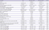
This study included HIV-infected patients who underwent dual energy X-ray absorptiometry (DEXA) scanning at our 560-bed medical center located in Seoul, Korea, between December 2010 and September 2011. Using computerized hospital records, we retrospectively selected patients fulfilling the following criteria: 1) those who were at the age of ≥ 20 yr; 2) those who received in- or outpatient services; 3) those who were on ART or were naïve to ART; and 4) those who were not treated with calcium or bisphosphonate. Patients' characteristics analyzed were sex, age, BMI, date of HIV diagnosis, AIDS stage according to the US Centers for Disease Control and Prevention (CDC) classification, nadir CD4 count, current CD4 count, peak HIV RNA titer, current HIV RNA titer, ART regimen and duration of ART. Among acquired risk factors for osteoporosis, smoking status, alcohol consumption, the types and treatment durations of glucocorticoid or antiepileptic drugs, past history of tuberculosis treatment, current tuberculosis state, and serological statuses of hepatitis B and C viruses were identified. Smoking status was categorized as who has never smoked (never smoker) and who is either current or exsmoker (ever smoker). Alcohol consumption was also categorized into two groups: non-drinkers or current drinkers. BMD of the lumbar spine and hip were measured by dual energy X-ray absorptiometry (Prodigy advance, GE-lunar, Madison, WI, USA). The PA view of L1-L4 was used for spine BMD measurement. Anatomically abnormal vertebrae were excluded from analysis when there was a T-score difference of more than 1.0 between the vertebra in question and adjacent vertebrae. For the hip region, the femoral neck or total proximal femur was used, whichever was the lowest BMD. The World Health Organization (WHO) classification was used for diagnostic purposes. Osteopenia was defined as a T-score between -1 SD and -2.5 SD, and osteoporosis was defined as a T-score of less than -2.5 SD.
All patients were included in the analysis to determine the prevalence and risk factors of low BMD. To compare the effects of ABC and ZDV on bone loss, we analyzed patients without changes in the dual NRTI regimen by the time of BMD measurement. The prevalence of osteopenia and osteoporosis were evaluated in terms of treatment duration and measurement areas.
Laboratory methods
HIV plasma RNA was measured by real-time PCR using an Abbott real time PCR system m2000sp/m2000rt (Abbott Park, IL, USA). CD4 cell counts were made by flow cytometry.
Statistical analysis
Comparisons were made between normal BMD and low BMD with the Mann-Whitney U test for quantitative variables as well as the chi square test or Fisher's exact test for qualitative variables. Multivariate logistic regression analysis assessed risk factors for low BMD. Variables with P < 0.2 in univariate analysis were included in the multivariate analysis. To evaluate the prevalence between ABC and ZDV-based regimens over time, linear by linear association and logistic regression including an interaction term were applied. The one year separation time point was chosen by visual inspection of data.
RESULTS
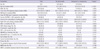
A total of 224 HIV-infected patients were included in the study. The characteristics of the HIV patients are described in Table 1. Of the 224 patients, 210 (93.8%) were male and 14 (6.2%) were female. The median age of the patients was 38 yr (interquartile range [IQR], 30-48 yr) for men and 45 yr (IQR, 34-51) for women. The median follow-up duration after the date of HIV infection diagnosis was 814 days (IQR, 339.0-1,566.0). The median peak HIV RNA was 4.5 log copies/mL (IQR, 3.8-5.0) and current HIV RNA was < 200 copies/mL in 149 patients (66.5%). The median nadir CD4 cell count was 226.0 cells/µL (IQR, 102.0-329.8), and the median current CD4 cell count was 427 cells/µL (IQR, 277.3-581.8). Clinical stages of AIDS were reported by 46 patients (20.5%). Ever smoker and current alcohol drinker were 133 patients (60.2%) and 138 patients (62.4), respectively. Histories of tuberculosis treatment or currently taking anti-tuberculosis medication were reported by 23 patients (10.3%). Viral hepatitis (HBV surface antigen positive or HCV RNA positive) occurred in 13 patients (5.8%). Eighteen patients (8%) had a history of taking glucocorticoid or antiepileptic drugs (valproic acid, benzodiazepine, carbamazepine or phenytoin) over 3 months. At the time of BMD measurement, 173 patients (77%) were on ART and 145 patients (64.7%) were on it for more than 3 months.
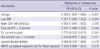
Osteopenia and osteoporosis were diagnosed in 93 (41.5%) and 29 (12.9%) of 224 patients, respectively. Of the male patients, 89 (42.4%) and 25 (11.9%) had osteopenia and osteoporosis, respectively. Of the female patients, 4 (28.6%) had osteopenia and another 4 (28.6%) had osteoporosis, respectively. Of the 171 patients aged < 50 yr, 76 (44.4%) were diagnosed with low BMD; osteopenia and osteoporosis were diagnosed in 65 patients (38.0%) and 11 (6.4%) of these patients, respectively. Of the 53 patients aged ≥ 50 yr, 46 (86.8%) were diagnosed with low BMD; osteopenia and osteoporosis was diagnosed in 28 (52.8%) and 18 (34%) of these patients, respectively. The prevalence were significantly higher patients aged ≥ 50 yr than in those aged < 50 yr (P < 0.001). Of the 49 male patients aged ≥ 50 yr, 27 (55.1%) and 15 (30.6%) were diagnosed with osteopenia and osteoporosis, respectively. Of the 4 female patients aged ≥ 50 yr, 1 (25.0%) and 3 (75.0%) were diagnosed with osteopenia and osteoporosis, respectively. Age, BMI, current HIV RNA < 200 copies/mL, ART for > 3 months, past history of tuberculosis or current anti-tuberculosis medication, use of steroid or antiepileptic drugs for > 3 months, current alcohol drinker and NNRTI cumulative exposure day for those exposed were included in the multivariate analysis (Table 1). Among them, HIV RNA < 200 copies was excluded because it was highly correlated with ART > 3 months. Older age (odds ratio [OR], 1.056; 95% CI, 1.028-1.084; P < 0.001), lower BMI (OR, 1.201; 95% CI, 1.089-1.323; P < 0.001) and ART > 3 months (OR, 2.197; 95% CI, 1.099-4.392; P = 0.026) were identified as risk factors for low BMD (Table 2).
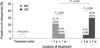
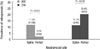
To compare the effect of the 2 different NRTI regimens, we analyzed 141 patients whose NRTI regimen had not been changed at the time of BMD measurement. Of the 141 patients, 96 (68.1%) were on an ABC-based regimen and 45 (31.9%) were on a ZDV-based regimen, respectively. There were no significant differences between the 2 groups in risk factors for low BMD, including percentage and cumulative days of administration of PI and NNRTI. BMD in the spine and femur were not significantly different between the 2 groups (Table 3). When the prevalence of low BMD over time and at different anatomical sites was compared according to NRTI regimen, there was no significant difference (data not shown). However, the prevalence of osteoporosis (21.1%, 8/38) was higher in patients on the ABC-based regimen for < 1 yr than in those on for ≥ 1 yr (6.9%, 4/58). In contrast, the prevalence was higher in patients on the ZDV-based regimen for ≥ 1 yr (29.6%, 8/27) than in those on < 1 yr (5.6%, 1/18). Osteoporosis was more frequent within 1 yr after ART initiation in patients on the ABC-based regimen, while it was more frequent 1 yr after ART initiation in those on the ZVD-based regimen (P = 0.017) (Fig. 1). We assessed the prevalence of osteoporosis at different anatomical sites between the 2 groups. In patients on the ABC-based regimen, the prevalence of osteoporosis was higher in the spine (11.5%, 11/96) than in the femur (2.1%, 2/96) (P = 0.010). However, in patients on the ZDV-based regimen, osteoporosis did not occur significantly in the spine (P = 0.245) (Fig. 2).
DISCUSSION
Our observational study evaluated the prevalence of low BMD in the population using ABC/3TC or ZDV/3TC as preferred dual-NRTI in Korea. The prevalence of osteopenia and osteoporosis were 41.5% and 12.9% of the total patients, respectively. These were higher in patients aged 50 yr and older (52.8% and 34.0%). The risk factors for low BMD were lower BMI, older age and ART for > 3 months. BMI and advancing age are conventional risk factors for low BMD. ART also has been associated with low BMD in several studies (1, 7, 8). The ABC- and ZDV-based regimens showed different prevalence of osteoporosis depending on the duration of drug administration and measurement areas. In patients on the ABC-based regimen, osteoporosis was mainly developed in the early phase of < 1 yr after ART began. In contrast, the prevalence of osteoporosis was higher in patients on the ZDV-based regimen for ≥ 1 yr than < 1 yr. Osteoporosis in patients on the ABC-based regimen was more common in the spine than in the femur.
The prevalence of low BMD in Korean HIV patients seemed to be higher than in HIV non-infected subjects. Although the prevalence of osteoporosis varies from 1.4% to 45.7% depending on the population studied, the method used to assess BMD and the different reference value (10), the recent study in a community-based cohort in Korea estimated that 12.9% for men and 24.0% for women over 50 yr of age (11). When compared with these results, the present study indicated a high prevalence of low BMD, especially in elderly patients. Furthermore it was also consistent with meta-analytic review of prevalence of osteoporosis in HIV-infected individuals.
Our results confirmed ART for > 3 months was significantly associated with low BMD, although the association between individual HIV drugs and low BMD was not confirmed. Both PI and NRTI tenofovir have been strongly associated with bone loss, whereas Brown et al. (12) described initiation of ART induces a significant loss of BMD, regardless of the initial choice of ART. An important finding in our study was that association with ART use and low BMD was confirmed under circumstances where tenofovir was not used.
The significant differences in both the prevalence of osteoporosis over time and affected areas between the 2 groups imply that bone loss may be caused by direct effects of ABC and ZDV on bone cells rather than common pathways, such as immune reconstitution or modification of vitamin D. In addition, it appears that ABC induce high bone turnover because the spine has a higher proportion of trabecular bone where bone remodeling is more rapid than the femoral head. We speculate that proinflammatory properties of ABC induce high bone turn over in early phase of treatment. A previous study of a plausible biological mechanism for the increased risk of cardiovascular diseases in patients receiving ABC has demonstrated that those on ABC have higher levels of inflammatory markers in the early phases of treatment, including cytokines and adhesion molecules which were proven to cause bone loss (13-16). ZDV can affect bone metabolism independently of the effects of HIV infection (17). ZDV can inhibit DNA polymerase gamma which leads to the depletion of mitochondrial DNA and drug toxicity. This effect increase serum lactate levels and cause bone demineralization (18). A recent study has indicated that ZDV-containing regimens cause significant bone loss in the late phases of treatment. These results correspond well with those of our study which reported osteoporosis occurred more frequently in the patients on the ZDV-based regimen for ≥ 1 yr after ART initiation.
Previous studies found that bone loss was induced within the first 6 months after ART initiation and recovered between 1 and 2 yr, regardless of regimen (8, 12). Some investigators have claimed that bone loss after ART initiation is a general phenomenon related to T-cell recovery initiated by all ART regimens rather than due to the direct effects of specific ART constituents on bone cells (19). In contrast, our study showed that the 2 NRTI groups had different prevalence of osteoporosis over time and that different areas were prone to osteoporosis. Therefore, it is thought that the unique mechanism of each NRTI may play a role in development of bone loss rather than universal characteristics such as immune reconstitution that all ART regimens share. More studies of the precise mechanisms of NRTIs on bone loss are essential for choosing initial ART regimen or switching it with another to preserve BMD.
The results of our study are subject to some limitations. First, the BMD results might be overestimated by using the T score for patients aged < 50 yr. However, our results were compatible with those of several cross-sectional studies by using T score in younger population (20, 21). Second, cross-sectional design of the study fails to measure BMD change after initiation of ART. Finally, dose-response relations between amount of smoking and/or alcohol between low BMD was not available.
In conclusion, our results show that low BMD may be common in HIV-infected patients on ABC and ZDV as preferred NRTIs, and older age, lower body mass index, and ART > 3 months were independent risk factors for low BMD. Osteoporosis was more prevalent in the early stages of treatment and was more prone to the spine area in the patients on ABC-based regimen, than in those on the ZDV-based regimen. Those findings suggest that ABC and ZDV may have different mechanisms which can affect bone mineral loss. Further studies are needed to determine the demineralization mechanism of NRTIs so as to prescribe antiretroviral drugs for reducing bone loss.
 XML Download