

 PDF
PDF ePub
ePub Citation
 Print
Print


INTRODUCTION
Erectile dysfunction is associated with cardiovascular disease risk factors such as smoking, hypercholesterolemia, and diabetes (1, 2). Diabetes is known to be a major risk factor for erectile dysfunction. Most diabetic model studies for erectile dysfunction have been conducted in animal models of type 1 diabetes. By contrast, few studies have examined the underlying mechanisms of erectile dysfunction in animal models of type 2 diabetes (3). The animal models of type 2 diabetes have shown that erectile dysfunction results from an impairment in endothelium-dependent cavernous smooth muscle relaxation (4, 5) and endothelial nitric oxide synthase (e-NOS) enzyme activity and a decrease in expression of vascular endothelial growth factor (VEGF) in the penis (6). Thus, augmentation of endogenous nitric oxide bioactivity and reestablishment of the structural and functional microvasculature may be a promising therapeutic strategy in patients with erectile dysfunction associated with diabetes. In this aspect, neovascularization is an emerging therapy for the treatment of erectile dysfunction with vascular causes.
Local intracavernous delivery of the VEGF gene or protein has been shown to restore erectile function in a rat model of type 1 diabetic erectile dysfunction induced by streptozotocin administration (7, 8). However, these studies did not demonstrate endothelial regeneration, although a restoration of smooth muscle architecture and partial or complete recovery of erectile function was noted.
Angiopoietin-1 (Ang1) is the ligand of the Tie2 receptor tyrosine kinase and is expressed on endothelial cells. Ang1 acts as an angiogenic growth factor that specifically functions to generate a nonleaky, stable, and functional vasculature (9-12). Ang1 and Tie2 signaling is thought to be involved in the branching and remodeling of the vascular network (11). Recently, Cho et al. (13) developed a soluble, more potent Ang1 variant than native Ang1, cartilage oligomeric matrix protein (COMP)-Ang1. They reported that COMP-Ang1 administered by adenoviral vector induced long-lasting, stable vascular enlargement resulting from endothelial proliferation (12).
Until now, studies have been lacking on the effect of angiopoietin on cavernosal angiogenesis in a rat model of type 2 diabetes. The Otsuka Long-Evans Tokushima Fatty (OLETF) rats develop a syndrome with multiple metabolic and hormonal disorders that shares many features with human obesity linked to hyperlipidemia and diabetes (14). In the present study, we investigated the effect of COMP-Ang1 on angiogenesis in the penile tissue in OLETF rats as a model of type 2 diabetes.
MATERIALS AND METHODS
COMP-Ang1 recombinant protein
COMP-Ang1 recombinant protein was provided by Dr. GY Koh (Korea Advanced Institute of Science and Technology, Korea).
Animal treatment
Male OLETF rats (1 yr old, n = 24) and sex- and age-matched Long-Evans Tokushima Otsuka (LETO) rats (n = 8) were obtained from the Otsuka Pharmaceutical Tokushima Research Institute (Tokushima, Japan). The OLETF rats were divided into 3 experimental treatment groups: vehicle only (phosphate-buffered saline [PBS] plus 0.1% bovine serum albumin; n = 8), 10 µg COMP-Ang1 (n = 8), and 20 µg COMP-Ang1 (n = 8). Animals received one half of the COMP-Ang1 dose in the corpus cavernosum on day 1 and the second half on day 7. During intracavernosal injection of COMP-Ang1, a constriction band was applied at the base of the penis, and the needle was left in place for 5 min and then removed to allow the medication to diffuse throughout the cavernous space. Rats were allowed access to standard rat feed and water. After 4 weeks, serum blood glucose levels were measured, and the penile tissues of the rats were obtained for histology and Western blot analysis. This investigation conformed to the guide for the care and use of laboratory animals published by the US National Institutes of Health. The animal studies were performed after receiving approval of the institutional animal care and use committee (IACUC) in Chonnam National University .
Tissue preparation for histology and immunoblotting
The middle segment of the penile tissue was dissected and immediately processed for fixation and protein extraction. Tissue sampling was identical in the three groups. For fixation, the tissue (n = 8 in each group) was placed in 4% paraformaldehyde at 4℃ in PBS for 6 hr and was then processed for washing and dehydration. The tissues were embedded in paraffin and cut into 5-µm sections, 10 sections for each animal. After dissection and washing, the samples were snap-frozen in liquid nitrogen for immunoblotting (n = 8 per group).
Immunohistochemistry
The penile tissue sections were deparaffinized in xylene, rehydrated in a graded series of ethanol washes, rinsed twice in PBS, and then treated with 3% H2O2 in 60% methanol for 30 min to quench endogenous peroxidase activity. After being washed twice (5 min) in PBS, the sections were blocked in PBS containing 5% normal horse serum for 1 hr. The sections were incubated for 12 to 14 hr with antibodies to platelet-endothelial cell adhesion molecule-1 (PECAM-1; Santa Cruz Biotechnology Inc., Santa Cruz, CA, USA, 1:200) and VEGF (Santa Cruz Biotechnology, 1;100) diluted in PBS with 0.3% bovine serum albumin. For control experiments, the primary antibody was omitted or replaced by pre-immune serum. The sections were then rinsed 3 times in PBS and incubated sequentially for 30 min each with the biotinylated secondary antibody and the Avidin Biotin Complex (ABC) reagents, followed by incubation (7 min) with the peroxidase substrate solution, diaminobenzidine, contained in the kit (Vectastain ABC Kit, Vector Laboratories, Burlingame, CA, USA). Finally, the tissue sections were examined and photographed with a light microscope.
Western blot
The tissue homogenates (50 µg of protein) were separated by 10% sodium dodecyl sulfate polyacrylamide gel electrophoresis and transferred to a polyvinylidene difluoride membrane (Amersham Pharmacia Biotech, Little Chalfont, United Kingdom). The blots were then washed with Tris-buffered saline Tween-20 (10 mM Tris-HCl, pH 7.6, 150 mM NaCl, 0.05% Tween-20). The membrane was blocked with 5% skim milk for 1 hr and incubated with the appropriate primary antibody. Monoclonal rabbit antibodies for PECAM-1 (Santa Cruz, 1:1,000) and VEGF (Santa Cruz Biotechnology, 1;1,000) and a monoclonal rabbit antibody against β-actin (Sigma Chemical Co., St. Louis, MO, USA).) were used. The membrane was then washed and detected with goat anti-rabbit-IgG conjugated to horseradish peroxidase. Antibody incubations were performed in a 4℃ incubator. The bands were visualized by using an enhanced chemiluminescence kit (Amersham Pharmacia Biotech).
RESULTS
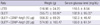
Body weight (g) was significantly higher in OLEFT rats than in LETO rats (555.73 ± 93.92 vs 516.96 ± 24.25). Also, fasting blood glucose concentrations (mg/dL) were significantly higher in the OLEFT rats than in the LETO rats (192.9 ± 95.03 vs 110.5 ± 8.54; P < 0.05; Table 1).
Immunohistochemistry
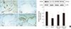
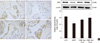
After 4 weeks of COMP-Ang1 injection, immunohistochemistry with the blood vessel endothelial cell marker PECAM-1 and VEGF showed decreased immunoreactivity of PECAM-1 and VEGF in the OLEFT rats (Fig. 1, 2) compared with the LETO rats (Fig. 1A, B). Furthermore, the immunoreactivity of PECAM-1 (Fig. 1C, D) and VEGF (Fig. 2C, D) was increased in rats treated with COMP-Ang1 compared with that in rats treated with vehicle only (Fig. 1B, 2B). Moreover, blood vessels and the expression of PECAM-1 and VEGF were notably augmented in rats treated with 20 µg/kg COMP-Ang1 (Fig. 1D, 2D) compared with that in rats treated with 10 µg/kg COMP-Ang1 (Fig. 1C, 2C).
Western blot
PECAM-1 protein expression decreased significantly in the OLETF group compared with the control LETO group (Fig. 1E). However, this expression increased significantly after intracavernosal injection of COMP-Ang1 (20 µg). VEGF protein expression was also significantly decreased in the OLETF group compared with the control LETO group (Fig. 2E). However, this expression was restored to the level of the control after intracavernosal injection of COMP-Ang1 in a dose-dependent manner.
DISCUSSION
In the present study, we investigated the effect of intracavernosal injection of COMP-Ang1 on angiogenesis in the cavernosal tissue in a diabetes mellitus-related animal model of erectile dysfunction by using OLETF rats (15). At 4 weeks after intracavernosal injection of COMP-Ang1, the immunoreactivity of PECAM-1 and VEGF was decreased in OLEFT rats compared with that in control LETO rats. Somewhat increased expression of PECAM-1 and VEGF was observed in rats treated with COMP-Ang1 compared with the OLEFT rats. Moreover, the expression of PECAM-1 and VEGF in penile tissue was notably augmented in rats treated with 20 µg/kg Comp-Ang1 compared with that in rats treated with 10 µg/kg Comp-Ang1. Western blot analysis showed that PECAM-1 and VEGF protein expression was significantly decreased in OLEFT rat penis compared with that in control LETO rats. However, this expression was restored to the level of the control after the intracavernosal injection of COMP-Ang1 in a dose-dependent manner. These results suggest that intracavernosal injection of COMP-Ang1 enhanced angiogenesis in the corpus cavernosum.
The penis has a specialized vascular bed, and not surprisingly, erectile dysfunction has a predominantly vasculogenic origin. Diabetes is well known to have a powerful effect on the development of both angiopathy and erectile dysfunction. Luttrell et al. (5) used an in vivo animal model of type 2 diabetes and demonstrated the dual impairments that contribute to the diminished erectile function in the type 2 diabetic mouse. They suggested that diabetic mice have a venoocclusive disorder that comes from a lack of tissue filling due to both altered vasoreactivity consistent with impaired cavernosal relaxation as well as impairments in tissue dispensability. The association between diabetes and erectile dysfunction is attributed to the impairment of endothelium-dependent relaxation in the smooth muscle cells of the corpus cavernosum (4, 5). In those studies, the animal model of type 2 diabetes mellitus showed that impairment of endothelium-dependent cavernosal smooth muscle relaxation and endothelial nitric oxide synthase activity induces diabetes mellitus-related erectile dysfunction. Also, decreased expression of VEGF and its receptors in the corpus cavernosum was found in this animal model.
Several current clinical studies have attempted to elucidate the effect of angiogenic factor on neovascularization in the ischemic vascular condition (16, 17). Also, the intracellular signaling pathways that mediate the proangiogenic effects of these angiogenic factors and vascular growth factors, such as VEGF, are being extensively investigated. Ryu et al. (18) reported that down-regulation of the expression of the angiogenic factors and their downstream signal molecules, and decreased endothelial content in the corpus cavernosum, was detected in erectile dysfunction induced by hypercholesterolemia in rats. Local intracavernous delivery of VEGF gene or protein has been shown to recover erectile function in rat and rabbit models of vasculogenic erectile dysfunction induced by traumatized iliac arteries (19) or hyperlipidemia (20), although those studies could not show increased endothelial content after therapy. Concerning the role of VEGF on vascular formation, it was reported that VEGF administration can induce leaky, immature, and unstable vessels (21). In comparison, Ang1, the ligand of the Tie-2 receptor, is an angiogenic factor that plays important roles in the stabilization and maturation of blood vessels in angiogenesis (10). Ang1 also counteracts VEGF-induced inflammation in endothelial cells while having an additive effect on vessel formation (9). Ang1 is a critical angiogenic factor for vascular maturation and enhances VEGF-induced angiogenesis in a complementary manner. Transgenic overexpression of Ang1 produces enlarged vessels with an increased number of endothelial cells, and gene delivery of Ang1 in ischemic tissue induces enlarged blood vessels (9, 22). Ang1-induced vascular remodeling is regarded as vascular enlargement resulting from endothelial cell proliferation (9, 22, 23).
Combined treatment with two angiogenic factors for enhancing therapeutic vascularization and angiogenesis has been tried, and it was shown that administration of Ang1 together with VEGF strongly promotes revascularization in ischemic animal models. Ryu et al. (24) reported that combined Ang1 and VEGF gene transfer promotes angiogenesis cooperatively in a rat model of hypercholesterolemic erectile dysfunction and results in the recovery of erectile function. Cho et al. (13) developed a soluble, stable, and more potent Ang1 variant, COMP-Ang1. They replaced the amino-terminal portion of Ang1 with the short coil-coil domain of cartilage oligomeric matrix protein. COMP-Ang1 is more potent than naïve Ang1, and COMP-ang1-induced vascular remodeling is mediated mainly through Tie2 receptor activation (13). In their consecutive study, they also found that sustained treatment with COMP-Ang1 can produce long-lasting vascular enlargement resulting from endothelial cell proliferation and an increase in blood flow in the trachea (12).
Until now, study of the expression of Comp-Ang1 and endothelial content in diabetes mellitus-induced erectile dysfunction has been lacking. Cho et al. (25) revealed that systemic treatment with COMP-Ang1 resulted in accelerated wound closure, enhanced angiogenesis and lymphangiogenesis, and higher blood flow in the wound region in the diabetic mouse model and indicated that COMP-Ang1 can promote wound healing in diabetes through enhanced blood flow and angiogenesis as measured by PECAM-1 expression. Jin et al. (3) also determined the effectiveness of intracavernosal injection of COMP-Ang1 in promoting angiogenesis and erectile function in a mouse model of type 2 diabetic erectile dysfunction. They reported that local delivery of COMP-Ang1 protein significantly increased eNOS activity and cGMP and cAMP expression. Furthermore, repeated intracavernous injections of COMP-Ang1 protein completely restored erectile function and cavernous endothelial content through enhanced cavernous neoangiogenesis with the increased feature of PECAM-1 expression. In the present study, PECAM-1 and VEGF protein expression was significantly decreased in the OLEFT group and tended to increase to the level of the control after the intracavernosal injection of COMP-Ang1 in a dose-dependent manner. These results suggest that efficient recovery of erectile function could be expected in the penile tissue of diabetic erectile dysfunction rats treated with COMP-Ang1 as a result of the enhanced angiogenesis in the corpus cavernosal region. Our results also suggest that the efficacy of COMP-Ang1 treatment may act in a dose-dependent manner. The promising results gained from this animal model of type 2 diabetes shed light on the application of therapeutic angiogenesis in curing human erectile dysfunction with a vascular origin.
This study had a limitation. We did not conduct a functional study to differentiate the functional changes in the erectile tissue of each experimental group before and after the COMP-Ang1 treatment, such as measuring penile blood flow and intracavernosal pressure. Further functional study is needed to confirm the effect of COMP-Ang1 on the treatment of erectile dysfunction in the diabetic rat model.
In conclusion, local administration of COMP-Ang1 in the corpus cavernosum enhanced angiogenesis in diabetic rats, and these effects of COMP-Ang1 were dose-dependent. These results suggest that penile angiogenic factors may improve endothelial function in diabetic erectile dysfunction. Further functional study is needed to confirm the effect of COMP-Ang1 on the treatment of erectile dysfunction in the diabetic rat model.
 XML Download