

 PDF
PDF ePub
ePub Citation
 Print
Print


INTRODUCTION
Osteogenesis imperfecta (OI) comprises a heterogeneous group of disorders characterized bone fragility, deformity of the spine and long bones, and other clinical symptoms such as short stature, blue sclera, and dentinogenesis imperfecta (1, 2). Almost all patients have presumed or proven defects in type I (pro)collagen biosynthesis (3). In 1979, David Sillence developed a four-type classification that is still in use for classifying clinical/radiological features: OI type I (mild OI with bone fragility and blue sclera), II (perinatal lethal), III (progressive deforming), and IV (normal sclera and mild deformity) (4, 5). Dominantly inherited mutations of COL1A1 or COL1A2, which encode collagen type I alpha chains, appear to be causative in the majority of OI types.
In 2004, OI types V and VI were added to this classification because of specific clinical/radiological or typical histological features, absence of abnormalities of type I (pro)collagen synthesis, and absence of causative variants COL1A1 or COL1A2 (6-8). With the discovery of the rare recessively inherited mutations of OI, an extension of the classification was proposed to include OI types VII and VIII (7). However, the classification and subdivision into different types of OI is still under discussion (3) because the phenotypic spectrum that results from mutations in some of these genes is almost as broad as that seen for mutations in type I collagen genes.
Recessively inherited OI genes have been reported, including CRTAP, LEPRE1, PPIB, FKBP10, PLOD2, SERPINF1, SERPINH1, BMP1, LRP5, and SP7. Recent research has demonstrated that OI type VI is caused by loss-of-function mutations in SERPINF1 (9). An earlier report had found inactivating mutations of SERPINF1 in severe OI, but the lack of bone histology prevented establishment of a link to OI type VI (10).
In the present study, we describe the clinical, radiological, and molecular findings of a Korean patient who presented with a novel compound heterozygous mutation of SERPINF1. The patient showed an initially mild and then progressively worsening form of OI, with severe deformities of the long bones.
CASE DESCRIPTION
Patient summary
The patient was born after 40 weeks of gestation by spontaneous vaginal delivery. Her birth height was 50 cm (50th percentile), and her birth weight was 3,400 g (50th percentile). No limb deformities or other abnormalities, including joint hyperlaxity or skin hyperelasticity, were noted at birth and both scleras were white. The patient's parents were non-consanguineous and healthy; her only sibling, an older sister, was unaffected. No family history of bone fragility was identified. She was able to sit independently at 9 months and walk independently at 16 months.
At 18 months (2005-05-02), she presented with a nondisplaced fracture in the right proximal femur diaphysis without any traumatic episode (Fig. 1A). At that time, her height was 78.6 cm (10-25th percentile), and her weight was 9.5 kg (3rd percentile). The data from biological tests (i.e., routine blood cell count, blood and urinary levels of calcium, phosphate, creatinine, serum alkaline phosphatase, 25-hydroxyvitamin D, parathyroid hormone, and urine analysis) appeared within normal ranges, while the serum level of the bone resorption marker type I collagen telopeptide was slightly above the normal values. After the first fracture, she frequently experienced nontraumatic fractures. No radiographic signs of rickets were observed. The history of the frequent fractures without apparent injury suggested bone fragility and the radiologic features, including osteopenia, enabled us to diagnose OI. Dual-energy x-ray absorptiometry showed the values of the lumbar vertebrae L1- to-L4 to be very low (0.204 g/cm2, T score -7.0) at age 2 yr 10 months. She had received monthly intravenous pamidronate (20 mg per m2 of body surface area) beginning at the age of 3 yr 1 month, because she had had frequent episodes of fractures. She showed no side effects; however, her growth remained very slow and she continued to have one or more long-bone fractures annually. After 12 cycles of pamidronate, the L1- to-L4 spine areal bone mineral density T-score was -6.1 (0.277 g/cm2).
At the time of her most recent evaluation, she was 8 yr old and had been receiving monthly intravenous pamidronate due to intermittent fractures. Growth was severely restricted; therefore, her height at the time of this writing was markedly less than the 3rd percentile (about 100 cm). The girl could not walk independently due to frequent lower-extremity fractures, resulting in severe deformity. She was only able to stand with assistance, and was dependent upon a wheelchair for mobility. No clinical signs were seen of hearing impairment, ophthalmological problems, cardiac murmur, respiratory difficulty, neurologic problems, or dentinogenesis imperfecta. Her intellectual development was normal.
She experienced more than ten fractures in her lifetime until 8 yr of age; six of these fractures required orthopedic surgical procedures. She had severe scoliosis (Fig. 1B), bilateral coxa vara, acetabular protrusion, severe bowing of all long bones, and "popcorn epiphyses" at the age of 8 yr (Fig. 1C).
The heterogeneity of OI hampers genetic diagnosis, particularly in autosomal recessive forms of OI. Given that many genes are involved in OI, and that the COL1A1 and COL1A2 genes are difficult to analyze by conventional Sanger sequencing, we performed whole exome sequencing (WES) to identify the mutations in our patient. Blood samples were collected from the patient and her parents after informed consent had been obtained.
Mutations in SERPINF1
Detailed methods of exome sequencing and validation of novel variants by Sanger sequencing are shown in the supplementary data. A mean coverage of 100.5X was achieved and 89.2% of the targeted bases were read >10 times by exome capture and massive parallel sequencing (Supplementary Table 1). We checked the OI-related genes, including COL1A1, COL1A2, CRTAP, P3H1, FKBP10, LEPRE1, PLOD2, PPIB, SERPINH1, and SP7, covering all exons and their flanking intronic regions, but found no disease-associated mutations in these genes.
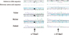
We ultimately focused on two deleterious variants, c.77dupC and c.421dupC, of SERPINF1 as the best candidates for further evaluation. Detailed view of individual sequencing reads is shown in Supplementary Fig. 1. They were validated by Sanger sequencing in the patient and her family members. We identified compound heterozygous insertions in exon 2 (c.77dupC; p.Glu27Glyfs*38) and in exon 4 (c.421dupC; p.Arg141Profs*5) of SERPINF1 in the patient (Fig. 2). The father and mother of the patient were confirmed to be heterozygous carriers of each variant. These variants were not found in 192 control chromosomes.
DISCUSSION
To date, only eight SERPINF1 mutations have been reported and are homozygous nonsense or frameshift mutations or in-frame duplications. We report the first compound heterozygous mutations (c.77dupC and c.421dupC) in the SERPINF1 gene that are associated with OI.
Patients with OI type VI usually do not have fractures or bone deformity at birth, but typically sustain their first fracture after the age of 6 months. Compared to other types of OI, bisphosphonate treatment appears to be less effective at reducing fracture rates in OI type VI (11). This type of OI type differs from other types in that a mineralization defect occurs in bone tissue, which leads to the accumulation of unmineralized osteoids, such as those seen in osteomalacia (8). These characteristic features of OI type VI are readily revealed by bone histology, but OI type VI has no distinguishing radiological signs and the parameters of calcium and phosphorus metabolism are within normal limits. However, because bone biopsy is invasive, we relied on genetic analysis to confirm our diagnosis.
Our patient appeared healthy at birth and did not suffer any fracture until 18 months of age. The absence of dentinogenesis imperfecta, lack of blue sclera, severe progression of the disease, frequent fractures without adequate trauma, markedly short stature, and inability to walk without assistance were symptoms similar to those recently reported for having homozygous mutations in SERPINF1 (9, 10, 12). We concluded that our patient has OI type VI, considering her normal condition at birth, frequent fracture history after 18 months of age, poor response to pamidronate treatment, and severe bony deformities.
SERPINF1 encodes PEDF (pigment epithelium-derived factor), a 50kDa secreted glycoprotein of 418 amino acid residues that shows high affinity for collagens of the extracellular matrix as well as multiple and varied biological properties (13-15). The SERPINF1 mutants described so far all display normal synthesis, posttranslational modification, and secretion of type I collagen (12). PEDF is believed to play a role in bone homeostasis as an inhibitor of bone resorption, since it upregulates osteoprotegerin, which inhibits osteoclast maturation (16). Further studies for a role of PEDF in the pathogenesis of OI type VI should be conducted.
In summary, we report novel compound heterozygous SERPINF1 mutations in a Korean patient who showed an initially mild and then progressively worsening form OI, with severe deformities of the long bones. Further investigations in other patients with SERPINF1 mutations are needed. The present study aids in further definition of the peculiar clinical features of this form of OI.

 XML Download