

 PDF
PDF ePub
ePub Citation
 Print
Print


INTRODUCTION
Hypoplasia is an elongated, tubular, homogeneous stenosis of a part of artery. Since the definition of hypoplasia is based on the macroscopic appearance of an affected vessel, it should be differentially diagnosed by histopathological characteristics. Fibromuscular dysplasia (FMD) is an uncommon, non-inflammatory, non-atherosclerotic arterial disease that can be associated with spontaneous dissection, aneurysm, or severe stenosis (1, 2). FMD most commonly involves the renal artery and internal carotid artery, less commonly the vertebral artery (VA). Typically, FMD appears 'string of beads' in angiography with dissection or aneurysm and fibrous or fibromuscular thickening in the tunica media. In this report, we describe a rare case of hypoplasia in intracranial VA associated with intimal type FMD in its extracranial and intracranial courses.
CASE DESCRIPTION
During a routine dissection at Jeju National University Medical School in 2011, a case of hypoplasia in left vertebral artery was observed in an 82-yr-old Korean woman cadaver, whose cause of death was 'unknown'. Hypoplastic VA was defined as a lumen diameter less than 2 mm in an anatomical study (3) or as with diameter less than 50% of the contralateral side in radiological study (4).
In this case, each VA originated from the subclavian artery with an external diameter of left 4.6 mm and right 5.0 mm; and entered to the foramen transversarium with a diameter of left 3.3 mm and right 5.0 mm (Fig. 1A). The extracranial parts of both VAs did not show macroscopic anomaly, but intracranial part of the left VA had hypoplasia with a total length of 21 mm (Fig. 1B). The external diameters of the intracranial parts of VAs were found to be 3.7 mm on the right side and 1.0 mm on the left side. The vertebrobasilar junction was found to be at the level of bulbopontine sulcus. The posterior inferior cerebellar artery bilaterally arose from the basilar artery 2.5 mm from the vertebrobasilar junction. The other brain vessels had no anomalies in their course or in the origin of their branches on either side.
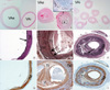
Histopathologically, FMD is heterogeneous with various degrees of collagen hyperplasia, internal elastic lamina rupture, and disorganization of the tunica media (1, 2). For histopathological classification of FMD, tissue sections were stained with hematoxylin and eosin (Fig. 2A-C), Orcein for elastic fiber (Fig. 2D-F), and immunohistochemistry with anti smooth muscle actin antibody for tunica media (Fig. 2G-I) and anti-CD31 antibody for endothelium (Fig. 2B). Compared to the right VA (Fig. 2A, D, G), both extracranial and intracranial left VA pathologically diagnosed as an intimal type of FMD. Left VA has long smooth narrowing of lumen with circumferential or eccentric deposition of collagen in the tunica intima and disrupted internal elastic lamina (Fig. 2E). The stenotic lumen was not caused by hypertrophy in medial smooth muscles. In extracranial VA, neovascularization (rectangle in Fig. 2B) in the hypertrophied intimal layer was seen with immunopositivities of smooth muscles (Fig. 2H). But the immunoreactivity with smooth muscles was not obvious in intracranial VA as compared to the extracranial VA (Fig. 2I).
DISCUSSION
We encountered hypoplasia in left VA associated with intimal type FMD during routine dissection course. The intracranial parts of the VAs, the basilar artery and their branches together form the vertebrobasilar system (VBS). Numerous anomalies can be seen in the VBS and also observed in this study: the posterior inferior cerebellar artery originated from the basilar artery, but this variation is not uncommon (22.5%) (5). Moreover, the rate of hypoplasia in VA shows increasing frequency due to the radiological development.
In an anatomical study (3), it was reported that there is usually a difference between the external diameter of left (3.02 ± 0.81 mm) and right (2.85 ± 0.99 mm) intracranial VA. The hypoplasia of VA was observed at rates of 20.2% on the right, 14.4% on the left and 4.8% bilaterally. In radiological study (4), the unilateral hypoplasia and/or aplasia of VA has been frequently reported in Koreans (about 27%, right > left) without the vertebrobasilar insufficiency symptoms. Hypoplasia and aplasia of extracranial or intracranial VA is classified as follows (4); type I denotes hypoplasia showing extracranial and/or intracranial VA, type II represents hypoplasia having any invisible part, and type III denotes aplasia both extracranial and intracranial VA. According to the radiological classification, our report correspond to type Ia of hypoplasia in intracranial VA based on macroscopic results, and type Ic of hypoplasia in extracranial and intracranial VA based on microscopic findings.
Due to a collagen deposition within the tunica intima, intimal type FMD often results in a concentric stenosis or long tubular lesion (1). It is coincided with the angiographic classification of unifocal FMD: tubular (> 1 cm) or focal (< 1 cm in length). These unifocal subtypes seem to be more commonly related to intimal type FMD of this report. Surprisingly, histopathologically proven involvement of the VA has not been reported to our knowledge. A few histologically confirmed cases are reported from intracranial vessels, but Kimura et al. (6) only confirmed the medial hyperplasia type of FMD associated with aneurysm of the VA and posterior inferior cerebellar artery. Both hypoplasia and FMD in same vessel was only reported from abdominal aorta (7), but there was medial hyperplasia type of FMD associated with aneurysm. This is the first report consists of hypoplasia and intimal type FMD of the VA.
Luminal narrowing in hypoplasia of VA may produce ischemic stroke both by the hemodynamic effects of atherosclerotic stenosis, thromboembolism, or FMD, since FMD of the internal carotid artery is related with focal cerebral ischemia by atherosclerotic stenosis or thromobotic occlusion (8). Ipsilateral hypoplasia of VA has a close relation to posterior inferior cerebellar artery infarction or lateral medullary infarction (3, 9), and the diagnosis of FMD is a rare case. Furthermore, patients with posterior circulation strokes showed a higher rate of hypoplasia of VA than those with anterior circulation strokes which had similar frequency compared to that of normal (3). The frequency of FMD in patients with ischemic stroke, transient ischemic attack, or subarachnoid hemorrhage is likely to be very small, since the prevalence of FMD ranged from 0.3% to 3.2% (2). VA affected 7% to 19% of patients with FMD, which is less common than renal artery or internal carotid artery involvement (1). However, the prevalence in the general population is not known. A report (10) performed at the Mayo Clinic only suggests presumptive evidence that FMD of the internal carotid artery was detected in only 0.02% of 20244 consecutive autopsies. Therefore, intracranial FMD of the VA is indeed rare than extracranial FMD.
Although the etiology is unknown, FMD can be inherited and intimal type FMD may result from arterial injuries (2). It should also be remembered that deficiency of protein S is associated with progressive intracranial occlusive disease of VA (11). Intracranial FMD of the VA seems to be extremely rare. But the rate of VA involvement in vertebrobasilar insufficiency was up to 26% (12). Intracranial FMD corresponds to an intracranial extension of extracranial FMD (2), and it is also true in this report that hypoplasia is only seen in intracranial VA while FMD observed in both intracranial and extracranial VA.
Taken together, signs and symptoms of vertebrobasilar insufficiency should be carefully evaluated since hypoplasia of VA is not uncommon even in normal adults.

 XML Download