

 PDF
PDF ePub
ePub Citation
 Print
Print


A myocardial ischemia/reperfusion (I/R) injury is a complex process involving the generation and release of reactive oxygen species and inflammatory cytokines (1). An urgent restoration of coronary blood flow to the ischemic area is the most effective strategy to improve outcomes of myocardial I/R injury. Upon resuming the perfusion, reperfusion injury salvage kinases (RISK) survival signaling pathways such as phosphatidylinositol-3-OH kinases (PI3K)-Akt, extra-cellular signal-regulated kinases (ERK1/2), and their downstream target glycogen synthase kinase (GSK)-3β may be activated to confer tissue protection (2). However, at the same time, the reperfusion may also activate 'stress-responsive' mitogen-activated protein kinase (MAPK) subfamilies (socalled cell death signaling pathway), i.e., c-Jun N-terminal kinases (JNK) and p38 MAPKs, resulting in necrosis (3). Therefore, the degree of I/R injury may be determined by a relative extent of death and survival kinases activation (4).
Sauchinone is a biologically active lignan isolated from Saururus chinensis which has been long used in folk medicine for the treatment of edema, jaundice, and several inflammatory diseases (5). It has been known to inhibit apoptotic death of C6 glioma cells induced by staurosporine (6) and to reduce ischemia-induced neuronal death via suppression of intracellular radical production (7). We have previously shown that sauchinone inhibits lipopolysaccaride-induced tumor necrosis factor-α (TNF-α) expression in macrophages via suppression of NF-κB activation (8).
The present study aimed at examining whether sauchinone would provide a protection against regional myocardial I/R injury. The expression of molecules related with cell survival such as Akt and ERK1/2/GSK-3β as well as that related with cell death such as JNK and p38 MAPKs was determined.
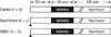
The procedures were carried out after the approval by the institutional animal care and use committee, Research Institute of Medical Science at Chonnam National University Medical School (Gwangju, Korea). Male Sprague-Dawley rats weighing 250-350 g were used. They were housed in a vivarium maintained at 20℃-23℃ with 12-hr light/dark cycle and given food and water ad libitum. On the experimental day, the rats were anesthetized with phenobarbital (100 mg/kg, intraperitoneal), and mechanically ventilated with an air-oxygen mixture. The polyethylene catheter was cannulated into the common carotid artery for the arterial blood pressure monitoring and into the internal jugular vein for drug administration (9). Core body temperature was monitored with a rectal temperature probe and maintained at 37.5℃-38.5℃ using heating pad. Left thoracotomy was performed at the fifth intercostal space, and 6-0 silk ligature was placed around left anterior descending coronary artery (LAD). After stabilization period of 20 min, the LAD was occluded for 20 min and then reperfused for 2 hr. LAD occlusion was confirmed by ST segment changes in electrocardiogram and the presence of regional cyanosis in the myocardial surface. There were three I/R groups as shown in Fig. 1: 1) control group, 2) sauchinone group which received sauchinone (10 mg/kg, dissolved in dimethyl subfoxide [DMSO]), and 3) DMSO group which received vehicle only. Sauchinone was isolated from the n-hexane fraction of Saururus chinensis by successive silica gel chromatography and reverse-phase high-pressure liquid chromatography, and was > 97% pure (10). The dosage of sauchinone adopted was effective to increase survival rates and to decrease the plasma level of TNF-α in mice administered lipopolysaccharide/D-galactosamine (10). Sauchinone and DMSO were administered intraperitoneally 30 min before the onset of ischemia. At the end of 2 hr reperfusion, the heart was quickly excised and mounted on a Langendorff apparatus. The LAD was reoccluded, and then fluorescent polymer microspheres were infused into the aorta to demarcate the risk zone as the tissue without fluorescence. The heart was sliced into 2 mm transverse sections, which were incubated at 37℃ for 20 min in triphenyl tetrazolium chloride in sodium phosphate buffer. The slices were immersed in 10% formalin to enhance the contrast between stained (viable) and unstained (necrotic) tissue and then squeezed between glass plates spaced exactly 2 mm apart. The myocardium at risk was identified by illuminating the slices with ultraviolet light. The infracted and risk zone regions were traced on a clear acetate sheet and quantified with Image Tool (San Antonio, TX, USA). The areas were converted into volumes by multiplying the areas by slice thickness. Infarct size is expressed as a percentage of the risk zone (11).
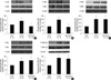
For immunoblotting, the heart was excised 5 min after reperfusion, and the risk area in the left ventricle was separated in each group. Myocardial samples were homogenized and equivalent amounts of proteins were loaded to 10% Tris-HCl SDS polyacrylamide gel. Proteins were electrotransferred to a polyvinylidene difluoride membrane. The membrane was then incubated overnight at 4℃ with rat polyclonal specific primary antibodies to phosphorylated p38, JNK, Akt, ERK1/2 and GSK-3β (Ser9) (Cell Signaling Technology; Danvers, MA, USA). It was subsequently incubated with anti-rabbit or anti-rat immunoglobulin HRP-coupled secondary antibodies. Immunoreactive bands were visualized using ECL Western blotting detection reagents. The membranes were then stripped using a stripping buffer, and reprobed with antibodies specific for total p38, JNK, Akt, ERK1/2, GSK-3β and β-actin (Cell Signaling Technology). Data were expressed as means ± SD. Differences in infarct size and immunoblot data between groups were tested by one-way analysis of variance (ANOVA). A Scheffé test was used for multiple pair-wise comparisons when a significant difference was indicated with ANOVA. P < 0.05 was considered to be statistically significant.
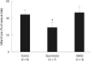
The heart rate and arterial pressure did not significantly differ among the groups at any time point (data not shown). Nor did the ratio of the area at risk to left ventricular mass differ among the groups. Sauchinone significantly reduced the infarct size (Fig. 2). Sauchinone significantly attenuated the degree of phosphorylation of p38 and JNK (P < 0.05), without affecting that of ERK1/2, Akt and GSK-3β (Fig. 3).
Our study demonstrated that sauchinone has a novel protective effect against regional myocardial I/R injury in an open-chest rodent model. The protective effect was associated with attenuated phosphorylation of cell death signaling pathways (p38 and JNK), without effects on that of RISK pathways (ERK1/2, Akt) and GSK-3β. Several recent studies have suggested that activation of p38 MAPK and JNK, known enhancers of the expression of adhesion molecules and the production of cytokines (12), plays a major role in cell death following I/R injury. The concept has been supported by the observations in which an inhibition of p38 activation reduced the caspase-3 activity and protected cardiac myocytes following I/R insult in vitro (13), and improved postischemic cardiac function in vivo (14). It was also shown that an inhibition of JNK decreased ischemia-induced necrosis and apoptosis in myocardial I/R injury (15, 16). However, an activation of p38 MAPK or JNK has been also shown to play a protective role in I/R injury (17, 18). The distinction of beneficial versus detrimental role of p38 and/or JNK signaling remains to be further addressed.
On the other hand, the extent of phosphorylation of Akt, ERK and GSK-3β induced by I/R was not affected by sauchinone in the present study, suggesting that RISK/GSK-3β pathways were not involved. The disturbance of ion homeostasis resulting from ATP depletion following the opening of the mitochondrial permeability transition pore (mPTP) can lead to necrotic and apoptotic cell death (19). Both ischemic/pharmacological pre- and post-conditioning are generally perceived to protect the heart against I/R injury via activation of the RISK pathway involving PI3K-Akt and ERK1/2 at the time of reperfusion (2). Its activation may prevent cellular damages by converging on the mPTP through GSK-3β, which in turn inhibits its opening (19). Sauchinone (EC50 = 1 µM) has been shown to prevent mitochondrial dysfunction and apoptosis induced by iron plus arachidonic acid in the hepatocyte, possibly through inhibition of reactive oxygen species production (20). Further studies defining the role of mPTP in sauchinone-induced myocardial protection against I/R injury may be needed.
In summary, the present study demonstrates a cardioprotective effect of sauchinone against regional I/R injury in the rat, which may be related to an attenuation of cell death signaling pathways involving p38 and JNK but not to activation of RISK/GSK-3β pathways.
 XML Download