

 PDF
PDF ePub
ePub Citation
 Print
Print


INTRODUCTION
Primary peritonitis constitutes less than 1% of peritonitis and spontaneous bacterial peritonitis usually occurs in patients with comorbid conditions. In absence of comorbid conditions, including liver cirrhosis, immunosuppression, or nephrotic syndrome, primary peritonitis is rare, particularly in cases of healthy young subjects. Primary peritonitis due to S. pyogenes is an unusual condition because Streptococcus pyogenes usually causes pharyngitis, erysipelas, and necrotizing fasciitis. In this report, we present the first case to the best of our knowledge of primary peritonitis due to S. pyogenes in Korea.
CASE DESCRIPTION
29-yr-old woman presented to the emergency room with lower abdominal pain on April 20, 2011. She had no travel history and had not taken any medication. On physical examination, she was alert and cooperative. Her body temperature was 38℃, blood pressure 80/60 mmHg, pulse 113 beats per minute, respiratory rate 20 breaths per minute, and oxygen saturation 99% while breathing ambient air. There was generalized abdominal tenderness with guarding and rebound tenderness. Rectal examination was not specific. Auscultation of the lungs revealed clear breathing sounds. A cardiac examination revealed rapid but regular heart sounds and no murmurs. There was no pretibial pitting edema. The neurologic examination was normal and no skin lesions were detected.
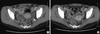
Results of gynecological examination from gynecologists were also normal and no history of abnormal vaginal discharge was known. Her last menstruation was 3 weeks ago and she was sexually active. Initial blood tests showed leukocyte count of 6,248/µL, platelet count of 133,000/µL, C-reactive protein (CRP) level of 12.36 mg/dL, and procalcitonin level of 36.9 ng/mL. Renal and liver function tests, clotting screen, and arterial blood gas were all within normal range. Urinary tests were normal with no microorganisms detected in the urine. Chest radiography showed unremarkable findings with normal results of sputum analysis. Abdominal CT revealed edematous swelling of the intestinal wall and ascites with peritoneal enhancement suggesting peritonitis (Fig. 1A).
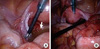
As physical, laboratory, and radiologic findings suggested acute peritonitis, laparoscopy searching for etiology was performed. Purulent ascites was found in the pelvic cavity but both ovaries and fallopian tubes were intact (Fig. 2). There were no intra-abdominal abnormalities such as bowel perforation, appendicitis, or necrosis. Reports of blood culture, ascites culture, and cervical swab culture showed gram-positive cocci. Final results of all cultures tested confirmed S. pyogenes. Initial empiric antibiotics (ampicillin/sulbactam 2 g/1 g every 6 hr) switched to penicillin G (300 MU every 4 hr) and metronidazole (500 mg every 8 hr). Subsequent abdominal CT showed improving peritonitis (Fig. 1B). She was discharged on day 16 without sequelae.
DISCUSSION
Streptococcus pyogenes is a usual cause of pharyngitis, erysipelas, and necrotizing fasciitis. Primary peritonitis caused by Streptococcus pyogenes is uncommon and rarely diagnosed in a healthy person without underlying diseases. Spontaneous bacterial peritonitis due to Streptococcus pyogenes in a cirrhotic child was reported in Korea (1) but S. pyogenes peritonitis in a healthy person has not yet been reported in Korea. Therefore, this is the first case of primary peritonitis due to Streptococcus pyogenes in a healthy person in Korea. It is reported that most instances of spontaneous primary peritonitis are due to Streptococcus pneumoniae (2). There are a few cases describing S. pyogenes peritonitis in healthy women, even though the entry site of S. pyogenes in peritonitis is not uncertain. Moskovitz et al. (3) suggested that in some women, S. pyogenes in peritonitis may be via the genital tract, despite lack of gynecological symptoms even though many studies have shown an absence of S. pyogenes as normal flora of the female genital tract. The hematogenous route may be an alternate, possibly from pharyngeal or cutaneous primary sites (3). In this case, ascending genital infections was considered to be the entry site because of the positive cervical swab culture.
The majority of primary peritonitis is diagnosed retrospectively when secondary causes are excluded after surgical approach (4). Preference for laparoscopy or laparotomy is established by the surgeon's choice and laparotomy is predominantly performed in previous cases. However, Farooq and Ammori (5) claimed that laparoscopy could be used as a diagnostic tool in the management of generalized peritonitis. We chose laparoscopy as a diagnostic tool in this case because no significant abnormalities suggesting secondary peritonitis were found on abdominal CT.
The severe infection of S. pyogenes requires a high index of suspicion, prompt diagnosis, and rapid initiation of appropriate antibiotics (6). In this case, reports of blood culture, abdominal ascites culture, and cervical swab culture showed gram-positive cocci, so ampicillin/sulbactam was applied. After final result of culture confirmed S. pyogenes, antibiotics were switched to penicillin G and metronidazole. It is reported that some S. pyogenes serotypes are more commonly associated with invasive group A streptococcal disease than other GAS isolates (7-9). We did not obtain the serotype of S. pyogenes, so further study on S. pyogenes serotypes in primary peritonitis is required.
In summary, we report the first case of primary peritonitis in Korea in a young, healthy woman due to S. pyogenes. The port of entry was thought to be via the genital tract, despite no local symptoms. Laparoscopy as a diagnostic tool was performed in this case and we recommend laparoscopy over laparotomy when no significant abnormalities suggesting secondary peritonitis are seen on abdomen computed tomography. An appropriate diagnostic approach and prompt antibiotic therapy is essential in primary GAS peritonitis.
 XML Download