

 PDF
PDF ePub
ePub Citation
 Print
Print


INTRODUCTION
During the last five decades, long-term therapy with immunosuppressive agents such as pulse cyclophosphamide in conjunction with high-dose corticosteroids has enhanced both patient survival and renal survival in patients with diffuse proliferative lupus nephritis (DPLN) (1-4). However, the beneficial effect of cyclophosphamide must be weighed against its considerable side effects, such as severe infectious complications. Infections are an important cause of treatment-related morbidity and mortality in systemic lupus erythematosus (SLE). It is estimated that at least 50% of them will suffer a severe infectious episode during the course of the disease (5). A broad spectrum of infections have been reported in SLE, including bacterial, mycobacterial, viral, fungal and parasitic infections (6). Although fungal infections account for a significant number of infections (6-8), aspergillosis, especially invasive aspergillosis, has seldom been described (9). Moreover, central nervous system (CNS) aspergillosis is extremely rare and no case of aspergillus cerebral aneurysm has been reported.
We describe here the first case of internal carotid artery (ICA) mycotic aneurysm due to aspergillosis in an immunocompromised DPLN patient who was successfully treated with combination of antifungal therapy, graft stent insertion and aneurysmal neck clipping.
CASE DESCRIPTION
A 46-yr-old woman was referred to our hospital for generalized edema. Her edema had started one week previously after a one-week history of mild febrile sense, headache and cough. The laboratory test on admission showed anemia (hemoglobin 9.1 g/dL, hematocrit 26.8%), thrombocytopenia 98 × 103/µL) with acute renal failure (blood urea nitrogen 31 mg/dL, creatinine 1.5 mg/dL) and her urinalysis showed moderate hematuria and proteinuria (4.6 g/day). Her serologic tests were abnormal with high ANA titer of 1:160, low C3 of 35 mg/dL (normal 70-150 mg/dL) and C4 of 6 mg/dL (normal 10-35 mg/dL), and elevated anti-dsDNA autoantibody titer of 146 IU/mL (normal < 10 IU/mL). She had a kidney biopsy and was diagnosed with DPLN (ISN/RPS Class IV-G(A)).
In the absence of any evidence of infection, she received two courses of steroid pulse therapy (1 g of methylprednisolone) followed by oral high dose steroid (1 mg/kg of prednisolone). Before receiving the third course of steroid pulse therapy, her chest radiograph showed a 4 cm-sized round opacity in her right upper lung field, although she had no respiratory symptoms. Computed tomography (CT) scan of the chest disclosed a mass with central necrosis surrounded by a halo of ground-glass attenuation. These images were highly suggestive of invasive pulmonary aspergillosis. Therefore, she underwent a percutaneous needle biopsy for the lesion, but no microorganisms were revealed by histological examinations including Grocott's methenamine silver staining, acid-fast staining, periodic acid-Schiff (Ed- the acronyms GMS and PAS are unnecessary as they are not used anywhere else in the paper) staining and Gram staining of the lung tissue specimen.
By clinical impression, the patient commenced a course of amphotericin B due to the possibility of other fungal infections, such as mucormycosis. After one month, the lesion was cleared up in the chest CT, amphotericin B was stopped, and chest radiograph did not show any lung lesion suggestive of active infection such as fungal infection. Subsequently, she received nine courses of cyclophosphamide pulse therapy (monthly 6 times and quarterly 3 times with total dose of 5.75 g) over 15 months. Oral steroid was tapered down from daily 5 mg/kg to daily 5 mg. At this time, her urinalysis showed no hematuria and proteinuria was around 100 mg/day with normal kidney function (0.9 mg/dL), indicating remission of DPLN.
While waiting for the next (10th) cyclophosphamide pulse therapy, the patient suffered two episodes of massive epistaxis and magnetic resonance imaging (MRI) of the paranasal sinuses (PNS) showed both maxillary sinusitis and a 1 cm-sized, strongly enhanced lesion in the left sphenoid sinus with focal bony defect in the posterolateral aspect of left sphenoid sinus, suggesting aneurysm of the left cavernous ICA.
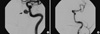
The patient was transferred to the interventional neuroradiology unit, where transfemoral cerebral angiography revealed one aneurysm in the cavernous segment of the left carotid artery (Fig. 1A). A graft stent was deployed in the cavernous ICA for parent vessel reconstruction, and there was no filling of contrast media into the aneurysm at delayed angiography (Fig. 1B). Endosinus sphenoidectomy was performed to remove the suspected infection source and to identify the pathogen. Histological examination of the sinus mucosa biopsy specimen revealed fungal hyphae that were morphologically consistent with Aspergillus, although the culture study for sinus secretion and repeated serum Aspergillus galactomannan antigen assay were negative. The patient underwent PNS MRI every two months while receiving voriconazole therapy. Since she was still on remission of DPLN with a minimal maintenance dose of steroid (daily 5 mg) and in the absence of any evidence of infection such as paranasal sinusitis and cerebral mycotic aneurysms on follow up MRI images, voriconazole was stopped seven months later.
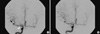
Three months after stopping anti-fungal therapy, one small aneurysm was suspected at the right middle cerebral artery bifurcation on routine follow up MRI images. Transfemoral cerebral angiography revealed two aneurysms: one at the right middle cerebral artery bifurcation, and one at the anterior choroidal artery that was not seen on MRI (Fig. 2A). Aneurismal neck clipping of the affected artery was done by a neurosurgeon (Fig. 2B).
Chest CT and brain MRI images were performed regularly afterwards and up to one-year follow-up there was no evidence of further infections such as cerebral aneurysms, sinusitis and pulmonary nodules.
DISCUSSION
We report here the first case of aspergillus-related cerebral aneurysm in a lupus patient. Maria et al. (9) first reported 23 cases of invasive aspergillosis in SLE, but there was no CNS infection. CNS aspergillosis in SLE patients is extremely rare. Uppin et al. (10) reported two cases of CNS aspergillosis, in the form of arteritis and thrombosis with bland infarct. One patients had disseminated aspergillosis in the lungs, kidneys and brain, while the other was immunocompetent and had sphenoidal sinusitis, as in our case. Both patients were diagnosed at autopsy, despite extensive imaging and laboratory studies. Lammens et al. (11) reported an immunocompromised lupus patient with persistent neutrophilic meningitis which was found at autopsy to be due to aspergillosis. Sugawara et al. (12) reported a DPLN patient with multiple brain abscess who was treated successfully by craniectomy and intraventricular antifungal agents.
While vascular invasion by Aspergillus species is common, true mycotic aneurysm formation due to Aspergillus species is rare (13). Mycotic cerebral aneurysm results from direct invasion of the vessel wall, either from the luminal or adventitial side of the vessel. Endovascular infection leads to initiation of infection from the luminal surface whereas infection from the adventitial aspect usually occurs in the context of an infectious process surrounding the vessel such as in meningitis. Progressive invasion of the vascular wall leads to a loss of a vascular integrity and aneurysm formation (14). In the current case, mycotic ICA aneurysm was attributed to direct invasion of the sphenoidal fungal sinusitis. It could be diagnosed as CNS aspergillosis since proven invasive aspergillosis at a contiguous site such as sinuses or orbit is direct evidence to implicate Aspergillus species (15).
Most infection-associated aneurysms are caused by bacterial pathogens, usually secondary to endocarditis. Fungal aneurysms, especially involving carotid artery, are extremely rare. In 2007, Hot et al. (15) reported a total of 12 cases worldwide of fungal ICA aneurysms since 1968. There were 8 cases with aspergillosis but no SLE patients. However, with the prolonged survival of severely immunocompromised patients, the incidence of fungal arterial infections is expected to increase, making the recognition of this devastating complication extremely important. Fungal aneurysms pose challenges for diagnosis and management because they are rare, unpredictable, and often occur in a clinical context that is neither specific nor alarming.
Amphotericin B was the standard therapy for invasive aspergillosis until Herbrecht et al. (16) showed that voriconazole had a better response and improved survival with fewer severe side effects. Voriconazole also showed encouraging efficacy in CNS aspergillosis (17). This is because voriconazole (349 Da.) displays more enhanced penetration against the blood-brain barrier (Ed- the acronym BBB is unnecessary as it is not used anywhere else in the paper) than other relatively large antifungal agents such as amphotericin B (700 Da.).
In The Infectious Disease Society of America (IDSA) 2008 guidelines for CNS aspergillosis, voriconazole is the primary recommendation for systemic antifungal therapy. Surgical resection of lesions maybe the definitive treatment and may prevent serious neurological sequelae. Treatment of contiguous infection of the PNS is necessary.
Open surgical procedure, such as ligation of the affected artery, has generally been accepted as the technique of choice for carotid aneurysms (18). However, recent reports have advocated the use of minimally invasive endovascular therapy (coil embolization or graft stent insertion) as an alternative approach for such aneurysms (19). The pitfall of endovascular therapy is that the coil of the graft could be a potential nidus for persistent infection (19). In our case, after graft stent insertion, two mycotic aneurysms recurred in distal sites of cerebral arteries, suggesting that the graft stent might have been the infection source. After surgical ligation for the aneurysms, subsequent infection had not occurred at one-year follow-up.
 XML Download