

 PDF
PDF ePub
ePub Citation
 Print
Print


INTRODUCTION
Scrub typhus, also known as tsutsugamushi disease, is caused by Orientia tsutsugamushi, which is considered to be an acute infectious disease and to be successfully cured with antibiotics. Studies using the polymerase chain reaction (PCR) reveal that O. tsutsugamushi DNA in blood disappears gradually over 1 month after antibiotic treatment (1-4). It has not been clarified whether the DNA detected by PCR after the disappearance of symptoms of scrub typhus represents dead or viable bacteria, and its relevance and clinical significance have not been investigated. Although isolation of O. tsutsugamushi beyond 1 month after scrub typhus in humans has been reported sometimes (5-7), these cases are thought to be exceptional. Contrast to the short duration of scrub typhus in humans, persistence of O. tsutsugamushi is a common finding in mice (8-14). Ability of O. tsutsugamushi to persist in cell culture assay is also documented (15). Chloramphenicol and tetracyclines show bacteriostatic effects against O. tsutsugamushi in mice (10, 16); therefore, theoretically antibiotics cannot eradicate the bacterium from the human body. Although the immune system is known to be involved in the recovery of an individual from scrub typhus and in protection from rechallenge of O. tsutsugamushi (17), it is never been proven that this system can completely eradicate O. tsutsugamushi from the human body. In the present study, we investigated the persistence of viable O. tsutsugamushi and its association with any clinical or laboratory abnormalities in patients who had recovered from scrub typhus.
MATERIALS AND METHODS
Patients
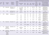
Blood specimens beyond 30 days after the onset of fever (defined as 'chronic phase') were available from 6 patients. Five patients (patient No. 1 to 5) were followed from the onset of scrub typhus diagnosed at our hospital between 2008 and 2009, and 1 patient (patient No. 6) was during follow up of other chronic illnesses after scrub typhus. Scrub typhus was diagnosed by the presence of fever and positive serology or blood nested PCR that was performed as described previously (3). Clinical and laboratory characteristics of the patients are summarized in Table 1.
In vitro culture and immunofluorescent staining
Details of cell culture and immunofluorescent (IF) staining were described previously (15). Briefly, 0.3 mL of EDTA-treated whole blood was inoculated into a monolayer of ECV304 cells (18) grown in a tissue culture flask (25 cm2). Twenty-four hours later, the inoculated blood was washed out with phosphate buffered saline, and culture was maintained in M199 medium (Gibco BRL, Gaithersburg, MD, USA) supplemented with 10% heat-inactivated fetal bovine serum (Gibco BRL) at 37℃ in a humidified atmosphere containing 6% CO2. Media were changed every 3 to 4 days, and cultures were maintained for 7 months without subculture. During the maintenance of the cell culture, cell cultures were inspected daily with optical microscopy (Olympus, Tokyo, Japan). After cytopathic changes were identified, a small amount of the ECV304 monolayer was aspirated with a pipette, and IF staining of the aspirate was performed with mouse polyclonal hyperimmune serum against the Boryong strain of O. tsutsugamushi. For negative control, blood from 5 healthy persons and 5 patients with unrelated illnesses (cerebrovascular accident [n = 2]; acute myocardial infarction [n = 2]; and diabetes mellitus [n = 1]) were processed as the same manner.
Polymerase chain reaction and sequencing of 56-kDa gene
To identify the genotype of cultured isolates and to compare nucleotide sequences of the isolates, the 56-kDa type-specific antigen (TSA56) gene of O. tsutsugamushi was amplified using PCR and then sequenced. Genomic DNA from the infected ECV304 cells was prepared with QIAamp DNA Mini kit (QIAGEN, Hilden, Germany) according to the manufacturer's instruction. PCR in detail was described previously (19). The primer set used for the amplification of the first half TSA56 gene of O. tsutsugamushi were primer 1 (forward) (5'-TTTCGAACGTGTCTTTAAGC-3'; corresponding to nucleotide position -266 to -285 from the start codon of the 56-kDa gene based on the Gilliam strain) and primer 2 (reverse) (5'-ACAGATGCACTATTAGGCAA-3'; 847-865). Primer 3 (forward) (5'-ATGCTAATAAACCTAGCGCT-3'; 731-749) and primer 4 (reverse) (5'-CTAGAAGTTATAGCGTACACCTGCACTTGC-3'; 1546-1575) were used for amplification of the second half one. Primer 9 (5'-GTTTAGAATGGTTACCAC-3'; -36 to -53) and primer 7 (5'-AGCGCTAGGTTTATTAGCAT-3'; 731-749) were used for sequencing of the amplified first half of the TSA56 gene, and primer 8 (5'-TCCACATACACACCTTCAGC-3'; 1459-1478) and primer 10 (5'-CCTAGCGTTACTCCTGTCAAAG-3'; 742-763) were used for the amplified second half of the TSA56 gene. The first half of the TSA56 gene was amplified in a 50-µL reaction mixture consisting of 5 µL of template DNA, 250 µM (each) deoxynucleotide triphosphate, 200 µM each of primers 1 and 2, 1.25 U of Taq polymerase and 5 µL of 10 × PCR buffer (Takara Shuzo Co., Kyoto, Japan) in a Perkin-Elmer model 9600 thermocycler (Perkin-Elmer Cetus Instruments, Norwalk, CT, USA). The mixture was incubated in the thermal cycler at 94℃ for 5 min, then cycled 30 times at 94℃ for 1 min, at 55℃ for 1.5 min, at 72℃ for 2 min, and finally once at 72℃ for 7 min. Amplification of the second half of the TSA56 gene was performed as described above except that primers 3 and 4 were used and the annealing period was shortened to 1 min instead of 1.5 min. The amplified products were electrophoresed in 1.5% agarose gels, purified with the QIAquick kit (QIAGEN), and sequenced by the single-pass sequencing method using the BigDye terminator cycle sequencing reaction kit and ABI PRISM 3700 & 3730xl automatic sequencer (Applied Biosystems, Foster City, CA, USA). All sequencing reactions were performed at least twice. Genotypes of the isolates were determined by identifying a representative strain revealing a maximum pairwise identity score with the TSA56 gene from the GenBank (National Institutes of Health, Bethesda, MD, USA), and sequence homology of the isolates were examined by pairwise comparisons using BLAST (National Institutes of Health).
RESULTS
All six patients revealed O. tsutsugamushi in blood culture in the afebrile state at 1 to 18 months after scrub typhus (Table 1). The genotypes of isolates were the Boryong in five patients (patients 2, 3, 4, and 6) and non-Boryong genotypes in two patients (Yonchon from patient 1 and Karp from patient 5). Nucleotide sequences of isolates serially collected from each patient were identical in all five patients in whom nucleotide sequences were compared. Nucleotide sequences of 9 isolates typed as the Boryong were 99% to 100% homologous, thus it is less helpful to differentiate each other; however, 2 isolates of the Yonchon and 2 isolates of the Karp types were distinctively different from other isolates. From 5 healthy persons and 5 patients with unrelated illnesses, no organism was isolated.
Patient 1, who presented with shock and was managed with steroid in addition to azithromycin, experienced a relapse 2 days after completion of 7-days' azithromycin therapy; treatment with doxycycline abated fever. Patients 1 and 2 complained of weakness in the absence of fever for 1 to 2.5 months after the illness and revealed positive blood nested PCRs; patient 1 showed abnormal aminotransferase level on day 31; and patient 2 revealed leukopenia on day 73, and retreatment with doxycycline resulted in improvement of weakness over 5 days. Patient 4 showed elevated erythrocyte sedimentation rate and positive PCRs 2 times over 4 months and experienced a transient ischemic attack (TIA) 8 months after scrub typhus. Patient 6, who had been managed with angina pectoris, experienced dizziness of cardiac origin and underwent percutaneous coronary angioplasty 6 months after scrub typhus.
DISCUSSION
Asymptomatic harboring of O. tsutsugamushi or its DNA is frequently observed in animals, and these data are regarded as to represent an indirect evidence of persistent O. tsutsugamushi infection. For example, Jackson et al. (8) isolated O. tsutsugamushi from 12 (13.2%) among 91 wild rodents in Korea. Using PCR, O. tsutsugamushi DNA was detected in 12 (9.5%) among 127 wild rodents (9). In studies with experimentally infected mice, persistence of O. tsutsugamushi was more clearly demonstrated (10-13). Using mouse inoculation, persistence of viable O. tsutsugamushi is documented for up to 100 (10), 270 (11), and 610 days (12), irrespective of antibiotic therapy. Nested PCR also revealed persistence of O. tsutsugamushi DNA until 64 days after infection (13).
On the contrary, there have been few studies on the persistence of O. tsutsugamushi in human. Hayashi and Watanabe (5) detected O. tsutsugamushi bacteremia in a patient who was at 34 days after defervescence. The Pescadores strain of O. tsutsugamushi was isolated from a patient at 5 months after scrub typhus (6). Smadel et al. (7) recovered O. tsutsugamushi from the lymph nodes of 1 person among 12 who had experienced scrub typhus 1 to 2 yr previously. Because these 3 reports were performed before the introduction of modern technology, identification of isolates is not sufficient to affirm the homology of isolates. In volunteers experimentally infected by laboratory-reared chiggers, rickettsemia was present until 22 days after the infection (20). Thereafter, attempts to isolate viable O. tsutsugamushi from patients have seldom been tried.
Instead, in recent years, PCR has been used to show that O. tsutsugamushi DNA disappeared gradually over 5 to 27 days after administration of antibiotics and reaffirmed short duration of O. tsutsugamushi bacteremia in human (1-4). These PCR data were contrasted to the result of the present study, and the main reason for the difference is thought to be low sensitivity of PCR. Nested PCR is very sensitive enough to detect 10 attogram (10-18g), i.e., 1 copy, of O. tsutsugamushi DNA, if it is performed by an experienced person and using Southern blot hybridization. However, if agarose gel electrophoresis is used, which was used in the above PCR studies, the sensitivity is lowered by 103. Additionally, if clinical specimens such as blood or spleen are examined, it can lower the sensitivity of PCR further (13). Among the above PCR studies, only 1 study described that the detection threshold of the study method was 100 femtogram (10-15g), i.e., 104 copies, of O. tsutsugamushi DNA (2). The quantity of O. tsutsugamushi in blood during scrub typhus, which was evaluated by real-time PCR, varies from undetectable level during days 1-4 to up to 106 copies per mL during days 9-12; and the detection limit of the real-time PCR was 1,062 target gene copies per mL of EDTA blood (21). We assume that the quantity of O. tsutsugamushi in blood during the chronic phase is below the quantity during days 1 to 4. Thus, nested PCR employed in clinical practice cannot detect O. tsutsugamushi DNA during the chronic phase in most scrub typhus patients. More importantly, the reason that the PCR studies did not detect O. tsutsugamushi DNA beyond 1 month of scrub typhus is that there has been little attempt to detect DNA beyond 1 month after the onset of scrub typhus (1-4, 21). Our study confirmed that the remaining DNA beyond the symptomatic period of scrub typhus was due to viable O. tsutsugamushi.
We used in vitro culture technique in addition to PCR. Cell culture has several disadvantages, such as requirement of long incubation, infectious hazard, and inability to detect dead organism; however, theoretically it can detect even 1 viable organism in blood. Regardless of which method is more sensitive, our study revealed a high isolation rate of O. tsutsugamushi during the chronic phase. One concern is laboratory contamination of cell culture during long incubation; however, this possibility can be excluded by the following reasons: firstly, our laboratory has about two decades' experiences in cultivating O. tsutsugamushi and maintains the contamination rate as minimal. Cell culture is not intrinsically prone to contamination as PCR, and if contamination occurs, bacterial contamination is more likely, and in turn culture is grossly spoiled. Secondly, nucleotide sequences of the isolates were identical within each patient, but different between patients infected by different genotypes. Lastly, culture from control persons did not revealed any organism.
Although the persistence of O. tsutsugamushi and its clinical significance in humans are firstly documented in the present study, persistence of Rickettsia prowazekii, a bacterium closely related to O. tsutsugamushi, in humans is well known, and Brill-Zinsser disease is a relapse of latent R. prowazekii infection. Likewise, persistent O. tsutsugamushi infection is associated with certain illnesses. It is already known that "early relapse", i.e., recurrence of fever within several days after completion of antibiotic therapy, is occasionally observed when antibiotic therapy is discontinued before the development of the protective immunity. In addition, "prolonged convalescence", i.e., persistent vague symptoms such as weakness in the absence of fever, as shown in patients 1 and 2, was clarified to be associated with the presence of O. tsutsugamushi in blood. This complication seems to occur as a result of slow development of the immunity. "Late relapse", i.e., recurrence of scrub typhus several months or years after the initial O. tsutsugamushi infection, is surely possible. Reactivation of dormant O. tsutsugamushi infection is documented in mice after inoculation of heterologous strain of O. tsutsugamushi or treatment with cyclophosphamide (14); in humans, coinfection of scrub typhus and leptospirosis (22), especially in patients not accompanied with eschar, may represent reactivation of scrub typhus triggered by leptospirosis.
Although it is not certain whether occurrence of the TIA in patient 4 and aggravation of coronary artery disease in patient 6 are related with persistent O. tsutsugamushi infection, these cases raise the necessity of long-term follow-up of scrub typhus patients, especially if they have multiple risk factors for atherosclerosis. O. tsutsugamushi is a representative bacterium infecting vascular endothelium, and infection may be one of risk factors for progression of atherosclerosis (23). Furthermore, antibiotics cannot eradicate O. tsutsugamushi, so once O. tsutsugamushi infection occurs, its effect may persists for a long time.
 XML Download