Abstract
Cardiac resynchronization therapy is known to reduce morbidity and mortality in patients with advanced heart failure as a result of dyssynchrony and systolic dysfunction of the left ventricle. Placement of the left ventricular (LV) lead via the coronary sinus can be difficult. When LV lead implantation is difficult, a video-assisted epicardial approach can be a good alternative. Although there are several reports of video-assisted epicardial LV lead implantation, mini-thoracotomy and lead implantation under direct vision have been used in most series. A 49-yr-old woman with dilated cardiomyopathy underwent the video-assisted epicardial LV lead implantation because percutaneous transvenous approach was difficult due to small cardiac veins. The patient was discharged without problems and showed improved cardiac function at the 3 follow-up months. We report the first successful total thoracoscopic LV lead implantation (without mini-thoracotomy) in Korea.
The incidence of chronic heart failure is increasing due to an aging population, and approximately 30% of patients with heart failure develop conduction disturbance such as atrioventricular (AV) block or left bundle branch block (1-4). Recently, cardiac resynchronization therapy (CRT) has become standard for the treatment of patients with heart failure and conduction disturbances. The mechanism underlying CRT is resynchronization of the ventricular activation sequence and improved coordination of atrioventricular timing and pumping efficiency. Biventricular stimulation requires lead positioning at the left ventricle (LV). The posterior wall of the LV appears to be an ideal area for LV lead positioning (5, 6) and has two technical approaches: percutaneous access via the coronary sinus and the coronary venous tree or surgical access via a left lateral mini-thoracotomy. The less invasive percutaneous approach appears to be more favorable. However, myocardial scar formation, difficult coronary venous anatomy, and phrenic nerve stimulation can limit the overall success rate of LV lead implantation, which has a failure rate of approximately 8% (7). Due to the invasiveness of surgical access, minimally invasive endoscopic approaches have also been attempted (7-10). However, mini-thoracotomy and lead implantation under direct vision have been used in most series.
We attempted LV lead implantation using video-assisted thoracic surgery (VATS) and report the first successful epicardial implantation of the left ventricular lead without mini-thoracotomy in Korea.
A 49-yr-old woman with dilated cardiomyopathy was transferred to our hospital from a local hospital because of heart failure (NYHA functional class IV) on 27 July 2011. She had a history of DDD pacemaker insertion for a complete atrioventricular block 2 yr previously.
On echocardiography, the LVEF was 17% with global hypokinesia. The LV end diastolic dimension was 78 mm. No regional wall motion abnormality was found. The severity of mitral regurgitation was mild to moderate, and the other valves were normal. The right ventricle was dyssynchronous. On an electrocardiogram the QRS duration was increased to 240 ms.
The first attempt at implantation of a CRT defibrillator (ProtectaXT; Medtronic, Minneapolis, MN, USA) was performed using the intravenous approach. The atrial lead and right ventricular lead were implanted without problems; however, LV lead insertion failed because of very small cardiac veins. The generator was positioned in the left subclavicular pocket, and the pacemaker leads except the LV lead were connected. The pocket was closed, and epicardial LV lead insertion via VATS was planned.
The operation was performed under general anesthesia using a double lumen tube for unilateral lung ventilation. The patient was placed in the right lateral decubitus position with sufficient posterior tilt. The left arm was draped and positioned over the patient's head to allow easy access to the pacemaker within the sterile field. The subclavicular pocket for the generator was reopened before one-lung ventilation.
After one-lung ventilation, a 2-cm incision for a 15-mm port was made in the sixth intercostal space at the posterior axillary line. A 30° thoracoscope was inserted for inspection of the thoracic cavity. Two additional 5-mm ports were positioned in the fourth intercostal space anterior to the anterior axillary line (second port) and mid-axillary level (third port). The thoracoscope was moved to the third port.
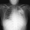
A 2-cm pericardiotomy was performed anterior to the phrenic nerve to expose the lateral wall of the left ventricle. A screw-in bipolar lead was implanted via the 15-mm port into an area of the myocardium that had been targeted on preoperative chest computed tomography. The proximal end was directed toward the cephalad. After threshold measurements, the proximal end of the lead was tunneled up to the subclavicular pocket and connected to the generator. A 20-cm loop of the lead was loosely placed in the thoracic cavity to facilitate re-expansion of the lung without exerting traction to the lead. A chest tube was inserted through the mid-axillary (third) port and guided cranially under thoracoscopic vision. After completion of the operation, the position of the LV lead was verified by chest X-ray (Fig. 1).
The threshold and impedance were 1.0/0.4 ms and 361 Ω, respectively. The QRS duration changed from 240 ms preoperatively to 156 ms postoperatively (Fig. 2). After a follow-up of 3 months, LVEF had improved to 30%, and LV end diastolic dimension had decreased to 69 mm. NYHA functional class was II.
LV lead implantation via VATS was simple, safe, and effective. Furthermore, the preselected position of the LV lead could be easily accessed. On the other hand, a percutaneous transvenous approach for CRT is dependent on several factors, such as coronary sinus anatomy, and can be time consuming (11). In the case of small coronary veins, it may even be unfeasible, whereas in the case of large coronary veins, it is often associated with changes in pacing threshold. Furthermore, life-threatening complications such as coronary sinus perforation may occur (12). Suboptimal LV lead positioning may lead to unfavorable clinical outcomes following CRT. Clinical studies have shown that a sizable number of patients, approximately 30%, fail to respond to CRT.
Surgical epicardial LV lead positioning has the advantage that direct visualization aids selection of the most suitable surface and helps to avoid areas of epicardial fat or fibrosis that can cause changes in pacing thresholds. However, thoracotomy is associated with a considerable rate of related morbidity and pain. VATS has become a routine procedure in thoracic surgery, with the advantages of clear visualization of the operation field, less invasiveness, less pain, and a better cosmetic result. VATS enables accurate positioning of the lead as a result of greater freedom of access to lateral and posterobasal areas of the left ventricle.
The VATS approach has the added advantage that it obviates the need for fluoroscopy and contrast. According to Perisinakis et al. (13), radiation risks associated with fluoroscopically guided CRT procedures can be considerable. For similar reasons and having demonstrated the efficacy and feasibility of surgical epicardial lead placement, Mair et al. (14) recommend that coronary sinus (CS) lead implantation should be stopped if the procedure exceeds 2 hr. Jutley et al. (15) suggest earlier surgical placement rather than persisting for several hours with the patient in the supine position, which may compromise the failing heart. In our case, it took only 25 min from skin incision to completion of LV lead implantation, and we found that the third port could be omitted.
In terms of lead function, the pacing threshold in our patients was lower or comparable to that of CS leads (14). We acknowledge that the pacing threshold of epicardial leads may increase over time due to myocardial fibrosis, and that this may lead to early battery replacement. However, there is little information on long-term follow-up of LV lead threshold in CRT. Further studies with a longer follow-up are essential.
On the other hand, VATS has some disadvantages compared with the transvenous technique. It requires general anesthesia, placement of the patient in the lateral decubitus position, and maintenance of single-lung ventilation for the operation. Therefore, patients who do not tolerate single lung ventilation may be unsuitable for standard VATS.
In a multipurpose interventional hybrid operating room equipped with digital imaging diagnostics, CRT could easily be performed by cardiologists and surgeons working together. The surgeon would oversee the sterile technique, including the draping and preparation of the patient, and create the pocket, and the cardiologist would percutaneously place the leads. If the LV lead could not be positioned, the surgeon would perform epicardial LV lead implantation via VATS. Or, all procedures could be performed by the cardiac surgeon at the same time.
In conclusion, an epicardial approach via VATS using two ports seems to be an excellent alternative procedure for LV lead implantation. This approach has the advantages of ready availability, good pacing results, a short intervention time, and tolerable stress for the patient.
Figures and Tables
References
1. Moss AJ, Zareba W, Hall WJ, Klein H, Wilber DJ, Cannom DS, Daubert JP, Higgins SL, Brown MW, Andrews ML. Prophylactic implantation of a defibrillator in patients with myocardial infarction and reduced ejection fraction. N Engl J Med. 2002. 346:877–883.
2. Grines CL, Bashore TM, Boudoulas H, Olson S, Shafer P, Wooley CF. Functional abnormalities in isolated left bundle branch block. The effect of interventricular asynchrony. Circulation. 1989. 79:845–853.
3. Xiao HB, Lee CH, Gibson DG. Effect of left bundle branch block on diastolic function in dilated cardiomyopathy. Br Heart J. 1991. 66:443–447.
4. Erlebacher JA, Barbarash S. Intraventricular conduction delay and functional mitral regurgitation. Am J Cardiol. 2001. 88:A783–86.
5. Cazeau S, Leclercq C, Lavergne T, Walker S, Varma C, Linde C, Garrigue S, Kappenberger L, Haywood GA, Santini M, et al. Effects of multisite biventricular pacing in patients with heart failure and intraventricular conduction delay. N Engl J Med. 2001. 344:873–880.
6. Auricchio A, Stellbrink C, Sack S, Block M, Vogt J, Bakker P, Huth C, Schondube F, Wolfhard U, Bocker D, et al. Long-term clinical effect of hemodynamically optimized cardiac resynchronization therapy in patients with heart failure and ventricular conduction delay. J Am Coll Cardiol. 2002. 39:2026–2033.
7. Kleine P, Gronefeld G, Dogan S, Hohnloser SH, Moritz A, Wimmer-Greinecker G. Robotically enhanced placement of left ventricular epicardial electrodes during implantation of a biventricular implantable cardioverter defibrillator system. Pacing Clin Electrophysiol. 2002. 25:989–991.
8. Fernandez AL, Garcia-Bengochea JB, Ledo R, Vega M, Amaro A, Alvarez J, Rubio J, Sierra J, Sanchez D. Minimally invasive surgical implantation of left ventricular epicardial leads for ventricular resynchronization using video-assisted thoracoscopy. Rev Esp Cardiol. 2004. 57:313–319.
9. Jansens JL, Jottrand M, Preumont N, Stoupel E, de Canniere D. Robotic-enhanced biventricular resynchronization: an alternative to endovenous cardiac resynchronization therapy in chronic heart failure. Ann Thorac Surg. 2003. 76:413–417.
10. Morgan JA, Ginsburg ME, Sonett JR, Morales DL, Kohmoto T, Gorenstein LA, Smith CR, Argenziano M. Advanced thoracoscopic procedures are facilitated by computer-aided robotic technology. Eur J Cardiothorac Surg. 2003. 23:883–887.
11. Alonso C, Leclercq C, d'Allonnes FR, Pavin D, Victor F, Mabo P, Daubert JC. Six year experience of transvenous left ventricular lead implantation for permanent biventricular pacing in patients with advanced heart failure: technical aspects. Heart. 2001. 86:405–410.
12. Cleland JG, Daubert JC, Erdmann E, Freemantle N, Gras D, Kappenberger L, Tavazzi L. The effect of cardiac resynchronization on morbidity and mortality in heart failure. N Engl J Med. 2005. 352:1539–1549.
13. Perisinakis K, Theocharopoulos N, Damilakis J, Manios E, Vardas P, Gourtsoyiannis N. Fluoroscopically guided implantation of modern cardiac resynchronization devices: radiation burden to the patient and associated risks. J Am Coll Cardiol. 2005. 46:2335–2339.
14. Mair H, Sachweh J, Meuris B, Nollert G, Schmoeckel M, Schuetz A, Reichart B, Daebritz S. Surgical epicardial left ventricular lead versus coronary sinus lead placement in biventricular pacing. Eur J Cardiothorac Surg. 2005. 27:235–242.
15. Jutley RS, Waller DA, Loke I, Skehan D, Ng A, Stafford P, Chin D, Spyt TJ. Video-assisted thoracoscopic implantation of the left ventricular pacing lead for cardiac resynchronization therapy. Pacing Clin Electrophysiol. 2008. 31:812–818.


 PDF
PDF ePub
ePub Citation
 Print
Print


 XML Download